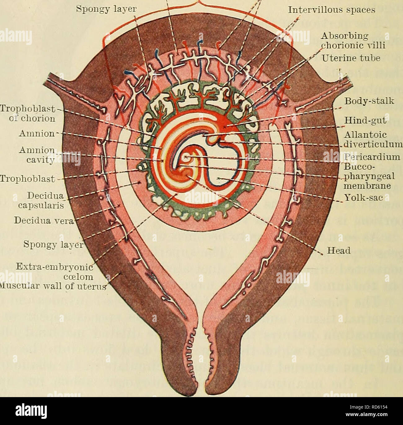
. Cunningham's Text-book of anatomy. Anatomy. ondary villus â Amnion cavity Amnion -Body-stalk Allantoic diverti- culum rimitive streak eurenteric canal Cavity of entoderm sac Extra-embryonic celom Decidua capsularis Decidua vera Embryonic are Fig. 75.âSchema of a Section of a Pregnant Uterus after the formation of the Intervillous Spaces. parts. (1) The parts which lie between adjacent blood spaces, the primary chorionic villi. (2) The parts which lie in con- tact with the mesoderm of the chorion, and which form with the mesoderm the chorion plate. (3) The parts which cover the maternal tissu
Image details
Contributor:
The Book Worm / Alamy Stock PhotoImage ID:
RD6154File size:
7.1 MB (419.8 KB Compressed download)Releases:
Model - no | Property - noDo I need a release?Dimensions:
1592 x 1569 px | 27 x 26.6 cm | 10.6 x 10.5 inches | 150dpiMore information:
This image is a public domain image, which means either that copyright has expired in the image or the copyright holder has waived their copyright. Alamy charges you a fee for access to the high resolution copy of the image.
This image could have imperfections as it’s either historical or reportage.
. Cunningham's Text-book of anatomy. Anatomy. ondary villus â Amnion cavity Amnion -Body-stalk Allantoic diverti- culum rimitive streak eurenteric canal Cavity of entoderm sac Extra-embryonic celom Decidua capsularis Decidua vera Embryonic are Fig. 75.âSchema of a Section of a Pregnant Uterus after the formation of the Intervillous Spaces. parts. (1) The parts which lie between adjacent blood spaces, the primary chorionic villi. (2) The parts which lie in con- tact with the mesoderm of the chorion, and which form with the mesoderm the chorion plate. (3) The parts which cover the maternal tissues and form the outer boun- daries of the blood spaces, the basal layer. The blood spaces themselves are called the in- tervillous spaces (Figs. 76, 79). After a time each primary villus differenti- ates into a cellular core and plasmodial periphery, and thereafter the villi are invaded by the mesoderm of the chorion and are thus converted into secondary villi (Fig. 76). The first-formed villi are non-vascular, but by the time the secondary villi have developed the um- bilical arteries have grown through the body-stalk (allantoic stalk) into the meso- derm of the chorion, and branches from them enter the meso- dermal cores of the villi, which thus be- come vascular. When the second- ary villi are fully developed each con- sists of a vascular mesodermal core con- tinuous with the mesoderm of the chorion Thp mpsn / 6.âSchema of a Frontal Section of a Pregnant Uterus at the vj uxi. j.ire luesu- period of the Formation of the Embryo. Xote extension of amnion dermal core IS Covered as contrasted with stage shown in Fi°- 75 Unchanged layer Maternal blood-vessels "i Placental area Spongy layer âV"^, , , "" "--^/â / Intervillous spaces Absorbing horionic villi Uterine tube Body-stalk Trophoblast-. Decidua capsularis Decidua v Spongy lay. Extra-embryon ecelom Muscular wall of uterus-'. Please note that these images are extracted from scanned page images tha