The Plant Vascular System: Evolution, Development and FunctionsF
The Plant Vascular System: Evolution, Development and FunctionsF
The Plant Vascular System: Evolution, Development and FunctionsF

You also want an ePaper? Increase the reach of your titles
YUMPU automatically turns print PDFs into web optimized ePapers that Google loves.
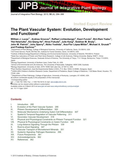
Journal of Integrative <strong>Plant</strong> Biology 2013, 55 (4): 294–388<br />
Invited Expert Review<br />
<strong>The</strong> <strong>Plant</strong> <strong>Vascular</strong> <strong>System</strong>: <strong>Evolution</strong>, <strong>Development</strong><br />
<strong>and</strong> Functions F<br />
William J. Lucas1∗, Andrew Groover2 , Raffael Lichtenberger3 , Kaori Furuta3 , Shri-Ram Yadav3 ,<br />
Ykä Helariutta3 , Xin-Qiang He4 , Hiroo Fukuda5 , Julie Kang6 , Siobhan M. Brady1 ,<br />
John W. Patrick7 , John Sperry8 , Akiko Yoshida1 , Ana-Flor López-Millán9 , Michael A. Grusak10 <strong>and</strong> Pradeep Kachroo11 1Department of <strong>Plant</strong> Biology, College of Biological Sciences, University of California, Davis, CA 95616, USA<br />
2US Forest Service, Pacific SW Res Stn, Institute for Forest Genetics, Davis, CA 95618, USA<br />
3Institute of Biotechnology/Department of Biology <strong>and</strong> Environmental Sciences, University of Helsinki, FIN-00014, Finl<strong>and</strong><br />
4State Key Laboratory of Protein <strong>and</strong> <strong>Plant</strong> Gene Research, College of Life Sciences, Peking University, Beijing 100871, China<br />
5Department of Biological Sciences, Graduate School of Science, <strong>The</strong> University of Tokyo, 7-3-1 Hongo, Bunkyo-ku, Tokyo 113-0033,<br />
Japan<br />
6Biology Department, University of Northern Iowa, Cedar Falls, IA, USA<br />
7School of Environmental <strong>and</strong> Life Sciences, <strong>The</strong> University of Newcastle, Callaghan, NSW 2308, Australia<br />
8Biology Department, University of Utah, 257S 1400E Salt Lake City, UT 84112, USA<br />
9<strong>Plant</strong> Nutrition Department, Aula Dei Experimental Station (CSIC), P.O. Box 13034, E-50080, Zaragoza, Spain<br />
10USDA-ARS Children’s Nutrition Research Center, Department of Pediatrics, Baylor College of Medicine, 1100 Bates Street, Houston, TX<br />
77030, USA<br />
11Department of <strong>Plant</strong> Pathology, College of Agriculture, University of Kentucky, Lexington, KY 40546, USA<br />
All authors contributed equally to the preparation of this manuscript.<br />
∗<br />
Corresponding author<br />
Tel: +1 530 752 1093; Fax: +1 530 752 5410; E-mail: wjlucas@ucdavis.edu<br />
F Articles can be viewed online without a subscription.<br />
Available online on 5 March 2013 at www.jipb.net <strong>and</strong> www.wileyonlinelibrary.com/journal/jipb<br />
doi: 10.1111/jipb.12041<br />
Contents<br />
I. Introduction 295<br />
II. <strong>Evolution</strong> of the <strong>Plant</strong> <strong>Vascular</strong> <strong>System</strong> 295<br />
III. Phloem <strong>Development</strong> & Differentiation 300<br />
IV. Molecular Mechanisms Underlying Xylem Cell Differentiation 307<br />
V. Spatial & Temporal Regulation of <strong>Vascular</strong> Patterning 311<br />
VI. Secondary <strong>Vascular</strong> <strong>Development</strong> 318<br />
VII. Physical <strong>and</strong> Physiological Constraints on Phloem Transport Function 321<br />
VIII. Physical & Physiological Constraints on Xylem Function 328<br />
IX. Long-distance Signaling Through the Phloem 339<br />
X. Root-to-shoot Signaling 347<br />
XI. <strong>Vascular</strong> Transport of Microelement Minerals 351<br />
XII. <strong>System</strong>ic Signaling: Pathogen Resistance 356<br />
XIII. Future Perspectives 361<br />
XIV. Acknowledgements 362<br />
XV. References 362<br />
C○ 2013 Institute of Botany, Chinese Academy of Sciences
William J. Lucas<br />
(Corresponding author)<br />
Abstract<br />
Insights into <strong>Plant</strong> <strong>Vascular</strong> Biology 295<br />
<strong>The</strong> emergence of the tracheophyte-based vascular system of l<strong>and</strong> plants<br />
had major impacts on the evolution of terrestrial biology, in general,<br />
through its role in facilitating the development of plants with increased<br />
stature, photosynthetic output, <strong>and</strong> ability to colonize a greatly exp<strong>and</strong>ed<br />
range of environmental habitats. Recently, considerable progress has<br />
been made in terms of our underst<strong>and</strong>ing of the developmental <strong>and</strong><br />
physiological programs involved in the formation <strong>and</strong> function of the<br />
plant vascular system. In this review, we first examine the evolutionary<br />
events that gave rise to the tracheophytes, followed by analysis of the<br />
genetic <strong>and</strong> hormonal networks that cooperate to orchestrate vascular<br />
development in the gymnosperms <strong>and</strong> angiosperms. <strong>The</strong> two essential<br />
functions performed by the vascular system, namely the delivery of resources (water, essential mineral<br />
nutrients, sugars <strong>and</strong> amino acids) to the various plant organs <strong>and</strong> provision of mechanical support are<br />
next discussed. Here, we focus on critical questions relating to structural <strong>and</strong> physiological properties<br />
controlling the delivery of material through the xylem <strong>and</strong> phloem. Recent discoveries into the role of<br />
the vascular system as an effective long-distance communication system are next assessed in terms<br />
of the coordination of developmental, physiological <strong>and</strong> defense-related processes, at the whole-plant<br />
level. A concerted effort has been made to integrate all these new findings into a comprehensive picture<br />
of the state-of-the-art in the area of plant vascular biology. Finally, areas important for future research<br />
are highlighted in terms of their likely contribution both to basic knowledge <strong>and</strong> applications to primary<br />
industry.<br />
Keywords: <strong>Evolution</strong>; vascular development; phloem; xylem; nutrient delivery; long-distance communication; systemic signaling.<br />
Lucas WJ, Groover A, Lichtenberger R, Furuta K, Yadav SR, Helariutta Y, He XQ, Fukuda H, Kang J, Brady SM, Patrick JW, Sperry J,<br />
Yoshida A, López-Millán AF, Grusak MA, Kachroo P (2013) <strong>The</strong> plant vascular system: <strong>Evolution</strong>, development <strong>and</strong> functions. J. Integr. <strong>Plant</strong><br />
Biol. 55(4), 294–388.<br />
Introduction<br />
<strong>The</strong> plant vascular system carries out two essential functions,<br />
namely the delivery of resources (water, essential mineral<br />
nutrients, sugars <strong>and</strong> amino acids) to the various plant organs,<br />
<strong>and</strong> provision of mechanical support. In addition, the vascular<br />
system serves as an effective long-distance communication<br />
system, with the phloem <strong>and</strong> xylem serving to input information<br />
relating to abiotic <strong>and</strong> biotic conditions above <strong>and</strong> below<br />
ground, respectively. This combination of resource supply <strong>and</strong><br />
delivery of information, including hormones, peptide hormones,<br />
proteins <strong>and</strong> RNA, allows the vascular system to engage in the<br />
coordination of developmental <strong>and</strong> physiological processes at<br />
the whole-plant level.<br />
Over the past decade, considerable progress has been<br />
made in terms of our underst<strong>and</strong>ing of the developmental <strong>and</strong><br />
physiological programs involved in the formation <strong>and</strong> function of<br />
the plant vascular system. In this review, we have made every<br />
effort to integrate these new findings into a comprehensive<br />
picture of the state-of-the-art in this important facet of plant<br />
biology. We also highlight potential areas important for future<br />
research in terms of their likely contribution both to basic<br />
knowledge <strong>and</strong> applications to primary industry.<br />
<strong>Evolution</strong> of the <strong>Plant</strong> <strong>Vascular</strong> <strong>System</strong><br />
Why the need for a vasculature system?<br />
For plants as photosynthetic autotrophs, the evolutionary step<br />
from uni- to multi-cellularity conferred an important selective<br />
advantage in terms of division of labor; i.e., functional specialization<br />
of tissues/organs to more effectively extract, <strong>and</strong><br />
compete for, essential resources in aquatic <strong>and</strong> terrestrial<br />
environments. Successful colonization of terrestrial environments,<br />
by plants, depended upon positioning of organs in both<br />
aerial <strong>and</strong> soil environments to meet their autotrophic requirements.<br />
For example, for photosynthetic efficiency, sufficient<br />
levels of light only co-occur with a supply of CO2 in aerial
296 Journal of Integrative <strong>Plant</strong> Biology Vol. 55 No. 4 2013<br />
environments, whereas water <strong>and</strong> mineral requirements are<br />
primarily acquired from soil environments. Thus, aerial <strong>and</strong> soil<br />
organs of early l<strong>and</strong> plants were nutritionally interdependent<br />
<strong>and</strong>, consequently, there was intense selection pressure for the<br />
evolution of an inter-organ transport system to allow access to<br />
the complete spectrum of essential resources for cell growth<br />
<strong>and</strong> maintenance.<br />
An important feature of early multicellular plants was the<br />
acquisition of plasmodesmata (PD), whose cytoplasmic channels<br />
established a symplasmic continuum throughout the body<br />
of the plant (Lucas et al. 1993). This symplasm allowed for<br />
the exchange of nutrients between the different plant organs.<br />
However, this symplasmic route, in which intracellular cytoplasmic<br />
streaming is arranged in series with intercellular diffusion<br />
through PD, is effective only over rather short distances. For<br />
example, a PD-mediated sucrose flux of 2 × 10 −4 mol m −2 s −1<br />
into heterotrophic cells would satisfy their metabolic dem<strong>and</strong>.<br />
Maximum reported permeability coefficients for sucrose diffusion<br />
through PD are on the order of 6 × 10 −6 ms −1 (Fisher<br />
<strong>and</strong> Wang 1995). Using this value, diffusion theory predicts that<br />
a significant sucrose concentration drop would be required,<br />
across each adjoining cell wall interface, to sustain this flux<br />
of sucrose from the autotrophic (photosynthetic) cells into the<br />
heterotrophic (water <strong>and</strong> mineral nutrient acquiring) cells. Thus,<br />
the path length would be limited to only a few cells, arranged in<br />
series, <strong>and</strong> the size of the organism would be limited to a few<br />
millimeters.<br />
In order for multicellular autotrophs to overcome these<br />
diffusion-imposed size constraints, a strong selection pressure<br />
existed to evolve an axially-arranged tissue system, located<br />
throughout the plant body, with a greatly increased conductivity<br />
for intercellular transport. <strong>The</strong> solution to this problem began<br />
over 470 Mya <strong>and</strong>, in combination with prevailing global climate<br />
change, including dramatic changes in atmospheric CO2 levels,<br />
gave rise to the development of the cuticle <strong>and</strong> stomata,<br />
important adaptations that both reduced tissue dehydration<br />
<strong>and</strong> increased the capacity for exchange of CO2, thereby<br />
enhancing the rates of photosynthesis (Franks <strong>and</strong> Brodribb<br />
2005; Ruszala et al. 2011; but see Duckett et al. 2009).<br />
Following acquisition of these two traits, early l<strong>and</strong> plants<br />
evolved cells specialized for long-distance transport of food<br />
<strong>and</strong> water (Ligrone et al. 2000, 2012; Raven 2003; van Bel<br />
2003; Pittermann 2010). Irrespective of plant group, these cells<br />
became arranged end-to-end in longitudinal files having a simplified<br />
cytoplasm <strong>and</strong> modified end walls designed to increase<br />
their intra- <strong>and</strong> intercellular conductivities, respectively.<br />
In l<strong>and</strong> plants, the degree of cellular modifications of transport<br />
cells increases from the bryophytes (pretracheophytes—also<br />
termed non-vascular plants—the liverworts, mosses <strong>and</strong> hornworts),<br />
to the early tracheophytes, the vascular cryptogams (lycophytes<br />
<strong>and</strong> pterophytes), on through to seed plants (Ligrone<br />
et al. 2000, 2012; Raven 2003; van Bel 2003). <strong>The</strong>se cell<br />
specializations neatly scale with maximal sizes attained by<br />
each group of l<strong>and</strong> plants. Interestingly, impacts of enhancing<br />
conductivities of cells transporting sugars converges with a<br />
greater influence imposed by evolving water conducting cells<br />
to sustain hydration of aerial photosynthetic tissues.<br />
<strong>Evolution</strong>ary origins <strong>and</strong> diversification of food<br />
<strong>and</strong> water transport systems<br />
Studies based on fossil records <strong>and</strong> extant (living) bryophytes<br />
have established that developmental programs evolved to form<br />
specialized water <strong>and</strong> nutrient conducting tissues. Based on<br />
the fossil record, early pretracheophyte l<strong>and</strong> plants appeared<br />
to have developed simple water-conducting conduits having<br />
smooth walls with small pores, likely derived from the presence<br />
of PD. Similar structures are present, for example, in some of<br />
the mosses, the most ancient being termed water-conducting<br />
cells (WCCs) <strong>and</strong> the more advanced being the hydroids of the<br />
peristomate mosses (Mishler <strong>and</strong> Churchill 1984; Kenrick <strong>and</strong><br />
Crane 1997; Ligrone et al. 2012).<br />
Hydroids often form a central str<strong>and</strong> in the gametophyte<br />
stem/sporophyte seta in the mosses (Figure 1A, B). During<br />
their development, these hydroid cells undergo various structural<br />
modifications to the cell wall <strong>and</strong> are dead at maturity<br />
(Figure 1C, D). Although in some cases the hydroid wall may<br />
become thickened, these are considered to be primary in<br />
nature <strong>and</strong> lack lignin. However, recent studies have indicated<br />
that bryophyte cell walls contain lignin-related compounds,<br />
but these do not impart mechanical strengthening properties<br />
(Ligrone et al. 2012). Although this absence of mechanical<br />
strength served as an impediment to an increase in body size,<br />
it allowed for hydroid collapse during tissue desiccation, <strong>and</strong><br />
rapid rehydration following a resupply of water (Figure 1E, F), a<br />
feature that likely minimized cavitation of these WCCs (Ligrone<br />
et al. 2012) (see also later section). This trait may also have<br />
allowed peristomate mosses to exp<strong>and</strong> into dryer habitats.<br />
<strong>The</strong> evolution of hydroids could have involved modification of<br />
existing WCCs. However, based on the distribution of WCCs<br />
in the early l<strong>and</strong> plants (Figure 2), it seems equally probable<br />
that they arose through an independent developmental<br />
pathway after the loss of perforate WCCs (Ligrone et al.<br />
2012).<br />
<strong>The</strong> fossil record contains less information on the evolution<br />
of specialized food-conducting cells (FCCs), due in large<br />
part to their less robust characteristics that limited effective<br />
preservation. However, insights can be gained from studies<br />
on extant bryophyte species. As with WCCs, early FCCs were<br />
represented by files of aligned elongated cells in which the<br />
cytoplasmic contents underwent a series of positional <strong>and</strong><br />
structural modifications (Figure 3). Here, we will use moss as an<br />
example; in some species (members of the order Polytrichales),
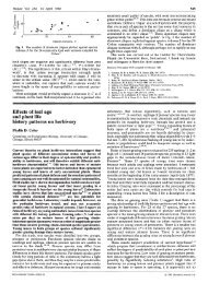
Figure 1. Water-conducting cells of early non-vascular (pretracheophyte)<br />
l<strong>and</strong> plants.<br />
(A) Light micrograph of a leafy stem from the moss Plagiomnium<br />
undulatum illustrating the prominent central str<strong>and</strong> of hydroid cells<br />
(h) surrounded by parenchyma cells (p).<br />
(B–D) Transmission electron micrographs illustrating hydroids having<br />
unevenly thickened walls in the leaf shoot of the moss Polytrichum<br />
juniperinum (B), a differentiating hydroid (C) <strong>and</strong> mature<br />
hydroids (D) in the leafy shoot of Polytrichum formosum.<br />
(E, F) Transverse sections of moss hydroids in the hydrated (E)<br />
<strong>and</strong> dehydrated condition (F); note that in the presence of water,<br />
dehydrated hydroids become rehydrated <strong>and</strong> functional. Images<br />
(A–D) reproduced from Ligrone et al. (2000), with permission of<br />
<strong>The</strong> Royal Society London; (E, F) reproduced from Ligrone et al.<br />
(2012), with permission of Oxford University Press. Scale bars:<br />
50 µm in(A), 5µm in(B–D) <strong>and</strong> 5 µm in(E), common to (F).<br />
the FCCs gave rise to a group of more specialized cells, termed<br />
leptoids <strong>and</strong> associated specialized parenchyma cells. During<br />
development, leptoids undergo a series of cytological changes,<br />
including cytoplasmic polarization <strong>and</strong> microtubule-associated<br />
Insights into <strong>Plant</strong> <strong>Vascular</strong> Biology 297<br />
alignment of plastids, mitochondria <strong>and</strong> the endoplasmic reticulum<br />
(ER), in a longitudinal pattern. At maturity, the FCCs of<br />
the bryophytes generally lack a large central vacuole <strong>and</strong>, in<br />
some species, there is partial degradation of the nucleus. In<br />
addition, the end walls of cells within these files of aligned FCCs<br />
develop a high density of PD (Figure 4), presumably to optimize<br />
symplasmic continuity for cell-to-cell diffusion of photosynthate<br />
(Ligrone et al. 2000, 2012; Raven 2003). Finally, FCCs/leptoids<br />
often develop in close proximity to WCCs/hydroids; in<br />
some species, the WCCs/hydroids are centrally located<br />
in the tissue/stem being ensheathed by FCCs/leptoids<br />
(Figure 3E).<br />
As with hydroids, the evolutionary events leading to the<br />
development of FCCs, <strong>and</strong> leptoids in particular, appear to<br />
have been driven by the necessity to withst<strong>and</strong> periods in<br />
which the early l<strong>and</strong> plants underwent desiccation. Insight sinto<br />
the presence <strong>and</strong> importance of such traits were offered by<br />
recent physiological <strong>and</strong> anatomical studies performed on the<br />
desiccation-resistant moss Polytrichum formosum. Here, the<br />
unique resilience of the lepoids to an imposed dehydration was<br />
shown to be associated with the unique role of microtubules<br />
in control over the special cytological features of these FCCs<br />
(Pressel et al. 2006). <strong>The</strong> properties of cavitation-resistant<br />
hydroids <strong>and</strong> desiccation-tolerant leptoids would likely have<br />
had an important impact on these mosses in terms of the ability<br />
to penetrate into diverse ecological niches.<br />
Emergence of xylem with lignified tracheids<br />
<strong>and</strong> vessels<br />
As indicated in Figure 2, xylem tissues may well have evolved<br />
independently from WCCs/hydroids. Although hydroids have a<br />
number of similar features to the early tracheary elements,<br />
including functioning after death, there are many important<br />
differences. Perhaps the most critical was the acquisition<br />
of a developmental program for the deposition of patterned<br />
secondary cell wall material. Of equal importance was the<br />
development of lignin <strong>and</strong> its deposition within the secondary<br />
wall of tracheary cells. Collectively, these evolutionary events<br />
imparted biomechanical support <strong>and</strong> compressive strength,<br />
with an ability to withst<strong>and</strong> tracheid collapse when the water column<br />
was placed under tension (see later section). Acquisition of<br />
biomechanical strength afforded the opportunity for an increase<br />
in plant height, with the benefit of enhanced competition for<br />
sunlight.<br />
A further defining feature of the early vascular plants was that<br />
their tracheary (water conducting) elements had pits of varying<br />
architecture that spanned the secondary wall. In contrast to<br />
the WCCs of the bryophytes, the formation of these pits is not<br />
dependent upon the dissolution of PD (Barnett 1982; Lachaud<br />
<strong>and</strong> Maurousset 1996), <strong>and</strong> in the early tracheary element, the<br />
tracheid (Edwards et al. 1992), the primary cell wall remains
298 Journal of Integrative <strong>Plant</strong> Biology Vol. 55 No. 4 2013<br />
Figure 2. Cladogram illustrating the distribution of water-conducting cells (WCCs) in early l<strong>and</strong> plants.<br />
Note that hydroids in the peristomate mosses lack perforate cell walls. Reproduced from Ligrone et al. (2012), with permission of Oxford<br />
University Press.<br />
imperforate. However, in the advanced form, the vessel element<br />
or vessel member, the primary wall is removed in discrete<br />
regions between adjacent members, thereby giving rise to a<br />
perforation plate. This evolutionary adaptation allows water to<br />
flow through many mature vessel members that collectively<br />
form a vessel, unimpeded by the primary cell wall; i.e., the<br />
perforation plate reduces the overall resistance to water flow<br />
through vessels.<br />
<strong>Evolution</strong>ary relationship between FCCs <strong>and</strong> early<br />
tracheophyte sieve elements<br />
<strong>The</strong> cytological features of FCCs are widespread in the<br />
bryophytes <strong>and</strong> many are also present in the phloem sieve<br />
elements of the lycophytes, pterophytes <strong>and</strong> gymnosperms<br />
(Esau et al. 1953) (Table 1). It is also noteworthy that the ER is<br />
present in PD located in the adjoining transverse walls between<br />
FCCs, leptoids <strong>and</strong> the sieve elements of ferns (Evert et al.<br />
1989) <strong>and</strong> conifers (Schulz 1992). Furthermore, both leptoids<br />
<strong>and</strong> early sieve elements, termed sieve cells, have supporting<br />
parenchyma cells. <strong>The</strong>se features, held in common between<br />
the more advanced FCCs <strong>and</strong> the phloem sieve elements of<br />
the early tracheophytes, raise the possibility of a developmental<br />
program having components shared between these nutrient<br />
delivery systems of the plant kingdom.<br />
<strong>Evolution</strong> of molecular mechanisms regulating<br />
vascular development<br />
Significant progress has been made in elucidating the molecular<br />
mechanisms regulating vascular development. In most<br />
cases, a modest number of angiosperm model species have<br />
been the focus of molecular-genetic <strong>and</strong> genomic analysis<br />
of vascular development. At present, individual genes<br />
regulating specific aspects of vascular development have<br />
been characterized in detail. In addition, models of how<br />
vascular tissues are initiated, patterned, balance proliferation<br />
<strong>and</strong> differentiation, <strong>and</strong> acquire polarity have been<br />
developed.<br />
<strong>Vascular</strong> development is currently being modeled at new<br />
levels of complexity in Arabidopsis <strong>and</strong> Populus, using computational<br />
<strong>and</strong> network biology approaches that make use<br />
of extensive genomic gene expression <strong>and</strong> gene regulation<br />
datasets. While incomplete, new models representing important<br />
phylogentic positions in l<strong>and</strong> plant evolution are also being<br />
developed, <strong>and</strong> will provide important insights into the origins<br />
<strong>and</strong> diversification of mechanisms regulating vascular development.<br />
Importantly, many of the key gene families that regulate<br />
vascular development predate tracheophytes. Thus, one major<br />
challenge for underst<strong>and</strong>ing the evolution of vascular development<br />
will be to determine the evolutionary processes by which
Figure 3. Cytological details of moss food-conducting cells.<br />
(A) Cytoplasmic polarity in a leafy stem of Plagiomnium undulatum;<br />
most of the organelles are in the top end of the lower cell.<br />
(B) Sphorophyte seta of Mnium hornum showing the longitudinal<br />
alignment of elongated plastids (p) <strong>and</strong> the highly elongated nucleus<br />
(n).<br />
(C, D) Longitudinal arrays of microtubules associated with tubules<br />
<strong>and</strong> vesicles in a leafy stem of Plagiomnium undulatum (C) <strong>and</strong><br />
Polytrichum juniperinum (D).<br />
(E) Transverse section of leptoids <strong>and</strong> adjacent hydroids (h) in a<br />
stem of Polytrichum commune.<br />
Scale bars: 4 µm in(A), 2µm in(B, E), 0.5µm in(C, D).<br />
Reproduced from Ligrone et al. (2000), with permission of <strong>The</strong> Royal<br />
Society London.<br />
regulatory genes <strong>and</strong> modules were duplicated, modified, or<br />
directly co-opted to function in vascular development (Pires <strong>and</strong><br />
Dolan 2012). Even more challenging will be determining the<br />
evolutionary steps underlying the many biochemical processes<br />
required for the production of vascular tissues <strong>and</strong> lignified<br />
secondary cell walls.<br />
Insights into <strong>Plant</strong> <strong>Vascular</strong> Biology 299<br />
Figure 4. Abundant plasmodesmata in the trumpet-shaped end<br />
walls between food-conducting cells in the moss Sphagnum<br />
cuspiatum.<br />
Scale bar: 10 µm. Reproduced from Ligrone et al. (2000), with<br />
permission of <strong>The</strong> Royal Society London.<br />
Table 1. Comparison of cytological features present in moss<br />
food-conducting cells <strong>and</strong> sieve cells in ferns <strong>and</strong> conifers<br />
Similaritiesa Differences (in sieve cells)<br />
Absence of vacuoles No cytoplasmic polarization<br />
Nacreous wallsb Apparent lack of polyribosomes<br />
Nuclear degenerationb Presence of endoplasmic<br />
reticulum (ER) within<br />
plasmodesmata (PD)<br />
Callose associated with PDc a Modified after Ligrone et al. (2000).<br />
b Restricted to the Polytrichales in mosses.<br />
c Restricted to the Polytrichales in mosses, absent in some lower<br />
tracheopytes.<br />
Auxin is an evolutionarily ancient regulator of vascular<br />
development<br />
In the following sections we present some examples of the<br />
genes <strong>and</strong> mechanisms regulating specific aspects of vascular<br />
development. This is not a complete review of the literature,<br />
but rather we aim to highlight some of the molecular-genetic<br />
models of vascular development. We begin with the enigmatic<br />
plant hormone auxin, which has been known to play<br />
fundamental roles in vascular development for decades, but<br />
only recently have insights been gleaned at the moleculargenetic<br />
level as to how it exerts its many influences on vascular<br />
development. To underst<strong>and</strong> the myriad of ways that auxin<br />
influences plant development, it is necessary to underst<strong>and</strong><br />
its synthesis, conjugation, transport, perception, <strong>and</strong> effects<br />
on gene expression. Fundamental insights into all of these<br />
processes have been gained, <strong>and</strong> have been summarized
300 Journal of Integrative <strong>Plant</strong> Biology Vol. 55 No. 4 2013<br />
in recent reviews. Importantly, auxin can apparently be synthesized<br />
by all plants (Johri 2008; Lau et al. 2009) in which<br />
it plays various roles in promoting growth, <strong>and</strong> has thus<br />
been recruited to participate in vascular development in the<br />
tracheopytes. Here, we consider one specific role for auxin<br />
during vascular development: how auxin transport proteins act<br />
during the establishment <strong>and</strong> propagation of interconnected<br />
vascular str<strong>and</strong>s.<br />
<strong>Vascular</strong> str<strong>and</strong>s consist of interconnected files of cells. It<br />
is critical that vascular str<strong>and</strong>s be properly spaced <strong>and</strong> patterned<br />
within tissues, <strong>and</strong> that functional cell types (e.g. water<br />
conducting tracheary elements) coordinate differentiation to<br />
produce interconnected conduits for transport. Auxin has long<br />
been known to play fundamental roles in both the induction of<br />
vascular str<strong>and</strong>s <strong>and</strong> in the differentiation of vascular cell types<br />
(Sachs 1991). How auxin is transported through tissues has<br />
been recognized as a primary factor in determining the location<br />
<strong>and</strong> propagation (or canalization) of vascular str<strong>and</strong>s. Major<br />
insights into how auxin transport is regulated have been gained<br />
through the identification <strong>and</strong> characterization of PIN-FORMED<br />
(PIN) proteins, which include plasma membrane spanning<br />
auxin efflux carriers. PIN proteins act through asymmetric<br />
subcellular localization to direct routes of auxin flow through<br />
cells <strong>and</strong> tissues (Petráˇsek et al. 2006, Petráˇsek <strong>and</strong> Friml<br />
2009).<br />
Importantly, PIN proteins are found in all l<strong>and</strong> plants (Krecek<br />
et al. 2009) including the pretracheophytes, <strong>and</strong> polar auxin<br />
transport (PAT) already existed in the Charophyta (Boot et al.<br />
2012), suggesting that ancestral functions of PIN proteins were<br />
not in vascular development, per se. <strong>The</strong> fossil record also<br />
provides insights into the role of auxin transport in the evolution<br />
of vascular tissues. Routes of auxin transport can be indirectly<br />
inferred from anatomical changes in extant angiosperms <strong>and</strong><br />
gymnosperms, in which circular patterns of tracheary element<br />
develop in secondary xylem above branches, which impede<br />
auxin transport. Amazingly, fossils of wood from 375-millionyear-old<br />
Archaeopteris (a progymnosperm) also show this<br />
pattern (Rothwell <strong>and</strong> Lev-Yadun 2005).<br />
A general expectation is that the auxin-related mechanisms<br />
regulating vascular differentiation are shared (i.e. are homologous)<br />
among vascular plants, but this remains to be verified<br />
through functional studies in all the major vascular plant lineages.<br />
Perhaps more intriguing will be the characterization of<br />
ancestral auxin-related mechanisms in non-vascular plants <strong>and</strong><br />
the determination of the evolutionary steps through which they<br />
were co-opted <strong>and</strong> modified during vascular plant evolution.<br />
CLASS III HD-ZIP transcription factors<br />
Gene transcription is a major mechanism for regulating<br />
vascular development. One increasingly well characterized<br />
transcriptional module is defined by the Class III homeodomain-<br />
leucine zipper (HD-ZIP) transcription factors. <strong>The</strong> genes encoding<br />
these transcription factors are evolutionarily ancient, <strong>and</strong><br />
are found in all l<strong>and</strong> plants (Floyd et al. 2006). Interestingly,<br />
transcript levels of Class III HD ZIPs are negatively regulated by<br />
miRNAs which are also highly conserved (Floyd <strong>and</strong> Bowman<br />
2004). <strong>The</strong> Class III HD-ZIP gene family exp<strong>and</strong>ed <strong>and</strong> diversified<br />
in l<strong>and</strong> plant lineages, acquiring new expression patterns<br />
<strong>and</strong> functions along the way. Indeed, the functions of Class<br />
III HD-ZIPs in regulating vascular tissues are undoubtedly<br />
derived, since Class III HD-ZIPs predate the appearance of<br />
vasculature in l<strong>and</strong> plant evolution.<br />
In Arabidopsis, the Class III HD-ZIP gene family is comprised<br />
of five genes, REVOLUTA (REV), PHABULOSA (PHB),<br />
PHAVOLUTA (PHV), ATHB8, <strong>and</strong> CORONA/ATHB15. Phylogenetic<br />
<strong>and</strong> functional relationships among them support the<br />
conclusion that ATHB8 <strong>and</strong> ATHB15, <strong>and</strong> REV, PHAB, <strong>and</strong><br />
PHAV represent subclades (Prigge et al. 2005, 2006; Floyd<br />
et al. 2006). Functional relationships among the Class III HD-<br />
ZIPs are complex, <strong>and</strong> different family members have been<br />
implicated in shoot apical meristem formation, lateral organ<br />
initiation, embryo patterning, <strong>and</strong> leaf polarity (Floyd et al.<br />
2006). However, all five Arabidopsis Class III HD-ZIPs have<br />
been implicated in some aspect of vascular development. <strong>The</strong><br />
role for the Class III HD-ZIPs in regulating vascular polarity will<br />
be discussed in depth later in the review.<br />
<strong>The</strong> interplay of transcription <strong>and</strong> hormones is just one of<br />
the many areas ripe for exploration in terms of the evolution<br />
<strong>and</strong> development of vascular biology. Powerful new tools are<br />
available or quickly being developed that will result in dramatic<br />
changes to both the scope <strong>and</strong> level of complexity that can<br />
be addressed in future studies. For example, new sequencing<br />
technologies now allow for comprehensive cataloguing of gene<br />
expression in vascular tissues from virtually any species. <strong>The</strong>se<br />
<strong>and</strong> related genomic technologies are also being used to<br />
provide massive datasets for new computational approaches,<br />
including network biology, which can model the complex interactions<br />
of genes that together regulate fundamental features<br />
of vascular development. Importantly, these new technologies<br />
must be integrated within the framework of paleobotany, plant<br />
anatomy, <strong>and</strong> plant physiology to provide meaningful models of<br />
the evolutionary steps that occurred, at the molecular-genetic<br />
level, to provide the diversity of vascular biology that we see in<br />
extant plants.<br />
Phloem <strong>Development</strong> & Differentiation<br />
Recently, several novel regulatory mechanisms that control the<br />
specification of vascular patterning <strong>and</strong> differentiation have<br />
been uncovered. Through the use of novel genomics <strong>and</strong><br />
molecular techniques in several model plant systems, such as<br />
Arabidopsis, Populus <strong>and</strong> Zinnia, new insights have become
available regarding the regulation of vascular development.<br />
<strong>The</strong> importance of signaling in the control of vascular morphogenesis<br />
has become increasingly apparent. <strong>The</strong> primary agents<br />
involved include well-known phytohormones such as auxin,<br />
cytokinin <strong>and</strong> brassinosteroids, as well as other small regulatory<br />
molecules. This level of underst<strong>and</strong>ing also involves the<br />
transporters <strong>and</strong> receptors of these factors. <strong>The</strong>re is increasing<br />
evidence for the notion that xylem <strong>and</strong> phloem development<br />
are highly coordinated. While many of the factors which will be<br />
described in this section of the review act cell-autonomously,<br />
the emerging importance of mobile regulatory factors will also<br />
be highlighted.<br />
Embryonic provascular <strong>and</strong> shoot vascular<br />
development<br />
During embryogenesis, the progenitor cells that will eventually<br />
become the vascular tissues are first established as undifferentiated<br />
procambial tissues. Surrounded by the epidermal <strong>and</strong><br />
ground tissue layers, this procambial tissue forms the innermost<br />
domain of the plant embryo. By the late globular embryonic<br />
stage, the four procambium cells have undergone periclinal<br />
divisions to generate the future pericycle <strong>and</strong> the vascular<br />
primordium. <strong>The</strong> complete root promeristem with all initials <strong>and</strong><br />
derived cell types is contained already in the early torpedo<br />
stage embryo (Scheres et al. 1995). Asymmetric cell divisions<br />
within the vascular primordium go on to establish the number<br />
of vascular initials present in the seedling root meristem.<br />
In the aerial parts of a seedling, the protovascular elements<br />
are first specified <strong>and</strong> start to differentiate in the cotyledons <strong>and</strong>,<br />
only subsequently, in the axis (Bauby et al. 2007). Protophloem<br />
differentiation can first be observed in the midvein by the<br />
typical cell elongation, followed by extension to the distal<br />
loops <strong>and</strong> cotyledonary node. Before vasculature differentiation,<br />
continuous procambial str<strong>and</strong>s can already be observed<br />
(Figure 5). <strong>The</strong> main agent in the establishment of this pattern<br />
is the phytohormone auxin. <strong>The</strong> accumulation of auxin, through<br />
polar transport mechanisms, as shown by the synthetic auxin<br />
reporter DR5 <strong>and</strong> the auxin-induced pre-procambial marker<br />
AtHB8, precedes the formation of vascular str<strong>and</strong>s in leaves<br />
(Scarpella et al. 2004). Facilitating this highly localized auxin<br />
accumulation is the auxin transporter PIN1, which channels<br />
auxin to the provascular regions. PIN1 is already expressed<br />
before procambium formation.<br />
After the initial differentiation, the vasculature develops in<br />
bundles (Esau 1969). <strong>The</strong>se bundles have a very distinct<br />
radial pattern of abaxial phloem <strong>and</strong> adaxial xylem divided by<br />
an active procambium (Figure 6). <strong>The</strong> radial patterning of the<br />
vascular bundles is already established during embryogenesis<br />
by the main factors of radial patterning, KANADI (KAN) <strong>and</strong> the<br />
Class III HD-ZIPs, PHB, PHV, REV <strong>and</strong> CORONA/ATHB15<br />
(McConnell et al. 2001; Emery et al. 2003). Auxin also plays<br />
Insights into <strong>Plant</strong> <strong>Vascular</strong> Biology 301<br />
a role in this determination of the radial vascular patterning<br />
(Izakhi <strong>and</strong> Bowman 2007). PIN1 localization is affected in kan<br />
mutants, showing the integration of the auxin transport pathway<br />
<strong>and</strong> KAN signaling. <strong>The</strong> triple mutant phb phv rev has radialized<br />
as well as abaxialized leaves <strong>and</strong> vascular bundles. In contrast,<br />
gain-of-function Class III HD-ZIP mutants with faulty microRNA<br />
(miRNA) regulation <strong>and</strong> kan1 kan2 kan3 have radialized <strong>and</strong><br />
adaxialized leaves <strong>and</strong> bundles. In addition, the Class III HD-<br />
ZIP genes also appear to regulate vascular tissue proliferation.<br />
As will be described in more detail later, the phloem translocation<br />
stream, or phloem sap, contains not only photosynthate<br />
but also a wide array of macromolecules, such as mRNA,<br />
small RNAs <strong>and</strong> proteins. Both in animals <strong>and</strong> plants, small<br />
RNAs have already been identified as important regulatory<br />
factors controlling cell fate. A bidirectional cell-to-cell communication<br />
network involving the mobile transcription factor<br />
SHORTROOT (SHR) <strong>and</strong> microRNA165/166 species specifies<br />
the radial position of two types of xylem vessels in Arabidopsis<br />
roots (Carlsbecker et al. 2010; Miyashima et al. 2011). Since<br />
microRNA165/166 is a factor restricting PHB activity, it also<br />
regulates phloem development.<br />
Recent studies have shown that CALLOSE SYNTHASE<br />
3 (CALS3), a membrane-bound enzyme which synthesizes<br />
callose, a β-1,3-glucan (Verma <strong>and</strong> Hong 2001; Colombani<br />
et al. 2004), appears to be involved in regulating the cell-to-cell<br />
movement of microRNA165/166 (Vatén et al. 2011). CALS3 is<br />
expressed both in phloem <strong>and</strong> meristematic tissues. Gain-offunction<br />
mutations in CALS3 result in increased accumulation<br />
of PD callose, a decrease in the PD aperture, multiple defects<br />
in root development, <strong>and</strong> reduced intercellular trafficking of<br />
various molecules. Using an inducible expression system for a<br />
modified version of CALS3 (CALS3m), Vatén et al. (2011) were<br />
able to show that increased callose deposition inhibited SHR<br />
<strong>and</strong> microRNA165 movement between the stele <strong>and</strong> the endodermis.<br />
This interesting result suggested that regulated callose<br />
biosynthesis, at the PD level, may be essential for control over<br />
cell-to-cell communication <strong>and</strong> cell fate determination.<br />
Root vascular development<br />
In the root, protophloem initially differentiates from an independent<br />
differentiation locus in the upper hypocotyl; only later<br />
does protophloem differentiation from the root apical meristem<br />
begin. As the root grows, the cellular pattern is established <strong>and</strong><br />
maintained by the self-renewal of pluripotent root meristem<br />
cells. Different cell identities are initiated from the stem cells<br />
around the quiescent center (QC): the provascular initials of<br />
the stele, the cortex/endodermal initials, the epidermal/lateral<br />
root initials, <strong>and</strong> the columella initials. In the Arabidopsis root,<br />
a central vascular cylinder (consisting of xylem, phloem <strong>and</strong><br />
procambium) is surrounded by radially symmetric layers of<br />
pericycle, endodermis, cortex <strong>and</strong> epidermal cells. In this root
302 Journal of Integrative <strong>Plant</strong> Biology Vol. 55 No. 4 2013<br />
Figure 5. Schematic of the primary phloem organization in Arabidopsis shoot <strong>and</strong> root.<br />
(A) Longitudinal section through the shoot apex, shoot <strong>and</strong> root.<br />
(B) Cross section of an early developing leaf showing the preprocambial bundles which precede the vascular bundles.<br />
(C) Cross section of a leaf showing established vascular bundles where primary phloem <strong>and</strong> xylem differentiate asymmetrically from a<br />
separating layer of procambium.<br />
(D) Cross section of the stem showing primary vascular bundles.<br />
(E) Cross section of the root showing the primary vascular patterning with two phloem poles consisting of sieve elements <strong>and</strong> companion<br />
cells flanking the xylem axis.<br />
(F) Cross section of the root tip showing two poles of protophloem sieve elements flanking the xylem axis.<br />
system, the vascular tissue is comprised of a central axis of<br />
water-conductive xylem tissue that is flanked by two poles of<br />
photoassimilate-conductive phloem tissue.<br />
In the root apical meristem, the phloem cell lineages arise<br />
from two domains of initials through asymmetric cell divisions<br />
(Mähönen et al. 2000). Periclinal divisions establish companion<br />
cells (CCs) <strong>and</strong> tangential divisions establish sieve elements<br />
(SEs). This asymmetry allows these initials to give rise to<br />
multiple cell lineages with different fates; in addition to the<br />
phloem lineage, they also precede undifferented procambial<br />
cell lineages. As opposed to the invariant pattern of cell<br />
lineages in the endodermis <strong>and</strong> outer layers, the number<br />
<strong>and</strong> exact pattern of these procambial divisions vary between<br />
individual seedlings.<br />
Mähönen et al. (2000) have described these initial asymmetric<br />
cell divisions in great detail through sequential<br />
cross-sections of the region immediately above the quiescent<br />
center, allowing the precise determination of the first true<br />
phloem domains. At first, although cell divisions <strong>and</strong> early xylem<br />
specification can be observed (though not yet the complete
Insights into <strong>Plant</strong> <strong>Vascular</strong> Biology 303<br />
Figure 6. <strong>Vascular</strong> patterning is regulated by KANADI <strong>and</strong> Class III HD-ZIP genes <strong>and</strong> the phytohormones auxin <strong>and</strong> cytokinin.<br />
(A) In shoot vascular bundles, the default radial pattern has phloem located abaxially <strong>and</strong> xylem adaxially.<br />
(B) In the phb phv rev mutant, phloem surrounds xylem.<br />
(C) Conversely, in Class III HD-ZIP gain-of-function mutants <strong>and</strong> kan1 kan2 kan3, xylem surrounds phloem.<br />
(D) Class III HD-ZIP genes are regulated by miR165/166 <strong>and</strong> interact with auxin <strong>and</strong> brassinosteroids.<br />
(E) In the root, auxin is restricted to the xylem axis by the presence of cytokinin.<br />
(F) In mutants with defective cytokinin signaling such as wol, auxin is abundant throughout the stele, leading to ubiquitous protoxylem<br />
differentiation <strong>and</strong> loss of phloem identity.<br />
xylem axis), phloem identity is not observable directly above<br />
the QC. <strong>The</strong> first newly formed cell walls associated with<br />
phloem development are only visible 27 µm above the QC.<br />
At a distance of 69 µm, the first protophloem SEs are clearly<br />
present.<br />
Hormonal balance determines the development<br />
of vascular poles in the root<br />
Cytokinin, an essential phytohormone for development in the<br />
root, is required for vascular patterning <strong>and</strong> the differentiation<br />
of all cell types except the protoxylem. Recently, it has been<br />
shown that the root vascular pattern is defined by a mutually<br />
inhibitory interaction between cytokinin <strong>and</strong> auxin (Bishopp<br />
et al. 2011a, 2011b). If cytokinin signaling is disturbed, as<br />
in the WOODEN LEG mutant, wol or the triple cytokinin<br />
receptor mutant ahk2 ahk3 ahk4 (ARABIDOPSIS HISTIDINE<br />
KINASE), or if cytokinin levels are reduced, as is found in<br />
transgenic plants overexpressing CYTOKININ OXIDASE, the<br />
effect is always an increased number of protoxylem cell files<br />
<strong>and</strong> the loss of other cell types in the root vasculature. <strong>The</strong><br />
domain of cytokinin activity is restricted by the action of<br />
the cytokinin signaling inhibitor, ARABIDOPSIS HISTIDINE<br />
PHOSPHOTRANSFER PROTEIN 6 (AHP6). Only through this<br />
mechanism does protoxylem differentiation occur in a spatially<br />
specific manner, allowing for the proper development of the<br />
phloem cell types.
304 Journal of Integrative <strong>Plant</strong> Biology Vol. 55 No. 4 2013<br />
Phloem differentiation<br />
Sieve elements comprise the main conductive tissue of the<br />
phloem. Like the CCs, they originate from phloem precursor<br />
cells in the procambium. However, very early in primary phloem<br />
development, they undergo dramatic changes in their morphology.<br />
As the SEs mature, they experience extensive degradation<br />
of their organelles. <strong>The</strong> nucleus, vacuoles, rough endoplasmic<br />
reticulum (ER) <strong>and</strong> Golgi are degraded in a process which<br />
has not yet been characterized at the molecular level. This<br />
reduction in cellular contents establishes an effective transport<br />
route through the sieve tubes. However, the SEs still remain<br />
living, as they retain a plasma membrane <strong>and</strong> a reduced<br />
number of other organelles, such as smooth ER, plastids <strong>and</strong><br />
mitochondria. <strong>The</strong> residual ER is localized near the PD which<br />
interconnect the SEs to their neighboring CCs.<br />
<strong>The</strong> cell walls of the SEs also undergo drastic changes in<br />
structure. <strong>The</strong> first observable process is an increase in callose<br />
that is deposited in platelet form around the PD of the SEs,<br />
replacing the already present cellulose. <strong>The</strong> cell walls which<br />
form the interface to adjoining SEs contain a high density<br />
of these callose-ensheathed PD. As these SEs mature, both<br />
these callose deposits <strong>and</strong> the middle lamella in these regions<br />
of the cell wall are removed, thereby forming a sieve plate<br />
with enlarged pores (Lucas et al. 1993). <strong>The</strong> lateral cell walls<br />
of SEs also develop specialized areas of PD-derived pores,<br />
which are called lateral sieve areas. Recent studies have<br />
shown that CALS3 <strong>and</strong> CALS7 are involved in depositing PDcallose<br />
during this developmental process (Vatén et al. 2011;<br />
Xie et al. 2011). <strong>The</strong> newly formed sieve plates, in combination<br />
with the lateral sieve areas, enable each individual SE to<br />
become a component of an integrated sieve tube system that<br />
can facilitate effective fluid transport by bulk flow. It is also<br />
noteworthy that these pores increase considerably in size as<br />
tissues age, thus increasing the transport potential of the more<br />
mature vasculature (Truernit et al. 2008).<br />
<strong>The</strong> survival <strong>and</strong> differentiation of SEs depends on a close<br />
association with their neighboring CCs, a specialized type of<br />
parenchyma cell. <strong>The</strong> cytoplasm of the CC is unusually dense,<br />
due in part to an increased number of plastids, mitochondria<br />
<strong>and</strong> free ribosomes (Cronshaw 1981). <strong>The</strong> CCs are connected<br />
to their adjacent SEs by numerous branched PD. Through<br />
these connections, the enucleate SEs are supplied with energy,<br />
assimilates <strong>and</strong> macromolecular compounds, such as proteins<br />
<strong>and</strong> RNA (Raven 1991; Lough <strong>and</strong> Lucas 2006). <strong>The</strong> size<br />
exclusion limit of these PD connections usually lies between<br />
10 <strong>and</strong> 40 kDa (Kempers <strong>and</strong> van Bel 1997), giving credence<br />
to the concept of protein transport from CCs to SEs.<br />
<strong>The</strong> morphological <strong>and</strong> physiological uniqueness of the<br />
phloem cell types described above is also a result of specific<br />
gene expression patterns, as shown by recent transcriptome<br />
studies (Lee et al. 2006; Brady et al. 2007, 2011). <strong>The</strong>se<br />
transcriptional programs are exquisitely controlled in space<br />
<strong>and</strong> time. To underst<strong>and</strong> how these unique cell identities<br />
are acquired, a deeper underst<strong>and</strong>ing of these programs is<br />
absolutely essential. Microarray analyses of a high-resolution<br />
set of developmental time points, <strong>and</strong> a comprehensive set of<br />
cell types within the root, has resulted in the most detailed root<br />
expression map to date.<br />
Numerous distinct expression patterns have been identified<br />
through these analyses, several of which are specific to SEs<br />
or to SEs <strong>and</strong> CCs together. More than a thous<strong>and</strong> genes<br />
have been identified as having phloem-specific expression,<br />
highlighting the phloem as a highly specialized tissue within<br />
the stele. <strong>The</strong> data from these microarray studies has been<br />
made publicly available in the AREX LITE: <strong>The</strong> Arabidopsis<br />
Gene Expression Database (arexdb.org), providing an invaluable<br />
resource for future studies on phloem function.<br />
Bauby et al. (2007) identified several phloem markers, which<br />
they named Phloem Differentiation 1–5 (PD1-PD5) by screening<br />
the Versailles collection of gene trap mutants for plant lines<br />
expressing the uidA reporter gene in immature vascular tissues.<br />
PD1-4 were restricted to protophloem cells, as determined by<br />
their cell shape. However, PD5 was expressed in both protophloem<br />
<strong>and</strong> metaphloem cells. Using these markers, these<br />
authors could track the onset of phloem development directly<br />
after embryogenesis. PD4 is expressed in the tips of leaf<br />
primordia some 3 days after germination (dag). Spreading from<br />
there, by 7 dag, PD4 has already traced out the entire future<br />
leaf vasculature. <strong>The</strong> first expression of PD1 <strong>and</strong> PD3 was<br />
detected 3 dag in the proximal protophloem of leaf primordia.<br />
<strong>The</strong>se results establish that PD1 <strong>and</strong> PD3 are expressed<br />
during the differentiation of protophloem. PD4 <strong>and</strong> PD5 gene<br />
reporter-based expression was also detected in the location<br />
of the midveins <strong>and</strong> higher order veins before procambium<br />
differentiation, thereby defining the pre-patterning of the future<br />
veins.<br />
Regulation of phloem differentiation<br />
Currently, only two factors are known which specify phloem<br />
identity: ALTERED PHLOEM DEVELOPMENT (APL) <strong>and</strong> OC-<br />
TOPUS (OPS). APL is a MYB coiled-coil transcription factor<br />
essential for the proper differentiation of both SEs <strong>and</strong> CCs<br />
(Bonke et al. 2003). Additionally, APL contributes to the spatial<br />
limiting of xylem differentiation, is expressed both in SEs <strong>and</strong><br />
CCs, <strong>and</strong> has been shown to be nuclear localized. Loss of<br />
APL function has an extraordinary effect on the phloem, as<br />
can be observed in the loss-of-function mutant apl1 (Figure 7).<br />
This mutant is seedling-lethal <strong>and</strong> results in short-rooted plants.<br />
Neither CCs nor SEs can be detected in cross-sections of<br />
apl1 plants. Furthermore, phloem-specific reporters, such as<br />
the CC-specific sucrose transporter, SUC2, or the proto SE<br />
reporter, J0701, cease to be expressed entirely in these mutant
Figure 7. Model of OCTOPUS (OPS) <strong>and</strong> ALTERED PHLOEM DEVELOPMENT (APL) action.<br />
Insights into <strong>Plant</strong> <strong>Vascular</strong> Biology 305<br />
(A) OPS is located at the apical plasma membrane in the procambium <strong>and</strong> phloem lineage. OPS interprets vascular signals for phloem<br />
differentiation, such as APL.<br />
(B) In the ops mutant, phloem differentiation is delayed. Procambial cell number is increased <strong>and</strong> gaps of undifferentiated cells are visible in<br />
the protophloem str<strong>and</strong>.<br />
(C) In the apl mutant, the initiation of phloem differentiation is largely unperturbed, however proper protophloem fails to emerge. In their<br />
place, protophloem/protoxylem hybrid cells appear.<br />
plants. This dramatic effect is restricted to the phloem poles;<br />
other cell types appear similar to wild-type (WT) plants.<br />
As mentioned above, asymmetrical cell divisions establish<br />
the phloem poles in wild-type plants; periclinal divisions establish<br />
CCs <strong>and</strong> tangential divisions establish SEs. In the apl<br />
mutant, these cell divisions are often delayed, but they still<br />
take place, so APL does not appear to be required for these<br />
asymmetric cell divisions. However, since the subsequent<br />
differentiation of SEs cannot be observed, it can be concluded<br />
that APL is responsible for phloem differentiation, rather than<br />
the establishment of the phloem cell lineage.<br />
It has also been proposed that APL acts as an inhibitor to<br />
xylem differentiation. In the apl mutant, ectopic xylem str<strong>and</strong>s<br />
are seen in the place of the phloem poles. When APL is<br />
expressed ectopically in vascular bundles, xylem formation is<br />
inhibited. Recently, novel imaging techniques were employed<br />
to analyze the apl mutant in more detail (Truernit et al. 2008).<br />
With this increased resolution, it was discovered that protophloem<br />
differentiation proceeds normally in this mutant until<br />
2 dag. At this time, cells in the protophloem position display the<br />
normal characteristic shape <strong>and</strong> cell wall thickening. However,<br />
after this period, the previously described acquisition of xylem<br />
characteristics was observed, although the cells in place of<br />
the SEs still formed sieve plates. This finding suggests that<br />
these cells can be classified as hybrids between phloem <strong>and</strong><br />
xylem (Truernit et al. 2008). Thus, APL is absolutely required<br />
for the later stages of phloem development, although it is<br />
not the earliest factor acting in this process. <strong>The</strong> identity of<br />
such a factor, or factors, has yet to be determined. Additional<br />
support for the presence of such factor(s) was provided by Lee<br />
et al. (2006), who pointed out that there are numerous phloem<br />
markers with earlier SE expression than APL.
306 Journal of Integrative <strong>Plant</strong> Biology Vol. 55 No. 4 2013<br />
In addition to APL, OPS has also been identified as a gene<br />
related to phloem cell differentiation; this gene is required<br />
for phloem continuity during phloem development (Truernit<br />
et al. 2012). Interestingly, OPS was first reported based on<br />
its vascular expression pattern (Nagawa et al. 2006). Further<br />
detailed analysis revealed that its expression is initially in the<br />
provascular cells at the heart stage of embryo development,<br />
<strong>and</strong> it subsequently becomes restricted to the phloem lineage<br />
cells following phloem cell-type specification (Bauby et al. 2007;<br />
Truernit et al. 2012). Unlike APL, OPS expression can be<br />
observed in the phloem <strong>and</strong> procambium initials near the QC.<br />
<strong>Vascular</strong> patterning of the cotyledons, in the mature embryo<br />
of the ops mutant, is delayed, <strong>and</strong> the number of completed<br />
vascular loops in the developing cotyledon is reduced (Truenit<br />
et al. 2012). In contrast, the progression of vascular patterning<br />
is accelerated by OPS overexpression in the cotyledons of<br />
mature embryos, <strong>and</strong> the number of completed vascular loops<br />
in the developing cotyledon is increased. This suggests that<br />
OPS is involved in promoting the progression of vascular<br />
patterning. Furthermore, Truernit et al. (2012) found that, in ops<br />
hypocotyls <strong>and</strong> roots, the phloem SE cell file was interrupted<br />
by undifferentiated SEs, which failed to undergo an increase<br />
in cell wall thickness, callose deposition, or nuclear degradation;<br />
these cells failed to acquire SE-specific PD1 marker<br />
expression (Truernit et al. 2012). <strong>The</strong>se cellular differentiation<br />
defects caused inefficient phloem transport in the root. <strong>The</strong>se<br />
phenotypes indicated that OPS was required for continuous<br />
phloem development.<br />
OPS encodes a membrane-associated protein (Benschop<br />
et al. 2007) specific to higher plants (Nagawa et al. 2006). <strong>The</strong><br />
function of OPS is currently unknown; no functional domain<br />
has been identified in this protein. However, a functionally<br />
complementing OPS-GFP protein was located at the apical<br />
end of the SEs (Truernit et al. 2012). Inductive cell-to-cell communication<br />
from differentiated to undifferentiated neighboring<br />
cells is known to occur during xylem differentiation; XYLO-<br />
GEN is a polar-localized proteoglycan-like factor required to<br />
direct continuous xylem differentiation in Zinnia elegans L. <strong>and</strong><br />
A. thaliana (Motose et al. 2004). It is thought that, in a similar<br />
manner, OPS contributes to longitudinal signaling, thereby<br />
inducing SE differentiation in undifferentiated SE precursor<br />
cells. Further study of OPS function <strong>and</strong> identification of factors<br />
relating to OPS function will advance our underst<strong>and</strong>ing of how<br />
phloem continuity is organized <strong>and</strong> how the phloem develops.<br />
<strong>The</strong> Arabidopsis LATERAL ROOT DEVELOPMENT 3<br />
(LRD3) is another important gene which has been reported<br />
to regulate early phloem development <strong>and</strong> to control transport<br />
function of phloem (Ingram et al. 2011). <strong>The</strong> LRD3 gene<br />
encodes a LIM-domain protein which is specifically expressed<br />
in the CCs. <strong>The</strong> normal function of LRD3 is to maintain a<br />
balance between primary <strong>and</strong> lateral root growth <strong>and</strong> phloemmediated<br />
resource allocation within the root system. <strong>The</strong> lrd3<br />
loss of function mutant has decreased primary <strong>and</strong> increased<br />
lateral root growth <strong>and</strong> density, without having a significant<br />
effect on sucrose uptake. Additionally, aniline blue staining<br />
of the lrd3 primary root shows an overall reduction in the<br />
callose level in root meristems, developing SEs, <strong>and</strong> the PD<br />
that connect CCs to SEs, suggesting a non-cell-autonomous<br />
role for LRD3 in early phloem development.<br />
A detailed analysis of long-distance transport using different<br />
transport assays, including 14 C-sucrose, the fluorescent tracer<br />
dye carboxyfluorescein diacetate (CFDA) <strong>and</strong> CC-driven GFP<br />
(AtSUC2::GFP), demonstrated that phloem translocation to<br />
the primary root tip is severely limited in young lrd3 plants,<br />
whereas phloem loading <strong>and</strong> export from the shoot appear to<br />
be normal. Notably, these phloem defects were subsequently<br />
rescued, spontaneously, in older plants, along with a subsequent<br />
increase in phloem delivery to <strong>and</strong> growth of the primary<br />
root. Importantly, continuous exogenous auxin treatment could<br />
rescue the early phloem developmental defects <strong>and</strong> transport<br />
function in the primary roots of lrd3. This finding suggested<br />
either that auxin functions downstream of LRD3, or that it may<br />
have an independent key role in early phloem development.<br />
Interestingly, this study of the effects of lrd3 on root system<br />
architecture <strong>and</strong> the pattern of phloem translocation in the root<br />
system suggests that there might be some tightly regulated<br />
mechanism(s) which selectively supports a biased phloemmediated<br />
resource allocation in the lateral roots when the<br />
primary root is compromised.<br />
Phloem: A conduit for delivery of photosynthate <strong>and</strong><br />
information molecules<br />
Phloem is an important organ, vital for more than just the wellestablished<br />
function of photoassimilate transport from the photosynthetic<br />
organs to the sink tissues. New transport functions<br />
continue to be discovered, such as the phloem-based transport<br />
of phytohormones, small RNAs, mRNAs <strong>and</strong> proteins. As will<br />
be discussed later, this transport of macromolecules appears<br />
to play a role in facilitating the coordinated developmental<br />
programs in meristematic regions located at various locations<br />
around the body of the plant.<br />
In addition to its specialized transport functions, phloem also<br />
st<strong>and</strong>s out by the highly distinctive morphology of its cell types.<br />
In the angiosperms, the interaction between the enucleate SEs<br />
<strong>and</strong> the CCs needs to be highly integrated in order to maintain<br />
the operation of the sieve tube system (van Bel 2003). This<br />
process likely involves the cell-to-cell trafficking of a wide range<br />
of molecules via the PD that interconnect the SE-CC complex.<br />
Future studies on the nature of this molecular exchange will be<br />
greatly assisted by the use of the modified CALS3m system<br />
(Vatén et al. 2011) which can serve as an effective tool to<br />
modulate transport between specific cell types.
Molecular Mechanisms Underlying Xylem<br />
Cell Differentiation<br />
In the shoot apical meristem, stem cells differentiate into various<br />
cell types that comprise the shoot, while still proliferating<br />
in order to maintain themselves (Weigel <strong>and</strong> Jürgens 2002).<br />
Similarly, in the vascular meristem, procambial <strong>and</strong> cambial<br />
cells differentiate into specific vascular cells, such as tracheary<br />
elements, xylem fiber cells, xylem parenchyma cells, SEs, CCs,<br />
phloem parenchyma <strong>and</strong> phloem fiber cells, while again maintaining<br />
activity to proliferate (Figure 8). <strong>The</strong>refore, procambial<br />
<strong>and</strong> cambial cells are considered as vascular stem cells (Hirakawa<br />
et al. 2010, 2011; Miyashima et al. 2012). Recent studies<br />
have revealed that local communication between vascular<br />
stem cells <strong>and</strong> differentiated vascular cells directs the wellorganized<br />
formation of vascular tissues (Lehesranta et al. 2010;<br />
Hirakawa et al. 2011). During this vascular formation, plant<br />
hormones, including auxin, cytokinin <strong>and</strong> brassinosteroids, act<br />
as signaling molecules that mediate in this process of cell-cell<br />
communication (Fukuda 2004). In addition, recently, a tracheary<br />
element differentiation inhibitory factor (TDIF), a small<br />
peptide, was found to function as a signaling molecule both inhibiting<br />
xylem cell differentiation from procambial cells <strong>and</strong> promoting<br />
procambial cell proliferation (Ito et al. 2006; Hirakawa<br />
et al. 2008, 2010). TDIF belongs to the CLAVATA3/EMBRYO<br />
SURROUNDING REGION-related (CLE) family, some of<br />
whose members are central players in cell-cell communication<br />
within meristems (Diévart <strong>and</strong> Clark 2004; Matsubayashi <strong>and</strong><br />
Sakagami 2006; Fiers et al. 2007; Fukuda et al. 2007; Jun et al.<br />
2008; Butenko et al. 2009; Betsuyaku et al. 2011).<br />
Further insight into the differentiation of procambial cells<br />
into xylem cells has been gained from recent comprehensive<br />
gene expression <strong>and</strong> function analyses (Kubo et al. 2005;<br />
Zhong et al. 2006; Yoshida et al. 2009; Ohashi-Ito et al. 2010;<br />
Yamaguchi et al. 2011). In particular, the discovery of master<br />
genes that induce differentiation of various xylem cells greatly<br />
enhanced our underst<strong>and</strong>ing of xylem formation (Kubo et al.<br />
2005; Zhong et al. 2006; Mitsuda et al. 2007). Further analysis<br />
of these master genes revealed transcriptional networks that<br />
control xylem cell differentiation, which involves specialized<br />
secondary wall formation. Tracheary element differentiation<br />
also involves programmed cell death (PCD) (Fukuda 2000). In<br />
this section of the review, we will evaluate advances in our underst<strong>and</strong>ing<br />
of xylem cell differentiation from procambial cells,<br />
with a focus on cell-cell signaling, the underlying transcriptional<br />
network <strong>and</strong> the onset of PCD.<br />
Intercellular signaling pathways regulating xylem<br />
differentiation<br />
<strong>The</strong> TDIF-TDR signaling pathway regulates vascular stem cell<br />
maintenance. TDIF is a CLE-family peptide composed of twelve<br />
Figure 8. Xylem cells.<br />
Insights into <strong>Plant</strong> <strong>Vascular</strong> Biology 307<br />
(A) Poplar vascular tissue in which tracheary elements (TE) <strong>and</strong><br />
xylem fiber (XF) cells are formed.<br />
(B) VND6-induced tracheary elements.<br />
(C) SND1-induced xylem fiber cells.<br />
Scale bars: 25 µm in(B) <strong>and</strong> (C).<br />
amino acids with hydroxylation on two proline residues (Ito<br />
et al. 2006). In the Arabidopsis genome, this TDIF sequence<br />
is encoded by two genes, CLE41 <strong>and</strong> CLE44 (Hirakawa<br />
et al. 2008). <strong>The</strong> TDIF RECEPTOR/PHLOEM INTERCALATED<br />
WITH XYLEM (TDR/PXY) is a receptor for TDIF, which belongs<br />
to the Class XI LEUCINE-RICH REPEAT RECEPTOR-LIKE<br />
KINASE (LRR-RLK) family (Hirakawa et al. 2008).<br />
Genetic <strong>and</strong> physiological analyses have revealed that the<br />
TDIF-TDR signaling pathway is crucial for vascular stem cell<br />
maintenance, by inhibiting xylem differentiation from procambial<br />
cells <strong>and</strong> promoting procambial cell proliferation (Hirakawa<br />
et al. 2008) (Figure 9). TDR is expressed preferentially in the<br />
procambium <strong>and</strong> cambium (Fisher <strong>and</strong> Turner 2007; Hirakawa<br />
et al. 2008), whereas CLE41 <strong>and</strong> CLE44 are expressed<br />
specifically in the phloem <strong>and</strong> more widely in its neighbors,<br />
respectively. Defects in TDR or CLE41 cause the exhaustion<br />
of procambial cells located between the phloem <strong>and</strong> xylem,<br />
resulting in formation of xylem vessels adjacent to phloem<br />
cells in the hypocotyl (Fisher <strong>and</strong> Turner 2007; Hirakawa et al.<br />
2008, 2010). Ectopic expression of CLE41, under either a
308 Journal of Integrative <strong>Plant</strong> Biology Vol. 55 No. 4 2013<br />
Figure 9. Regulation of procambial cell fates by the tracheary<br />
element differentiation inhibitory factor (TDIF) –TDIF receptor<br />
(TDR) signaling pathway.<br />
TDIF is produced in phloem cells, secreted from phloem cells, <strong>and</strong><br />
perceived by TDR in procambial cells. TDR signaling is diverged<br />
into two pathways: one promotes self-renewal via WOX4, <strong>and</strong> the<br />
other inhibits tracheary element (TE) differentiation from procambial<br />
cells, probably indirectly via the suppression of VND6/VND7.<br />
xylem-specific or 35S promoter, disrupts the normal pattern<br />
of vascular tissues, indicating the importance of the synthesis<br />
site for this signaling peptide (Etchells <strong>and</strong> Turner 2010). Thus,<br />
phloem-synthesizing TDIF regulates procambial cell fate in a<br />
non-cell-autonomous fashion.<br />
<strong>The</strong> TDIF peptide signal activates expression of WOX4,<br />
a member of the WUSCHEL-related HOMEOBOX (WOX)<br />
gene family, in procambial <strong>and</strong> cambial cells (Hirakawa et al.<br />
2010; Ji et al. 2010; Suer et al. 2011). Interestingly, WOX4<br />
is required for TDIF-dependent enhancement of procambial<br />
cell proliferation, but not for the TDIF-dependent suppression<br />
of xylem differentiation (Hirakawa et al. 2010). Ethylene/ERF<br />
signaling is reported to be another pathway to regulate procambial/cambial<br />
cell division <strong>and</strong> may function in parallel to the<br />
CLE41-TDR/PXY pathway, <strong>and</strong>, under normal circumstances,<br />
TDR/PXY signaling acts to repress the ethylene/ERF pathway<br />
(Etchells et al. 2012). Hence, at least two intracellular signaling<br />
pathways that diverge after TDIF recognition by TDR<br />
may regulate, independently, the behavior of vascular stem<br />
cells. Lastly, TDIF, which is produced mainly by CLE42, has<br />
also been shown to play a role in axillary bud formation in<br />
Arabidopsis, indicating that it is a multifunctional peptide signal<br />
in plants (Yaginuma et al. 2011).<br />
CLE peptides inhibit protoxylem vessel formation through<br />
activating cytokinin signaling. Cytokinin is a key regulator of<br />
xylem development (Mähönen et al. 2000, 2006; Mok <strong>and</strong> Mok<br />
2001; Matsumoto-Kitano et al. 2008; Bishopp et al. 2011b).<br />
Recent studies have revealed crosstalk between CLE peptide<br />
<strong>and</strong> cytokinin signaling, which regulates xylem differentiation<br />
(Kondo et al. 2011). In roots, TDIF does not significantly<br />
affect vascular development (Kondo et al. 2011). In contrast,<br />
treatment with some CLE peptides, including CLE9/CLE10,<br />
inhibits formation of protoxylem but not of metaxylem vessels in<br />
Arabidopsis roots. CLE9 <strong>and</strong> CLE10, which encode the same<br />
CLE peptide, are preferentially expressed in vascular cells of<br />
roots (Kondo et al. 2011). Microarray analysis revealed that the<br />
CLE9/CLE10 peptide specifically reduces expression of type-A<br />
ARABIDOPSIS RESPONSE REGULATERs (ARRs) which are<br />
known as negative regulators of cytokinin signaling (Kiba et al.<br />
2003; To et al. 2004, 2007).<br />
<strong>The</strong> ARR5 <strong>and</strong> ARR6 are particular CLE9/CLE10 targets<br />
<strong>and</strong>, consistent with this finding, in the root of arr5arr6 double<br />
mutant plants, protoxylem vessel formation is often inhibited<br />
(Kondo et al. 2011). Conversely, arr10arr12, a double mutant<br />
for two type-B ARRs, which function positively in cytokinin<br />
signaling, displayed ectopic protoxylem vessel formation. Furthermore,<br />
arr10arr12 was resistant to the CLE9/CLE10 peptide<br />
in terms of protoxylem vessel formation. Interestingly,<br />
other combinations of type-B ARR mutants, such as arr1arr12<br />
<strong>and</strong> arr1arr10, showed much weaker resistance against the<br />
CLE9/CLE10 peptide compared with arr10arr12. This result<br />
implies that ARR10 <strong>and</strong> ARR12 act as major Type-B ARRs.<br />
Thus, the CLE9/CLE10 peptide activates cytokinin signaling<br />
through the repression of ARR5 <strong>and</strong> ARR6, resulting in the<br />
inhibition of protoxylem vessel formation. Genetic analysis<br />
suggests that the CLV2 membrane receptor <strong>and</strong> its partner<br />
CRN/SOL2 kinase (Miwa et al. 2008; Müller et al. 2008)<br />
may act in protoxylem vessel formation, downstream of the<br />
CLE9/CLE10 peptide signaling (Kondo et al. 2011).<br />
For cell-to-cell communication, plant cells send signaling<br />
molecules via the symplasmic pathway. A GRAS-family transcription<br />
factor, SHR, is a signal that moves cell to cell selectively<br />
through PD. SHR proteins are known to move from the<br />
stele into the endodermis to induce another GRAS-family transcription<br />
factor, SCARECROW (SCR), <strong>and</strong> then, together with<br />
SCR, they up-regulate expression of target genes, including the<br />
miR165/166 genes (Levesque et al. 2006; Cui et al. 2007; Gallagher<br />
<strong>and</strong> Benfy 2009). <strong>The</strong> mature miR165/166 moves back<br />
from the endodermis into the pericyle <strong>and</strong> protoxylem vessel<br />
poles in the stele, most likely through PD. Here, miR165/166<br />
degrades the transcripts of PHB <strong>and</strong> its family of Class III HD-<br />
ZIP genes (Carlsbecker et al. 2010). <strong>The</strong>se transcripts within
xylem precursors specify central metaxylem vessels, at high<br />
levels, <strong>and</strong> peripheral protoxylem vessels, at low levels. This<br />
reciprocal signaling between the inner vascular tissues <strong>and</strong> the<br />
surrounding cell layers allows the domain of response to be<br />
confined to one of two tissue compartments (Scheres 2010).<br />
Genome-wide analysis revealed the presence of direct targets<br />
of SHR not only in the endodermis but also in the xylem <strong>and</strong><br />
pericycle, suggesting a complex function of this transcription<br />
factor in vascular development (Cui et al. 2012).<br />
<strong>The</strong>rmospermine, a structural isomer of spermine (Ohsima<br />
1979), has been shown to act as a suppressor of xylem development.<br />
ACAULIS 5 (ACL5) encodes a thermospermine synthase<br />
(Kakehi et al. 2008) that is expressed specifically in early<br />
developing vessel elements (Muñiz et al. 2008). ACL5 lossof-function<br />
mutants cause excessive differentiation of xylem<br />
cells (Hanzawa et al. 1997; Clay <strong>and</strong> Nelson 2005; Muñiz et al.<br />
2008). Exogenously applied themospermine suppresses xylem<br />
vessel differentiation in both Arabidopsis plants <strong>and</strong> a Zinnia<br />
xylogenic culture (Kakehi et al. 2010). Genetic analysis of acl5<br />
identified a suppressor of the acl5 phenotype, sac51, whose<br />
causal gene encodes a basic helix-loop-helix (bHLH) transcription<br />
factor (Imai et al. 2006). <strong>The</strong>rmospermine is considered<br />
to regulate translational activity of SAC51 mRNA, resulting<br />
in the suppression of xylem development (Imai et al. 2008).<br />
A recent chemical biology approach also indicated that the<br />
SAC51-mediated thermospermine signaling pathway can limit<br />
auxin mediated promotion of xylem differentiation (Yoshimoto<br />
et al. 2012). Thus, the possibility exists that ACL5 may control<br />
xylem specification through the prevention of premature cell<br />
death (Muñiz et al. 2008; Vera-Sirera et al. 2010).<br />
Transcriptional regulation of xylem cell differentiation<br />
<strong>The</strong> Class III HD-ZIP genes have been shown to regulate<br />
xylem differentiation. In the phb phv rev cna athb8 mutant<br />
background, procambial cells fail to differentiate into xylem<br />
cells, but proliferate actively to produce many procambium<br />
cells. However, every quadruple loss-of-function mutant of the<br />
five Class III HD-ZIP genes exhibits ectopic xylem formation<br />
in the roots (Carlsbecker et al. 2010). In contrast, a gain-offunction<br />
mutant of PHB induces ectopic metaxylem vessel<br />
formation (Carlsbecker et al. 2010), <strong>and</strong> overproduction of<br />
ATHB8 promotes xylem differentiation (Baima et al. 1995).<br />
<strong>The</strong>se findings indicate that the Class III HD-ZIP members function<br />
positively in xylem specification. However, the regulation<br />
of xylem differentiation by these genes is more complicated.<br />
In roots, miR165/166, which degrades Class III HD-ZIP transcripts,<br />
promotes protoxylem vessel differentiation. <strong>The</strong>refore,<br />
it is proposed that high transcript levels of these genes inhibit<br />
protoxylem vessel formation, but promote metaxylem vessel<br />
formation. Because exogenously applied brassinosteroids promote<br />
the expression of Class III HD-ZIP genes (Ohashi-Ito <strong>and</strong><br />
Insights into <strong>Plant</strong> <strong>Vascular</strong> Biology 309<br />
Fukuda 2003) <strong>and</strong> xylem cell differentiation (Yamamoto et al.<br />
1997), brassinosteroids may promote xylem differentiation, at<br />
least partly, through activation of these genes. <strong>The</strong>se Class III<br />
HD-ZIP <strong>and</strong> KANADI transcription factors were also reported to<br />
regulate cambium cell differentiation, in which KANADI might<br />
act by inhibiting auxin transport <strong>and</strong> Class III HD-ZIPs by<br />
promoting xylem differentiation (Ilegems et al. 2010; Robischon<br />
et al. 2011).<br />
Members of a subgroup of NAM/ATAF/CUC (NAC) domain<br />
proteins, namely the VASCULAR-RELATED NAC-DOMAINs<br />
(VNDs) <strong>and</strong> NAC SECONDARY WALL THICKENING PRO-<br />
MOTING FACTORs/SECONDARY WALL-ASSOCIATED NAC<br />
DOMAIN PROTEINs (NSTs/SNDs), function as master transcription<br />
factors that can induce xylem cell differentiation by<br />
their ectopic expression (Demura <strong>and</strong> Fukuda 2007; Zhong <strong>and</strong><br />
Ye 2007). VND6 <strong>and</strong> VND7 initiate metaxylem <strong>and</strong> protoxylem<br />
vessel differentiation, respectively (Kubo et al. 2005). Similarly,<br />
SND1/NST3 <strong>and</strong> NST1 induce xylem fiber differentiation<br />
(Mitsuda et al. 2005, 2007; Zhong et al. 2006). However, a<br />
single loss-of-function mutant of each gene shows no morphological<br />
defects, suggesting that other family members may<br />
have redundant functions to induce xylem differentiation, although<br />
each does not induce xylem cell differentiation when<br />
overexpressed (Kubo et al. 2005).<br />
<strong>The</strong> activity of these master transcription factors appears to<br />
be regulated by the following three mechanisms. (1) Expressions<br />
of VND7 <strong>and</strong> two genes for AS2/LBD domain-containing<br />
proteins, ASL20/LBD18 <strong>and</strong> ASL19/LBD30, form a positive<br />
feedback loop to amplify their expression (Soyano et al. 2008).<br />
This rapid amplification of the master transcription factor may<br />
drive xylem cell differentiation promptly <strong>and</strong> irreversibly. (2)<br />
VND7 activity is also regulated at the protein level by its<br />
proteasome-mediated degradation (Yamaguchi et al. 2008). (3)<br />
A NAC domain transcription repressor, VND-INTERACTING2<br />
(VNI2), represses VND activity by protein-protein interaction<br />
(Yamaguchi et al. 2010). VNI2 has an unstable property because<br />
of the PEST proteolysis target motif in its C-terminal<br />
region, which may allow VND7 to exert its function promptly<br />
when it is required.<br />
Xylem cells form characteristic secondary walls. <strong>The</strong>se morphological<br />
events are regulated by master regulators such as<br />
SND1/NST3, VND6 <strong>and</strong> VND7. Microarray experiments revealed<br />
that VND6, VND7 <strong>and</strong> SND1 induce a hierarchical gene<br />
expression network (Zhong et al. 2008; Ohashi-Ito et al. 2010;<br />
Yamaguchi et al. 2010). <strong>The</strong>se master transcription factors<br />
induce, probably directly, the expression of genes for other<br />
transcription factors, such as MYB46, MYB83, <strong>and</strong> MYB103.<br />
MYB46 <strong>and</strong> MYB83 regulate redundant biosynthetic pathways<br />
for all three major secondary wall components, namely cellulose,<br />
lignin, <strong>and</strong> xylan (Zhong et al. 2007; McCarthy et al.<br />
2009). Here, two NACs <strong>and</strong> 10 MYBs appear to act downstream<br />
of SND1 (Zhong et al. 2008; Zhou et al. 2009). Of them,
310 Journal of Integrative <strong>Plant</strong> Biology Vol. 55 No. 4 2013<br />
MYB58, MYB63 <strong>and</strong> MYB85, which might be target genes of<br />
MYB46 <strong>and</strong>/or MYB83, specifically upregulate genes related<br />
to the lignin biosynthetic pathway (Zhou et al. 2009). Some of<br />
these key transcription factors are also induced by VND6 <strong>and</strong><br />
VND7, suggesting that this hierarchal structure is also true in<br />
the case of VND6 <strong>and</strong> VND7 (Ohashi et al. 2010; Yamaguchi<br />
et al. 2010). However, each master regulator also induces the<br />
expression of distinct genes, including transcription factors.<br />
Thus, SND1/NST3, VND6 <strong>and</strong> VND7, as master regulators,<br />
switch at the top of the hierarchy to upregulate transcription<br />
factors such as MYBs, which, in turn, functioning as the second<br />
<strong>and</strong> third regulators, upregulate expression of genes encoding<br />
enzymes catalyzing secondary wall thickening during specific<br />
stages of xylem cell differentiation.<br />
Interestingly, VND6 <strong>and</strong> VND7 can directly upregulate the<br />
expression of genes for enzymes such as XCP1 <strong>and</strong> CESA4,<br />
which are ranked lowest, as well as genes for transcription<br />
factors, such as MYB46, which are ranked higher in the<br />
gene expression hierarchy (Ohashi-Ito et al. 2010; Yamaguchi<br />
et al. 2011). Similarly, genes for enzymes such as 4CL1 are<br />
direct targets of SND1 (Zhong et al. 2008; McCarthy et al.<br />
2009). <strong>The</strong>se findings indicate a sophisticated transcriptional<br />
regulatory network, by the master regulator, over a hierarchy.<br />
Tracheary elements <strong>and</strong> xylem fibers possess different characteristics,<br />
such as cell wall structure <strong>and</strong> PCD. In accordance<br />
with these characters, VND6, but not SND1, induces the<br />
expression of genes related to rapid PCD, such as XCP1<br />
<strong>and</strong> XCPs, while SND1, but VND6 preferentially, upregulates<br />
genes for lignin monomer synthesis, such as PAL1, 4CL3, <strong>and</strong><br />
CCoAOMT (Ohashi-Ito et al. 2010).<br />
It is well-established that an 11-bp cis-element named<br />
the tracheary-element-regulating cis-element (TERE), which<br />
is found in upstream sequences of many genes expressed<br />
in xylem vessel cells, is responsible for xylem vessel cellspecific<br />
expression (Pyo et al. 2007). VND6 binds the TERE<br />
sequence <strong>and</strong> activates the TERE-containing promoter, in<br />
planta, but not a mutated promoter having substitutions in the<br />
TERE sequence (Ohashi-Ito et al. 2010). VND7 also binds<br />
TERE (Yamaguchi et al. 2011). <strong>The</strong>se results demonstrate<br />
that TERE is one of the target sequences contained within<br />
the VND6 promoter. In contrast, SND1 specifically binds to a<br />
19-bp sequence named SECONDARY WALL NAC BINDING<br />
ELEMENT (SNBE), to activate its target genes (Zhong et al.<br />
2010).<br />
Cellular events underlying xylem cell formation<br />
Xylem cell differentiation involves temporal <strong>and</strong> spatial regulation<br />
of secondary cell wall deposition. A number of xylem<br />
cell types exist, such as those with annular <strong>and</strong> spiral patterns<br />
in protoxylem vessels, reticulate <strong>and</strong> pitted patterns in<br />
metaxylem vessels, <strong>and</strong> a smeared pattern in xylem fibers.<br />
<strong>The</strong> cortical microtubules regulate the spatial pattern of the<br />
secondary cell wall by orientating cellulose deposition. By<br />
using cultures expressing GFP-tubulin it was discovered that<br />
cortical microtubules became gradually bundled, which, in turn,<br />
was followed by secondary wall deposition (Oda et al. 2005,<br />
2006, 2010). It is important to find microtubule associated<br />
proteins (MAPs) involved in secondary wall formation <strong>and</strong> to<br />
know their function. In this context, some important secondary<br />
wall-related MAPs that regulate cortical microtubule orientation<br />
have been discovered. For example, AtMAP70 family proteins<br />
appear to be involved in the formation of the secondary wall<br />
boundary (Pesquet et al. 2010). <strong>The</strong> plant-specific microtubule<br />
binding protein MIDD1/RIP3 promoted microtubule depolymerization<br />
in the future secondary wall pit area, resulting in a<br />
secondary wall-depletion domain (Oda et al. 2010). Further<br />
analysis revealed that ROPGEF4 <strong>and</strong> ROPGAP3 mediate local<br />
activation of the plant Rho GTPase ROP11, <strong>and</strong> this activated<br />
ROP11 then recruits MIDD1 to induce local disassembly of<br />
cortical microtubules (Oda <strong>and</strong> Fukuda 2012b). Interestingly,<br />
<strong>and</strong> conversely, cortical microtubules eliminate active ROP11<br />
from the plasma membrane through MIDD1. Such a mutual<br />
inhibitory interaction between active domains of ROP <strong>and</strong><br />
cortical microtubules gives rise to distinct patterns of secondary<br />
cell walls. <strong>The</strong>se findings shed new insights into the microtubule<br />
organizing mechanism regulating secondary wall patterning<br />
(Oda <strong>and</strong> Fukuda 2012a).<br />
PCD is a genetically regulated cell suicide process involved<br />
in many aspects of plant growth, such as seed germination,<br />
vascular differentiation, aerechyma tissue formation, reproductive<br />
organ development <strong>and</strong> leaf senescence (Kuriyama <strong>and</strong><br />
Fukuda 2002). During xylem development, rapid <strong>and</strong> slow PCD<br />
occurs in tracheary elements <strong>and</strong> xylem fiber cells, respectively,<br />
in order to facilitate the removal of cellular content for the<br />
formation of dead cells with secondary walls (Bollhoner et al.<br />
2012). PCD during tracheary element differentiation has long<br />
been recognized as an example of developmental PCD in<br />
plants (Fukuda 2004; Turner et al. 2007). This process includes<br />
cell death signal induction, accumulation of autolytic enzymes<br />
in the vacuole, vacuole swelling <strong>and</strong> collapse, <strong>and</strong> degradation<br />
of cell contents followed by mature tracheary element formation<br />
(Fukuda 2000).<br />
It has been suggested that the signals for xylem cell death are<br />
produced early during xylem differentiation, <strong>and</strong> that cell death<br />
is prevented through the action of inhibitors <strong>and</strong> the storage<br />
of hydrolytic enzymes in the vacuole (Bollhoner et al. 2012).<br />
According to the morphological process, the death of xylem<br />
tracheary elements is defined as a vacuolar type of cell death<br />
(Kuriyama <strong>and</strong> Fukuda 2002; Van Doorn et al. 2011a). Vacuolar<br />
membrane breakdown is the crucial event in tracheary element<br />
PCD, <strong>and</strong> bursting of the central vacuole triggers autolytic<br />
hydrolysis of the cell contents, thereby leading to cell death<br />
(Bollhoner et al. 2012).
Microarray analyses of gene expression have revealed a<br />
simultaneous expression of many genes involved in both secondary<br />
wall formation <strong>and</strong> PCD (Demura et al. 2002; Milioni<br />
et al. 2002; Kubo et al. 2005; Pesquet et al. 2005; Ohashi<br />
et al. 2010). As mentioned above, recent results have demonstrated<br />
that a transcriptional regulatory system, composed of<br />
transcription factors such as VND6 <strong>and</strong> a TERE-cis sequence,<br />
regulates the simultaneous expression of genes related to<br />
both secondary wall formation <strong>and</strong> PCD in tracheary elements<br />
(Ohashi-Ito et al. 2010). <strong>The</strong>se findings indicate that tracheary<br />
element-differentiation-inducing master genes initiate at least<br />
a part of PCD directly by activating PCD-related genes through<br />
binding the TERE sequence in their promoters. This suggests<br />
that, in contrast to apoptosis in animals, in which a common<br />
intracellular signaling system induces PCD, in plants, various<br />
developmental processes involving PCD may be regulated independently<br />
involving their own specific developmental steps.<br />
Nitric oxide (NO) <strong>and</strong> polyamine have been suggested as<br />
signals involved in the cell death induction in xylem development.<br />
NO production is largely confined to xylem cells; removal<br />
of NO from the cultured Zinnia cells, with its scavenger PTIO,<br />
results in dramatic reductions in both PCD <strong>and</strong> in the formation<br />
of tracheary elements (Gabaldon et al. 2005). Thus, NO might<br />
well be an important factor mediating PCD during tracheary<br />
element differentiation.<br />
Execution of PCD in developing tracheary elements involves<br />
expression <strong>and</strong> vacuole-accumulation of several hydrolytic<br />
enzymes, such as the cysteine proteases XCP1 <strong>and</strong> XCP2<br />
(Zhao et al. 2000; Funk et al. 2002; Avci et al. 2008), the Zn 2+ -<br />
dependent nuclease ZEN1 (Ito <strong>and</strong> Fukuda 2002) <strong>and</strong> RNases<br />
(Lehmann et al. 2001). Ca 2+ -dependent DNases were also<br />
detected in secondary xylem cells, <strong>and</strong> their activity dynamics<br />
were closely correlated with secondary xylem development<br />
(Chen et al. 2012a). ATMC9 encodes a caspase-like protein,<br />
which does not function as a caspase but as an arginine/lysinespecific<br />
cysteine protease (Vercammen et al. 2004). ATMC9<br />
was specifically expressed in differentiating vessels but not<br />
in fully-differentiated vessels (Ohashi-Ito et al. 2010). This<br />
result suggested the involvement of ATMC9 in PCD. However,<br />
application of caspase inhibitors significantly delays the time of<br />
tracheary element formation <strong>and</strong> inhibits DNA breakdown <strong>and</strong><br />
appearance of TUNEL-positive nuclei in Zinnia xylogenic cell<br />
culture (Twumasi et al. 2010).<br />
Recently, the protease responsible for developing xylemrelated<br />
caspase-3-like activity was purified <strong>and</strong> identified to<br />
be 20S proteasome (Han et al. 2012). <strong>The</strong> fact that treatment<br />
with a caspase-3 inhibitor Ac-DEVD-CHO causes a defect in<br />
veins in Arabidopsis cotyledons, <strong>and</strong> the proteasome inhibitor<br />
clasto-lactacystin β-lactone delays tracheary element PCD in<br />
VND6-induced Arabidopsis xylogenic culture, strongly suggest<br />
that the proteasome is involved in PCD during this process of<br />
differentiation (Han et al. 2012). Consistent with this notion, the<br />
Insights into <strong>Plant</strong> <strong>Vascular</strong> Biology 311<br />
26S proteasome inhibitors lactacystin <strong>and</strong> MG132 also delay<br />
or block the differentiation of suspension-cultured tracheary<br />
elements (Woffenden et al. 1998; Endo et al. 2001). Autophagy<br />
has also been suggested to be involved in tracheary element<br />
PCD (Weir et al. 2005). A recent finding that a small GTP<br />
binding protein RabG3b plays a positive role in PCD during tracheary<br />
element differentiation by activating autophagy (Kwon<br />
et al. 2010) provided support for this notion.<br />
<strong>The</strong> PCD of xylem fibers is less well characterized compared<br />
to that of xylem tracheary elements, likely due to the fact that<br />
this process proceeds slowly in these cell types. Microarray<br />
analyses revealed that a large number of genes encoding<br />
previously-uncharacterized transcription factors, as well as<br />
genes involved in ethylene, sphingolipids, light signaling <strong>and</strong><br />
autophagy-related factors, are expressed preferentially during<br />
xylem fiber development (Courtois-Moreau et al. 2009). Further<br />
comparison of genes related to PCD between xylem fibers<br />
<strong>and</strong> tracheary elements, in a model species like poplar, may<br />
help in advancing our underst<strong>and</strong>ing of PCD as it occurs in<br />
plants.<br />
Control over master transcription factors <strong>and</strong> crosstalk<br />
between signaling pathways<br />
It is now well established that xylem cell differentiation is<br />
regulated by various factors, both at the cell-autonomous <strong>and</strong><br />
non-cell-autonomous level. Auxin, cytokinin, brassinosteroids<br />
<strong>and</strong> CLE peptides act, cooperatively, at different stages of<br />
xylem cell differentiation. Importantly, an as-yet-unidentified<br />
intracellular signaling system initiates the expression of genes<br />
for master transcription factors such as VND6, VND7 <strong>and</strong><br />
SND1/NST1, each of which in turn induces distinctive xylem<br />
cell-specific gene expression. Further advances in our underst<strong>and</strong>ing<br />
of the events underlying xylem differentiation will<br />
be gained by studies on the related intracellular signaling<br />
pathways <strong>and</strong> the nature of the crosstalk that occurs between<br />
these specific signaling pathways.<br />
Spatial & Temporal Regulation<br />
of <strong>Vascular</strong> Patterning<br />
<strong>Vascular</strong> organization in leaves<br />
<strong>The</strong> leaf vascular system is a network of interconnecting<br />
veins, or vascular str<strong>and</strong>s, consisting of two main conducting<br />
tissue types: the xylem <strong>and</strong> the phloem. While the specialized<br />
conducting elements are composed of tracheary or vessel<br />
elements in the xylem <strong>and</strong> SEs in the phloem, the vascular<br />
system also contains non-conducting supporting cells, such as<br />
parenchyma, sclerenchyma <strong>and</strong> fibers. Thus, the development<br />
of cell types within the radial arrangement of the vascular<br />
bundle must be precisely spatially coordinated along with the
312 Journal of Integrative <strong>Plant</strong> Biology Vol. 55 No. 4 2013<br />
temporal longitudinal vein pattern in order to efficiently carry<br />
out their function as the long-distance transport system of the<br />
plant (Dengler <strong>and</strong> Kang 2001).<br />
<strong>The</strong> spatial organization of the leaf vascular system is both<br />
species- <strong>and</strong> organ-specific. Despite the diverse vein patterns<br />
found within leaves, the one commonality that is present during<br />
the ontogeny of the vascular system is the organization of the<br />
vascular bundles into a hierarchical system. Veins are organized<br />
into distinct size classes, based on their width at the most<br />
proximal point of attachment to the parent vein (Nelson <strong>and</strong><br />
Dengler 1997). Primary <strong>and</strong> secondary veins are considered<br />
to be major veins, not only due to their width, but because<br />
they are typically embedded in rib parenchyma, whereas higher<br />
order, or minor, veins such as tertiary <strong>and</strong> quaternary veins are<br />
embedded in mesophyll (Esau 1965a). <strong>The</strong> highest order veins,<br />
the freely ending veinlets, are the smallest in diameter <strong>and</strong> end<br />
blindly in surrounding mesophyll (Figure 10A).<br />
<strong>The</strong> presence of this hierarchical system in leaves reflects the<br />
function of the veins such that larger diameter veins function<br />
in bulk transport of water <strong>and</strong> metabolites, whereas smaller diameter<br />
veins function in phloem loading (Haritatos et al. 2000).<br />
In both the juvenile <strong>and</strong> adult phase leaves of Arabidopsis,<br />
the vein pattern is characterized by the major secondary veins<br />
that loop in opposite pairs in a series of conspicuous arches<br />
along the length of the leaf (Hickey 1973). This looping pattern,<br />
termed brochidodromous, is present in both juvenile <strong>and</strong> adult<br />
phase leaves. However, the hierarchical pattern is well defined<br />
in the adult leaves; there is a higher vein density <strong>and</strong> vein order<br />
(up to the 6 th order) when compared with the juvenile leaves<br />
(Kang <strong>and</strong> Dengler 2004). Despite this increasing vascular<br />
complexity, the overall vein pattern within a given species is<br />
highly conserved <strong>and</strong> reproducible, yet the vasculature itself<br />
is highly amenable to changes <strong>and</strong> re-modification during leaf<br />
development (Kang et al. 2007).<br />
Longitudinal vein pattern—procambium<br />
As indicated above, the procambium is a primary meristematic<br />
tissue that develops de novo from ground meristem cells to<br />
form differentiated xylem <strong>and</strong> phloem. In a temporal sense, the<br />
longitudinal vein pattern in Arabidopsis develops basipetally.<br />
However, the individual differentiating str<strong>and</strong>s of the preprocambium,<br />
procambium, <strong>and</strong> xylem develop in various directions<br />
(basipetally, acropetally or perpendicular to/or from the<br />
leaf midvein) depending on the stage of vascular development,<br />
as well as the local auxin levels (Figure 10B). Based strictly<br />
on its anatomical appearance, procambium is first identifiable<br />
by its cytoplasmically dense narrow cell shape <strong>and</strong> continuous<br />
cell files that seemingly appear either simultaneously or<br />
progressively along the length of the vascular str<strong>and</strong> (Esau<br />
1965b; Nelson <strong>and</strong> Dengler 1997).<br />
Figure 10. Longitudinal <strong>and</strong> radial vein patterning in leaves.<br />
(A) Venation pattern in the lamina of a mature Arabidopsis leaf.<br />
Vein size hierarchy is based on diameter of the veins at their<br />
most proximal insertion point. Vein size classes are color coded<br />
as follows: Orange, mid (primary) vein; purple, secondary/marginal<br />
veins; blue, tertiary veins; red, quaternary/freely ending veinlets.<br />
(B) <strong>Development</strong> of vein pattern in young leaves, as indicated by<br />
AtHB-8 (Kang <strong>and</strong> Dengler 2004; Scarpella et al. 2004). Establishment<br />
of the overall vein pattern in Arabidopsis is basipetal (black<br />
arrow). Secondary pre-procambium of the first pair of loops develop<br />
out from the midvein (dotted pink arrows, arrow indicates direction<br />
of pre-procambial str<strong>and</strong> progression). Pre-procambium of the<br />
second pair of secondary vein loops progresses either basipetally or<br />
acropetally. Third <strong>and</strong> higher secondary vein loop pairs progress out<br />
from the midvein towards the leaf margin <strong>and</strong> reconnect with other<br />
extending str<strong>and</strong>s (dotted black arrows). Procambium differentiates<br />
simultaneously along the procambial str<strong>and</strong> (blue solid lines). Xylem<br />
differentiation occurs approximately 4 d later <strong>and</strong> can develop<br />
either continuously, or as discontinuous isl<strong>and</strong>s, along the vascular<br />
str<strong>and</strong> (purple lines, arrow indicates direction of xylem str<strong>and</strong><br />
progression).<br />
(C) Differentiation of procambial cells. Pre-procambium is isodiametric<br />
in cell shape <strong>and</strong> is anatomically indistinguishable<br />
from ground meristem cells (maroon cell). Cell divisions of<br />
the pre-procambium are parallel to the direction of growth<br />
(light blue cells) of the vascular str<strong>and</strong>, resulting in elongated<br />
shaped cells characteristic of the procambium (dark blue<br />
cell).<br />
(D) Radial vein pattern in leaves. (Left to right): Procambial cells<br />
(as indicated by AtHB-8) are present within the vascular bundle.<br />
In a typical angiosperm leaf, xylem cells are dorsal to phloem<br />
cells (collateral vein pattern). In severely radialized leaf mutants,<br />
vein cell arrangement also becomes radialized. In adaxialized<br />
mutants such as phabulosa (phb), phavoluta (phv), <strong>and</strong> revolute<br />
(rev), xylem cells surround phloem cells (amphivasal), whereas in<br />
abaxialized mutants, such as those in the KANADI gene family,<br />
phloem cells surround the xylem cells (amphicribral) (Eshed et al.<br />
2001; McConnell et al. 2001; Emery et al. 2003).
<strong>The</strong> elongated procambium cells develop through distinct<br />
cell division patterns in which they divide parallel to the vascular<br />
str<strong>and</strong> (Kang et al. 2007) (Figure 10C). Although the<br />
anatomical distinction of procambium is clearly evident by its<br />
elongated shape, the precursor cells, pre-procambium, are<br />
isodiametric <strong>and</strong> are anatomically indistinguishable from surrounding<br />
ground meristem cells. Due to the difficulty of clearly<br />
identifying pre-procambium <strong>and</strong> ground meristem through<br />
anatomy alone, the use of molecular markers such as Arabidopsis<br />
thaliana HOMEOBOX 8 (AtHB-8), MONOPTEROS<br />
(MP), <strong>and</strong> PINFORMED 1 (PIN1) to identify procambium <strong>and</strong><br />
pre-procambium has facilitated the visualization of these early<br />
stages of vascular development (Mattsson et al. 2003; Kang<br />
<strong>and</strong> Dengler 2004; Scarpella et al. 2004; Wenzel et al. 2007).<br />
Auxin has been shown to regulate many aspects of plant<br />
development <strong>and</strong> play a critical role during vascular patterning,<br />
specifically vascular cell differentiation <strong>and</strong> vascular str<strong>and</strong><br />
formation (Aloni 1987; Aloni et al. 2003; Berleth et al. 2000;<br />
Mattsson et al. 2003). Early classical experiments showed<br />
that auxin is capable of inducing new str<strong>and</strong>s in response to<br />
wounding by promoting the transdifferentiation of parenchyma<br />
cells into continuous cell files towards the basal parts of the<br />
plant (Sachs 1981). <strong>The</strong>se early observations led to the “auxin<br />
canalization hypothesis” which suggested that auxin is transported<br />
directionally through a cell, progressively narrowing into<br />
discrete canals <strong>and</strong> operating through self-reinforcing positive<br />
feedback (Sachs 1981).<br />
In recent years, it has been shown that auxin is synthesized<br />
predominantly in leaf primordia <strong>and</strong> transported unidirectionally<br />
from apical to basal regions of the plant. Polar auxin transport<br />
(PAT) is accomplished by translocating auxin in a targeted<br />
manner through the cell, via auxin influx <strong>and</strong> efflux carriers<br />
(Lomax <strong>and</strong> Hicks 1992). Several gene families are known<br />
to affect vascular str<strong>and</strong> formation by modulating auxin levels<br />
during leaf development. <strong>The</strong> Arabidopsis family of efflux<br />
carriers, PIN1, regulates the polarity <strong>and</strong> elevated auxin levels<br />
from the shoot apical meristem into developing leaf primordia<br />
(Reinhardt et al. 2000; Benkova et al. 2003). <strong>The</strong> subcellular<br />
epidermal localization <strong>and</strong> convergence of auxin flow to the<br />
tip of the leaf primordia <strong>and</strong> subsequent basal transport of<br />
auxin directs the location of the future midvein <strong>and</strong> the sites of<br />
vascular str<strong>and</strong> formation (Reinhardt et al. 2003; Petráˇsek et al.<br />
2006).<br />
In young leaf primordia, the lateral marginal convergence<br />
points of PIN1 are required for vascular str<strong>and</strong> positioning <strong>and</strong><br />
arrangement (Sieburth 1999; Wenzel et al. 2007). Specifically,<br />
the initial broad expression domain of auxin in the developing<br />
leaf converges <strong>and</strong> tapers to narrow cell files (presumptive vascular<br />
str<strong>and</strong>s) that are dependent on auxin transport (Scarpella<br />
et al. 2006; Sawchuk et al. 2008). <strong>The</strong> large family of auxin<br />
response factors, which include transcription factors such as<br />
MP/AUXIN RESPONSE FACTOR 5 (ARF5), plays a key role<br />
Insights into <strong>Plant</strong> <strong>Vascular</strong> Biology 313<br />
in vascular str<strong>and</strong> formation (Wenzel et al. 2007). It is now<br />
well documented that mp loss-of-function mutants have reduced<br />
vasculature, discontinuous veins, <strong>and</strong> also affect embryo<br />
polarity <strong>and</strong> root meristem patterning (Hardtke <strong>and</strong> Berleth<br />
1998; Hardtke et al. 2004; Wenzel et al. 2007; Schuetz et al.<br />
2008).<br />
MP regulates vascular formation by inducing PIN1 expression,<br />
<strong>and</strong> recently, MP has been shown to directly target AtHB-<br />
8 through an activator that binds to the TGTCTG element<br />
in the AtHB-8 promoter to induce pre-procambial expression<br />
(Donner et al. 2009). Expression of AtHB-8 is simultaneously<br />
present along with the expression of SHR to demarcate presumptive<br />
vascular cells (Wenzel et al. 2007; Donner et al.<br />
2009; Gardiner et al. 2011). Although AtHB-8 expression is<br />
specifically localized to <strong>and</strong> remains in pre-procambial <strong>and</strong><br />
procambial cells, the expression domain of SHR is localized<br />
beyond the vascular str<strong>and</strong>, suggesting an alternative function<br />
beyond vascular development in leaves (Gardiner et al. 2011).<br />
<strong>The</strong> Class III HD-ZIP family of transcription factors, which<br />
includes AtHB-8, act as known regulators of both longitudinal<br />
<strong>and</strong> radial vascular patterning. <strong>The</strong> AtHB-8 gene is one of the<br />
earliest expressed in pre-procambial <strong>and</strong> procambial str<strong>and</strong>s<br />
to set up vascular patterning (Baima et al. 1995; Kang <strong>and</strong><br />
Dengler 2004; Scarpella et al. 2004). In Arabidopsis leaves,<br />
longitudinal vein pattern is initiated early in development<br />
through the acquisition of pre-procambial cells along a presumptive<br />
procambial str<strong>and</strong> (Kang <strong>and</strong> Dengler 2004; Scarpella<br />
et al. 2004; Sawchuck et al. 2007). Here, AtHB-8 is expressed<br />
in pre-procambial cells that are genetically identifiable from<br />
surrounding ground meristem cells. <strong>The</strong> distinct spatial organization<br />
of the secondary loops, characteristic of Arabidopsis<br />
vein patterning, develop uniquely based on the position of the<br />
secondary loop.<br />
<strong>The</strong> pre-procambium of the first secondary loop pair develops<br />
progressively away from the point of origin, the central midvein,<br />
to form a continuous loop (Figure 10B) (Kang <strong>and</strong> Dengler<br />
2004; Scarpella et al. 2004; Sawchuck et al. 2007). Later<br />
formed secondary pre-procambial str<strong>and</strong>s (i.e. second or third<br />
secondary loop pairs) also develop away from the point of<br />
origin; however, the direction of the extending str<strong>and</strong> can<br />
develop either acropetally or basipetally (Kang <strong>and</strong> Dengler<br />
2004). Procambial str<strong>and</strong>s develop simultaneously along the<br />
entire length of the vascular str<strong>and</strong> (Sawchuck et al. 2007). This<br />
simultaneous occurrence of the procambial str<strong>and</strong> is commonly<br />
seen in the first secondary loop pair. Importantly, procambial<br />
str<strong>and</strong>s of later formed secondary loop pairs can differentiate<br />
towards the leaf margin <strong>and</strong> reconnect with other str<strong>and</strong>s<br />
(Figure 10B).<br />
Approximately four d after procambium differentiation, xylem<br />
begins to develop both continuously from a point of origin<br />
or as discontinuous isl<strong>and</strong>s that connect either acropetally<br />
or basipetally with other str<strong>and</strong>s (Figure 10B). To date, many
314 Journal of Integrative <strong>Plant</strong> Biology Vol. 55 No. 4 2013<br />
vascular pattern mutants have been identified (Scarpella <strong>and</strong><br />
Meijer 2004; Scarpella <strong>and</strong> Helariutta 2010), <strong>and</strong> invariably,<br />
these mutants have disrupted <strong>and</strong>/or discontinuous vascular<br />
str<strong>and</strong>s, suggesting that proper formation or continuity of vascular<br />
str<strong>and</strong>s occurs early during the pre-procambial stages of<br />
development (Scarpella et al. 2010).<br />
Radial vein pattern—polarity <strong>and</strong> cell proliferation<br />
<strong>The</strong> spatial <strong>and</strong> temporal coordination of both longitudinal <strong>and</strong><br />
radial vein pattern is essential for proper functioning of the vascular<br />
system. As leaves arise from the shoot apical meristem,<br />
the incipient leaf primordia are initially radialized, but internal<br />
tissues quickly become polarized acquiring adaxial (dorsal)<br />
<strong>and</strong> abaxial (ventral) cellular identities. <strong>The</strong> juxtaposition of<br />
adaxial <strong>and</strong> abaxial characteristics allows the leaf to grow<br />
out into a flattened lateral organ. In a typical eudicot leaf, the<br />
vascular tissues are arranged in a collateral pattern with xylem<br />
adaxial to the phloem. This differentiation must occur from<br />
a uniform procambium cell population (Figure 10D). Much of<br />
what we currently know concerning vascular polarity is derived<br />
from work conducted on leaf polarity mutants, as alterations in<br />
leaf polarity often result in vascular bundle defects (Scarpella<br />
<strong>and</strong> Meijer 2004; Husb<strong>and</strong>s et al. 2009). In a completely<br />
radialized polarity mutant, vascular tissue is also radialized in<br />
that either xylem tissue surrounds a central cylinder of phloem<br />
(amphivasal) or phloem tissue surrounds a central cylinder of<br />
xylem (amphicribral) (Figure 10D).<br />
<strong>The</strong> establishment of adaxial-abaxial polarity is temporally<br />
regulated in the shoot apical meristem. Early experiments<br />
showed that meristem-derived signals may act to promote<br />
adaxial cell fate, as leaf primordia that were altered surgically<br />
became abaxialized (Sussex 1954). However, it is unlikely that<br />
meristem-derived factors alone are sufficient in establishing<br />
organ (<strong>and</strong> vascular) polarity, <strong>and</strong> that patterning of adaxialabaxial<br />
cell fate requires a number of genetic inputs. Transcription<br />
factors, such as those in the Class III HD-ZIP family, were<br />
identified as adaxial determinants based on radialized mutant<br />
phenotypes in Arabidopsis. Of these, the gain-of-function mutants<br />
in PHB, PHV, <strong>and</strong> REV display radialized leaves with<br />
amphivasal vascular bundles (McConnell et al. 2001; Emery<br />
et al. 2003). <strong>The</strong>ir abaxial counterparts, such as the KAN genes<br />
(MYB-like GARP transcription factors) are expressed in abaxial<br />
tissues, promoting abaxial identity. Gain-of-function mutations<br />
in these genes can result in amphicribral (phloem cells outside<br />
of a ring of xylem cells) vascular bundles (Kerstetter et al. 2001;<br />
Emery et al. 2003), or in the most extreme case, result in the<br />
complete elimination of vascular tissue within the radialized<br />
organ (McConnell <strong>and</strong> Barton 1998; Sawa et al. 1999).<br />
<strong>The</strong> intimate connection between the development of the<br />
procambium <strong>and</strong> the surrounding ground meristem (mesophyll)<br />
during tissue histogenesis has yet to be deciphered. <strong>The</strong><br />
genetic mechanisms coupling vascular cell proliferation with<br />
organ formation during tissue histogenesis are also largely<br />
unknown. However, it is known that cell proliferation is a<br />
critical developmental process that is required during tissue<br />
histogenesis. Attaining proper cell numbers within the radial<br />
vascular bundle is essential in order for vein size hierarchy<br />
to be properly established during vascular development. <strong>The</strong><br />
organization of this vein hierarchy is controlled, at least in part,<br />
by cell cycle regulators such as CyclinB1;1 (Kang <strong>and</strong> Dengler<br />
2002). Expression of CyclinB1;1::GUS is modulated within the<br />
vein orders so that cell cycling is prolonged in larger vein size<br />
classes, such as the midvein, but ceases first in smaller order<br />
veins. Modification of cell proliferation in leaves established<br />
that vein patterning is tightly coordinated with maintenance of<br />
meristematic competency of ground meristem cells to regulate<br />
(higher order) vein architecture (Kang et al. 2007). Although<br />
the direct association between the cell cycle <strong>and</strong> vascular<br />
patterning has yet to be determined, genes known to play a<br />
role in cell proliferation <strong>and</strong>/or stem cell maintenance may aid<br />
in elucidating this mechanistic pathway (Ji et al. 2010; Vanneste<br />
et al. 2011).<br />
Spatio-temporal regulation of root vascular<br />
development<br />
A number of comprehensive reviews exist that cover different<br />
aspects of root xylem development (Cano-Delgado et al. 2010;<br />
Scarpella <strong>and</strong> Helariutta 2010). In this section of the review,<br />
we will focus first on the morphological evidence for the timing<br />
of events that control vascular specification <strong>and</strong> differentiation<br />
within the root. We will then assess progress in elucidating<br />
the molecular markers <strong>and</strong> regulatory factors that govern<br />
spatio-temporal aspects of root vascular development. Spatial<br />
regulation involves cellular mechanisms that determine the<br />
arrangement of vascular cell types relative to each other.<br />
<strong>The</strong> temporal regulation of vascular development comprises<br />
mechanisms that determine cell specification of vascular cell<br />
type lineages beyond the quiescent center (QC), as well as differences<br />
in the timing of these differentiation events (Mähönen<br />
et al. 2000). <strong>The</strong> developmental time at which various cell types<br />
differentiate can be read according to the cell’s distance from<br />
the QC, or position relative to the root meristematic, elongation<br />
or maturation zone (Figure 11A). As the majority of research<br />
into the regulation of the spatial or temporal aspects of vascular<br />
development has been performed in the Arabidopsis root, we<br />
will use this model system to highlight progress in this area.<br />
Morphological markers of root vascular development<br />
Vasculature in the Arabidopsis root, as discussed earlier, is<br />
composed of the radially symmetric pericycle cell layer that<br />
surrounds the diarch vasculature. <strong>The</strong> pericycle is differentiated
Figure 11. Spatio-temporal regulation of root vascular patterning.<br />
Insights into <strong>Plant</strong> <strong>Vascular</strong> Biology 315<br />
(A) Spatial markers of vascular differentiation. <strong>Development</strong>al time points at which morphological markers consistent with differentiation of<br />
vascular cell types are indicated relative to the position along the longitudinal axis of the root. Changes in differentiation are highlighted<br />
in a change in cell color. MeZ, meristematic zone; El, elongation zone; MaZ, maturation zone; PP, protophloem; MP, metaphloem; CC,<br />
companion cells; PX, protoxylem; MX, metaxylem.<br />
(B) Temporal regulation of vascular regulator gene expression. <strong>The</strong> distinct temporal patterns of different vascular regulators are demonstrated<br />
along with the cell type with which these markers are associated. If a gene has a much higher peak of gene expression, then only this peak<br />
is shown.<br />
(C) Examples of genes whose expression shows fluctuating peaks in developmental time (first row), or dynamic expression between roots<br />
(compare first <strong>and</strong> second rows).<br />
into two cell types, the xylem <strong>and</strong> phloem pole pericycle cells.<br />
<strong>The</strong> former are located at the poles of the xylem axis <strong>and</strong><br />
are the only cells competent to become lateral root primordia,<br />
whereas the latter occupy the position between the xylem<br />
poles. <strong>The</strong>re are no morphological markers for phloem pole<br />
pericycle differentiation, other than their position relative to<br />
xylem pole pericycle cells, <strong>and</strong> the function of these cells<br />
remains to be elucidated.<br />
Phloem tissue is positioned interior to the pericycle cell<br />
layer <strong>and</strong> is located at the opposing poles of the vascular<br />
cylinder, whereas the central xylem axis cells form a median<br />
line transecting the vascular cylinder, perpendicular to the two<br />
phloem poles. Procambial cells are positioned between the<br />
xylem <strong>and</strong> phloem tissues. Xylem tissue is composed of two<br />
different cell types: protoxylem <strong>and</strong> metaxylem vessels. In the<br />
Arabidopsis root, there are two outer protoxylem cells <strong>and</strong> three<br />
inner metaxylem cells that can be distinguished based on their<br />
secondary cell wall characteristics. Protoxylem cells have a<br />
helical or annular pattern of secondary cell wall deposition,<br />
whereas metaxylem cells have a pitted deposition pattern.
316 Journal of Integrative <strong>Plant</strong> Biology Vol. 55 No. 4 2013<br />
Protoxylem vessels in the root mature before the surrounding<br />
tissues elongate; during cell expansion of these surrounding<br />
cells, these protoxylem vessels are often destroyed. Thus, the<br />
metaxylem vessels act as the primary water conducting tissue<br />
throughout the main body of the plant (Esau 1965b). Metaxylem<br />
cell differentiation is temporally separated from protoxylem<br />
differentiation in that the outer metaxylem cells differentiate<br />
only after protoxylem cells differentiate <strong>and</strong> the surrounding<br />
tissues have completed their expansion. <strong>The</strong> inner metaxylem<br />
vessel differentiates later than the outer two metaxylem cells.<br />
Phloem tissue is composed of three cell types: protophloem<br />
SEs to the outside, <strong>and</strong> metaphloem SEs to the interior of<br />
the vascular cylinder, with CCs flanking the SEs. Protophloem<br />
SEs differentiate earlier than the metaphloem SEs <strong>and</strong> their<br />
associated CCs.<br />
Detailed anatomical studies of the Arabidopsis root tip have<br />
elucidated the earliest events in the timing <strong>and</strong> patterning of<br />
vascular initial cell divisions that give rise to all vascular cell<br />
types in the primary root (Mähönen et al. 2000). Just above<br />
the QC, asymmetric cell divisions of vascular initial cells give<br />
rise to the presumptive pericycle layer <strong>and</strong> protoxylem cells.<br />
At a position close to the QC (∼9 µm), five xylem cells are<br />
visible, <strong>and</strong> these will eventually differentiate into protoxylem<br />
<strong>and</strong> metaxylem vessels (Figure 11A). Two domains of vascular<br />
initials give rise to the phloem <strong>and</strong> procambial cell lineages,<br />
<strong>and</strong> they are located between 3 µm <strong>and</strong> 6 µm above the<br />
QC (Mähönen et al. 2000; Bonke et al. 2003). <strong>The</strong> number<br />
<strong>and</strong> exact pattern of future procambial cell divisions is variable<br />
between individual plants of the same species.<br />
<strong>The</strong> full set of phloem cells (protophloem, metaphloem<br />
<strong>and</strong> CC) can be observed at a distance above the QC<br />
(∼27 µm) (Mähönen et al. 2000) (Figure 11A). Protophloem<br />
<strong>and</strong> metaphloem SEs result from one tangential division of<br />
precursor cells, whereas CCs arise from one periclinal division<br />
of precursor cells (Bonke et al. 2003). At a further distance<br />
above the QC (∼70 µm), the first histological evidence of<br />
differentiation can be observed in protophloem SEs, as determined<br />
by staining with toluidine blue (Mähönen et al. 2000).<br />
Thus, protophloem SE differentiation occurs much earlier in<br />
developmental time compared to protoxylem vessel formation<br />
(Figure 11A). Metaphloem SEs <strong>and</strong> CCs differentiate at an<br />
approximately similar time to the outer metaxylem SEs. However,<br />
morphological analyses have determined that the spatial<br />
patterning of xylem cells occurs temporally prior to the spatial<br />
patterning of the phloem cells within the root.<br />
<strong>Vascular</strong> proliferation—cytokinin signaling<br />
<strong>Vascular</strong> initial cells or stem cells are the progenitor cell type for<br />
all vascular cells within the primary root. Regulation of vascular<br />
initial cell division is the first step in vascular development<br />
<strong>and</strong> is accomplished, in part, by the two-component cytokinin<br />
receptor WOL (Mähönen et al. 2000). WOL is expressed early<br />
in the Arabidopsis embryo during the globular stage <strong>and</strong> is<br />
present throughout the vascular cylinder during all subsequent<br />
stages of embryo <strong>and</strong> primary root development (Figure 11B).<br />
Interestingly, vascular defects within the embryonic root have<br />
not yet been reported. In the primary root of a wol mutant,<br />
there are fewer vascular initial cells, <strong>and</strong> the entire vascular<br />
bundle differentiates as protoxylem. Although this suggests<br />
that wol is deficient in procambial, metaxylem vessel <strong>and</strong><br />
phloem cell specification, a double mutant between wol <strong>and</strong><br />
fass (which results in supernumerary cell layers) produces<br />
phenotypically normal procambial <strong>and</strong> phloem cells, as well as<br />
both protoxylem <strong>and</strong> metaxylem vessels. This demonstrates<br />
that the role of WOL is in vascular initial cell proliferation, <strong>and</strong><br />
that any influence on cell specification is secondary to this<br />
defect.<br />
Transcriptional master regulators<br />
<strong>and</strong> xylem development<br />
Xylem cell differentiation, as marked by secondary cell<br />
wall synthesis <strong>and</strong> deposition, occurs much later in root<br />
developmental time relative to protophloem cell differentiation<br />
(Figure 11A). However, cells destined to become xylem cells<br />
are morphologically identifiable immediately after division of<br />
vascular initial cells. Based on gene expression data, a downstream<br />
regulator of cytokinin signaling, the AHP6, an inhibitory<br />
pseudophosphotransfer protein, is likely one of the earliest<br />
regulators of protoxylem cell specification (Mähönen et al.<br />
2006), but is unlikely to be the sole regulator (Figure 11B). AHP6<br />
functions to negatively regulate cytokinin signaling through<br />
spatial restriction of signaling within protoxylem cells. In a<br />
wol mutant, therefore, there is a lack of cytokinin signaling,<br />
a decrease in the asymmetric division of vascular initial cells<br />
<strong>and</strong> ectopic protoxylem cell differentiation in the few remaining<br />
vascular cells. AHP6 acts in a negative feedback loop with<br />
cytokinin signaling – cytokinin represses AHP6 expression,<br />
while AHP6 represses <strong>and</strong> spatially restricts cytokinin signaling<br />
(Mähönen et al. 2006). Cytokinin regulates the spatial domain<br />
of AHP6 expression in embryogenesis prior to when primary<br />
root protoxylem differentiation occurs. Thus, it appears that<br />
this negative regulatory feedback between cytokinin <strong>and</strong> AHP6<br />
occurs upstream of protoxylem specification in the primary root<br />
(Mähönen et al. 2006).<br />
<strong>The</strong> earliest marker of protoxylem cell specification in the primary<br />
root is achieved through a TARGET OF MONOPTEROS<br />
5 (TMO5) promoter:GFP fusion, named S4 (Lee et al. 2006;<br />
Schlereth et al. 2010). TMO5 is required for embryonic root<br />
initiation, <strong>and</strong> expression of this bHLH transcription factor is<br />
turned on shortly after division of vascular initial cells in the<br />
primary root <strong>and</strong> is turned off prior to secondary cell wall<br />
differentiation in protoxylem cells. This marker then turns on
in metaxylem cells <strong>and</strong> subsequently turns off again prior<br />
to secondary cell wall differentiation. MYB46 is one of the<br />
transcription factors partially required for synthesis of various<br />
components of the secondary cell wall in the protoxylem <strong>and</strong><br />
subsequent metaxylem (Lee et al. 2006; Zhong et al. 2008).<br />
This gene is expressed towards the end of the elongation<br />
zone in protoxylem cells <strong>and</strong> then later in metaxylem cells.<br />
Together, these findings suggest that, although there are no<br />
morphological markers of protoxylem cell specification early<br />
in developmental time, there are indeed molecular markers<br />
<strong>and</strong> two distinct developmental states for protoxylem <strong>and</strong><br />
metaxylem cells: an “early” state <strong>and</strong> a “late” state. Gene<br />
expression data support this observation, as there are distinct<br />
gene expression profiles in cells marked by TMO5 as compared<br />
to MYB46 (Brady et al. 2007).<br />
As mentioned earlier, PHB, PHV, REV, COR <strong>and</strong> ATHB-<br />
8 act redundantly to regulate xylem cell fate differentiation<br />
<strong>and</strong> are sufficient to regulate xylem patterning. In a dominant<br />
mutant background of PHB, phb-1d, which its mRNA is resistant<br />
to miRNA degradation, ectopic protoxylem is observed<br />
(Carlsbecker et al. 2010). PHB is therefore sufficient to specify<br />
protoxylem differentiation. <strong>The</strong> developmental time point at<br />
which the patterning of xylem cells is first regulated by these<br />
transcription factors remains unknown. Upstream regulators<br />
of SHR have yet to be identified, although SHR is expressed<br />
throughout the vascular cylinder during vascular development.<br />
Class III HD-ZIP transcription factors are also expressed in<br />
various cell types of the vasculature, primarily early in root<br />
developmental time in the meristematic zone; miR165a <strong>and</strong><br />
miR166b are also expressed with a peak in endodermis cells in<br />
this same zone (Carlsbecker et al. 2010). Based solely on the<br />
temporal nature of these expression patterns, the patterning<br />
of protoxylem <strong>and</strong> metaxylem cells appears to occur prior to<br />
the action of VND6 <strong>and</strong> VND7, although validation of this<br />
hypothesis requires further experimentation.<br />
Phloem cell patterning <strong>and</strong> differentiation<br />
Only a few factors are known to regulate phloem cell patterning<br />
<strong>and</strong> differentiation, despite protophloem being histologically<br />
evident quite early in development relative to xylem cell types<br />
(Mähönen et al. 2000). Mutations in OPS, ops-1 <strong>and</strong> ops-2,<br />
result in irregular phloem differentiation within the root. In<br />
WT roots, SE <strong>and</strong> CC differentiation is first marked by cell<br />
elongation followed by callose deposition <strong>and</strong> subsequent cell<br />
wall thickening in the longitudinal dimension. In the ops-1<br />
mutant, cell elongation, callose deposition <strong>and</strong> cell wall thickening<br />
fail to occur within the phloem cell lineage (Truernit<br />
et al. 2012). In addition, the ops-1 mutant has impaired longdistance<br />
phloem transport, likely because of gaps in phloem SE<br />
continuity. However, OPS is not sufficient to specify phloem cell<br />
differentiation. Overexpression of OPS results in precocious<br />
Insights into <strong>Plant</strong> <strong>Vascular</strong> Biology 317<br />
phloem cell differentiation within the root, but only within already<br />
specified protophloem <strong>and</strong> metaphloem lineages (Truernit et al.<br />
2012).<br />
In roots, APL is expressed later than OPS, that is, at a<br />
distance from the QC (Bonke et al. 2003; Truernit et al.<br />
2012) (Figure 11B). In an apl mutant, protophloem cells are<br />
misspecified as protoxylem cells, <strong>and</strong> there is a short root<br />
phenotype (Bonke et al. 2003). Contrary to an ops mutant<br />
phenotype, in an apl mutant background, no protophloem,<br />
metaphloem, or CCs are present anywhere in the phloem pole<br />
position within the vascular cylinder. APL plays a multifaceted<br />
role in phloem development. First, APL regulates the timing<br />
of the asymmetric cell divisions that would normally give rise<br />
to the SE <strong>and</strong> CC lineages. Second, APL is required for<br />
protophloem <strong>and</strong> metaphloem SE differentiation. Third, APL<br />
represses protoxylem differentiation within cells in the phloem<br />
pole position. However, despite all these roles, APL is not<br />
sufficient to specify phloem cell specification <strong>and</strong> differentiation.<br />
Clearly, additional factors remain to be identified in phloem<br />
cell development. <strong>The</strong> presence of a master regulator for<br />
phloem development like VND6/7 <strong>and</strong> the Class III HD-ZIP<br />
family in xylem development that is both necessary <strong>and</strong><br />
sufficient has not been identified. Protophloem cells differentiate<br />
earlier than metaphloem cells <strong>and</strong> CCs. APL protein<br />
localization reflects this temporal difference in differentiation,<br />
but the factor(s) that determines this temporal delay has yet to<br />
be isolated. Finally, factors that determine the spatial patterning<br />
of protophloem, metaphloem <strong>and</strong> CCs are similarly unknown.<br />
Pericycle cell specification <strong>and</strong> differentiation<br />
Pericycle cells have been divided into two populations based<br />
on gene expression <strong>and</strong> function. Only xylem pole pericyle cells<br />
are competent to become lateral root primordia. One marker of<br />
xylem pole pericycle cell differentiation is the J0121 enhancer<br />
trap that marks xylem pole pericycle cells after they exit from the<br />
meristematic zone <strong>and</strong> pass through the elongation zone (Parizot<br />
et al. 2008). A marker of intervening cells within the pericycle<br />
tissue layer helped identify phloem pole pericycle cells. <strong>The</strong>se<br />
cells are marked by expression of S17, a basic leucine zipper<br />
transcription factor. <strong>The</strong> function of phloem pole pericycle cells<br />
has not been determined, nor are there histological markers of<br />
phloem pole pericycle differentiation. However, phloem pole<br />
pericycle cells have a very distinct expression pattern <strong>and</strong><br />
underlying transcriptional signature from that of xylem pole<br />
pericycle cells, as determined by expression profiling of marked<br />
populations of both of these cells relative to other cell types<br />
in the Arabidopsis root (Brady et al. 2007). Interestingly, the<br />
expression pattern of phloem pole pericycle cells more closely<br />
reflects that of cells in the developing phloem cell lineage.<br />
No early markers of either phloem or xylem pole pericycle<br />
cells have been identified, nor have regulatory factors been
318 Journal of Integrative <strong>Plant</strong> Biology Vol. 55 No. 4 2013<br />
found that are both necessary <strong>and</strong> sufficient for pericycle<br />
cell specification <strong>and</strong> differentiation. Cytokinin is sufficient to<br />
suppress xylem pole pericycle differentiation, as marked by<br />
the J0121 enhancer trap line, <strong>and</strong> this, in part, requires AHP6<br />
(Mähönen et al. 2006). MiR165/166 repression of PHB is also<br />
required for pericycle differentiation (Miyashima <strong>and</strong> Nakajima<br />
2011). In WT roots, AHP6 is expressed in protoxylem cells<br />
<strong>and</strong> the two abutting xylem pole pericycle cells (Figure 11B).<br />
In the scr-3 <strong>and</strong> phb-1d mutants, AHP6 expression was either<br />
completely lost or detected in only one of the aforementioned<br />
three cells. In addition, expression of an additional xylem pole<br />
pericycle marker gene, STELAR K + OUTWARD RECTIFIER<br />
(SKOR), was greatly reduced in these phb-1d <strong>and</strong> scr-3 mutant<br />
lines. Finally, in response to external auxin, which is sufficient to<br />
induce periclinal divisions in xylem pole pericyle cells, reduced<br />
periclinal divisions were observed in the phb-1d mutant. Expression<br />
of a mutant form of miR165, which is able to target the<br />
phb-1d miRNA-resistant PHB transcripts, was able to rescue<br />
the AHP6 <strong>and</strong> SKOR expression patterns in phb-1d. However,<br />
lateral root primordium development was somewhat delayed,<br />
suggesting that SHR/miR165-dependent regulation of PHB is<br />
required for pericycle function.<br />
Dynamic regulation of gene expression within the root<br />
vasculature<br />
Dynamic gene expression patterns have also been identified<br />
in the root vasculature, <strong>and</strong> thus far, oscillatory expression<br />
patterns have been linked to lateral root initiation. First, oscillations<br />
in auxin responsiveness, as measured by the DR5:GUS<br />
synthetic auxin response reporter in xylem pole pericycle cells<br />
within the root meristematic zone, were shown to temporally<br />
correlate with lateral root initiation at regular intervals in the maturation<br />
zone (De Smet et al. 2007). Further work demonstrated<br />
that this oscillatory auxin responsiveness likely determines<br />
competence of xylem pole pericycle cells to become lateral<br />
root primordia (Moreno-Risueno et al. 2010).<br />
To identify additional factors that play a role in lateral root development<br />
<strong>and</strong> which may act at the same time or downstream<br />
of this fluctuating auxin responsiveness, microarray analysis<br />
was used to identify genes whose expression oscillates in<br />
phase with DR5 auxin responsiveness as well as antiphase with<br />
auxin responsiveness. Many of these oscillating genes were<br />
shown to regulate prebranch initiation site formation <strong>and</strong> lateral<br />
root number (Moreno-Risueno et al. 2010). Numerous other<br />
genes have been identified that regulate dynamic expression in<br />
root developmental time. <strong>The</strong>se were obtained by sectioning an<br />
individual Arabidopsis root into 12 successive sections, each<br />
representing a specific point in a developmental time (Brady<br />
et al. 2007). Sections of an independent root served as a<br />
biological replicate.<br />
Based on these data, genes were identified whose expression<br />
fluctuates over root developmental time; e.g., they showed<br />
peaks of expression in the meristematic zone <strong>and</strong> maturation<br />
zone, with downregulation of expression in the meristematic<br />
zone (Figure 11C). A rigorous statistical method was developed<br />
to identify cases of dynamic expression between roots<br />
(Figure 11C), <strong>and</strong> many of these were expressed specifically in<br />
root phloem cell types or in xylem pole pericycle cells. <strong>The</strong>ir<br />
function was inferred to be associated with energy capture<br />
<strong>and</strong> lateral root initiation, respectively (Orl<strong>and</strong>o et al. 2010).<br />
Together, these data indicate that oscillatory, rhythmic <strong>and</strong><br />
fluctuating gene expression within roots <strong>and</strong> between roots in<br />
the root vasculature serve to regulate patterning of vascular<br />
cells, <strong>and</strong> likely other vascular biological functions.<br />
Spatio-temporal regulation of root <strong>and</strong> shoot vascular<br />
development <strong>and</strong> connectivity<br />
Studies on Arabidopsis root mutants defective in cell proliferation<br />
<strong>and</strong> cell differentiation may provide insight into possible<br />
common genetic regulatory mechanisms controlling radial<br />
vascular development in shoots <strong>and</strong> leaves. Mutants such as<br />
wol show decreased cell proliferation in root procambium <strong>and</strong><br />
differentiate completely into protoxylem (Mähönen et al. 2000),<br />
whereas the apl mutant shows defects in phloem development<br />
(Bonke et al. 2003). Although these genes appear to affect<br />
root cells exclusively, recent studies revealed that SHR is also<br />
involved in regulating cell proliferation in leaves (Dhondt et al.<br />
2010). Furthermore, expression of SHR is tightly correlated with<br />
AtHB-8 expression during vascular str<strong>and</strong> formation (Gardiner<br />
et al. 2011). While the spatial <strong>and</strong> temporal patterns of cell<br />
proliferation during root vascular development are becoming<br />
clearer, underst<strong>and</strong>ing this mechanism in leaves has proved<br />
to be more challenging. It is likely that combinatorial control<br />
of both hormones <strong>and</strong> genetics are in effect, <strong>and</strong> that these<br />
components are tightly integrated in both tissue <strong>and</strong> organ<br />
(leaf) morphogenesis. <strong>The</strong>refore, isolating these developmental<br />
components will be critical in order to thoroughly underst<strong>and</strong><br />
the spatial <strong>and</strong> temporal control of vascular development in<br />
leaves.<br />
Secondary <strong>Vascular</strong> <strong>Development</strong><br />
<strong>The</strong> term “secondary growth” refers to the radial growth of<br />
stems, <strong>and</strong> is ultimately the result of cell division within a<br />
lateral meristem, the vascular cambium (Larson 1994). <strong>The</strong><br />
vascular cambium produces daughter cells towards the center<br />
of the stem which become part of the secondary xylem, or<br />
wood (Figure 12). <strong>The</strong> cambium also produces daughter cells<br />
towards the outside of the stem which become part of the inner<br />
bark. <strong>The</strong>re are two types of cambial initials: fusiform <strong>and</strong> ray.
Figure 12. Internal structure of a woody plant stem.<br />
<strong>The</strong> vascular cambium consists of a centrifugal layer of fusiform<br />
secondary phloem <strong>and</strong> a centripetal layer of secondary xylem cells<br />
surrounding a central zone comprising phloem <strong>and</strong> xylem transit<br />
amplifying cells with a central uniseriate layer of cambial stem cells.<br />
Most angiosperms <strong>and</strong> gymnosperm trees species also contain<br />
radial files of near isodiametric ray cells that play a role in nutrient<br />
transport <strong>and</strong> storage (reproduced from Matte Risopatron et al.<br />
(2010) with permission).<br />
Fusiform initials give rise to the vertically-oriented cells, including<br />
water-conducting tracheary elements of the secondary<br />
xylem, <strong>and</strong> nutrient- <strong>and</strong> molecular signaling-conducting SEs of<br />
the secondary phloem. Ray initials produce procumbent cells<br />
that serve to transport materials radially in the stem, <strong>and</strong> likely<br />
serve storage <strong>and</strong> other functions that are currently poorly<br />
defined.<br />
To produce a functional, woody stem, these <strong>and</strong> many<br />
other developmental processes must be coordinated (Du <strong>and</strong><br />
Insights into <strong>Plant</strong> <strong>Vascular</strong> Biology 319<br />
Groover 2010). Importantly, these developmental processes<br />
are also highly influenced by environmental cues. This is<br />
evident when observing annual rings in a tree stump, where<br />
favorable environmental conditions in the spring can lead to<br />
rapid growth <strong>and</strong> production of wood with anatomical <strong>and</strong><br />
chemical differences from wood produced under draught <strong>and</strong><br />
less favorable conditions later in the growing season. Another<br />
notable example of how environmental cues influence secondary<br />
growth is the formation of reaction wood in response<br />
to gravity <strong>and</strong> mechanical stresses, with reaction wood serving<br />
to right bent stems or to support horizontal branches (Du <strong>and</strong><br />
Yamamoto 2007).<br />
In this section of the review, we will highlight some of the more<br />
recent advances in the underst<strong>and</strong>ing of how secondary growth<br />
is regulated. This is an exciting period in the study of secondary<br />
growth, as genomic approaches applied to a number of species<br />
have yielded comprehensive lists of the genes expressed in<br />
the cambium <strong>and</strong> secondary vascular tissues. Additionally, the<br />
model forest tree genus Populus now has a fully sequenced<br />
genome (Tuskan et al. 2006), which, when paired with relatively<br />
efficient transformation systems for some Populus genotypes,<br />
has allowed detailed functional characterization of a modest<br />
number of regulatory genes.<br />
One emerging theme from these studies is that at least some<br />
of the major regulatory genes <strong>and</strong> mechanisms that regulate<br />
the cambium <strong>and</strong> secondary vascular development have been<br />
either directly co-opted from the shoot apical meristem, or else<br />
represent genes derived from duplication of an ancestral shoot<br />
apical meristem regulator (Spicer <strong>and</strong> Groover 2010). Thus,<br />
the study of the cambium <strong>and</strong> secondary growth also presents<br />
opportunities to underst<strong>and</strong> the evolution of meristems <strong>and</strong><br />
details as to how regulatory modules of genes can be reused<br />
<strong>and</strong> repurposed during plant evolution. Furthermore, since<br />
secondary growth in angiosperms, <strong>and</strong> perhaps in both angiosperms<br />
<strong>and</strong> gymnosperms, is likely homologous, advances<br />
in our underst<strong>and</strong>ing within model species like Populus can<br />
potentially greatly accelerate our underst<strong>and</strong>ing of secondary<br />
growth in less tractable species.<br />
<strong>The</strong> many values of secondary woody growth<br />
To better underst<strong>and</strong> the importance of the fundamental processes<br />
involved in secondary growth, it is worthwhile to first<br />
gain an underst<strong>and</strong>ing of the practical reasons of how research<br />
of secondary growth is important to ecosystems, societies <strong>and</strong><br />
industries. <strong>The</strong> wood produced by forest trees during secondary<br />
growth represents durable sequestration of the greenhouse gas<br />
CO2, which is ultimately incorporated into wood as byproducts<br />
of photosynthesis. Wood is primarily composed of fiber <strong>and</strong><br />
tracheary element cells which undergo complex processes of<br />
differentiation in which they synthesize thickened secondary<br />
cell walls before undergoing PCD to produce cell corpses.
320 Journal of Integrative <strong>Plant</strong> Biology Vol. 55 No. 4 2013<br />
Fibers impart mechanical strength to wood, whereas tracheary<br />
elements provide both mechanical strength as well as water<br />
conduction. Lignin <strong>and</strong> cellulose are primary components of<br />
secondary cell walls, <strong>and</strong> thus of wood, <strong>and</strong> impart mechanical<br />
strength <strong>and</strong> resistance to degradation. Together, cellulose <strong>and</strong><br />
lignin are the most abundant biopolymers on the planet.<br />
<strong>The</strong> secondary cell walls in wood ultimately reflect storage<br />
of energy <strong>and</strong> CO2 derived from photosynthesis. With regards<br />
to carbon sequestration, forests are second only to the oceans<br />
in the biological sequestration of carbon, <strong>and</strong> are thus central<br />
to the carbon cycle <strong>and</strong> to mediating the levels of atmospheric<br />
CO2. <strong>The</strong> energy stored in wood has played central roles in<br />
history by providing heating <strong>and</strong> cooking fuels, <strong>and</strong> continues<br />
to play these vital roles in developing countries today (Salim<br />
<strong>and</strong> Ullsten 1999; FAO 2008). Looking to the future, the woody<br />
tissues of trees are increasingly of interest as a net-carbon<br />
neutral source of bioenergy (FAO 2008). While wood wastes<br />
have long played roles in cogeneration plants to supplement<br />
coal or to provide energy to forest industry mills, more recently<br />
woody biomass is being utilized as a “next generation” biofuel.<br />
Wood from trees can be utilized as a feedstock to produce<br />
ethanol, syngas, or other biofuels. In addition, similar to a<br />
petroleum refinery, the utility <strong>and</strong> economics of biorefineries<br />
will be bolstered by production of value added products such<br />
as acetic acid <strong>and</strong> other chemicals (Naik et al. 2010). Fast<br />
growing woody perennial crops, including forest trees such<br />
Populus, are beginning to be utilized as a source of biofuels<br />
on industrial scales (Sannigrahi et al. 2010).<br />
Wood is also used to produce pulp, paper, lumber, <strong>and</strong><br />
countless derived forest products. Forest products have been<br />
estimated to represent three percent of the total world trade<br />
(FAO 2009). Harvesting <strong>and</strong> processing of wood products, as<br />
well as related follow-on industries, represent vital components<br />
of rural economies in forested regions worldwide, where few<br />
alternative industries exist. Importantly, forests <strong>and</strong> the woody<br />
bodies of trees provide numerous ecosystem services to which<br />
it is difficult to ascribe an economic value (Salim <strong>and</strong> Ullsten<br />
1999; FAO 2009). Forests provide unique habitats, underpin<br />
crucial ecosystems, provide clean water, <strong>and</strong> are the focus of<br />
tourism industries worldwide. Perhaps the most difficult to value<br />
are the aesthetic, cultural, <strong>and</strong> spiritual aspects of forests, trees,<br />
<strong>and</strong> wood. In short, wood produced by forests is central to the<br />
health of our planet <strong>and</strong> society.<br />
<strong>The</strong> biology <strong>and</strong> regulation of secondary growth<br />
Secondary growth represents the culmination of a number<br />
of fascinating developmental processes. Currently, there are<br />
mechanisms identified <strong>and</strong> partially characterized that regulate<br />
specific developmental processes, including cambium initiation<br />
<strong>and</strong> maintenance, tissue patterning, <strong>and</strong> the balance of cell<br />
division <strong>and</strong> cell differentiation. While our underst<strong>and</strong>ing of<br />
secondary growth is far from complete, past <strong>and</strong> ongoing<br />
genomic studies have provided exhaustive lists of all the genes<br />
expressed during secondary growth. Molecular genetics studies<br />
have provided insights into the function of a modest number<br />
of regulatory genes, primarily encoding transcription factors,<br />
signaling peptides, <strong>and</strong> receptors. <strong>Plant</strong> growth regulators,<br />
including cytokinin, ethylene, gibberellic acid <strong>and</strong> most notably<br />
auxin, have all been implicated in influencing some aspect of<br />
secondary growth.<br />
Here, we provide a brief analysis of some of the mechanisms<br />
identified that regulate secondary growth. First, we will focus<br />
on auxin, as it has profound influences on secondary growth.<br />
Auxin has long been known to be a critical regulator of cambium<br />
functions <strong>and</strong> secondary growth. For example, exogenous<br />
auxin applied to decapitated shoots can stimulate cambial<br />
formation <strong>and</strong> activity (Snow 1935). It is generally assumed<br />
that auxin is produced in leaves <strong>and</strong> apical meristems, <strong>and</strong><br />
transported down the cambium to stimulate growth. However,<br />
direct determinations of the actual routes of auxin transport in<br />
the stem <strong>and</strong> the relative amount of auxin synthesized in the<br />
stem are currently lacking. Indeed, looking at trees like the giant<br />
sequoia in which the canopy foliage can be a hundred feet from<br />
the active cambium at the base of the stem, bring into question<br />
models for auxin synthesis <strong>and</strong> transport that were developed<br />
in smaller <strong>and</strong> more tractable plant species.<br />
<strong>The</strong> role of auxin transport <strong>and</strong> auxin gradients during secondary<br />
growth have been researched directly in forest trees<br />
(Uggla et al. 1996; Schrader et al. 2003; Kramer et al. 2008;<br />
Nilsson et al. 2008), but remain inconclusive. A radial auxin gradient<br />
is present across secondary vascular tissues <strong>and</strong> peaks<br />
in the region of the cambium <strong>and</strong> nascent secondary xylem in<br />
both angiosperm <strong>and</strong> gymnosperm trees (Uggla et al. 1996;<br />
Tuominen et al. 1997). This observation spurred speculation<br />
that auxin could create a radial morphogen gradient, but that<br />
concept has been brought into question by studies showing<br />
that few genes that are auxin-responsive actually show peaks<br />
of expression in the cambial zone (Nilsson et al. 2008).<br />
Auxin has also been shown to be transported basipitally<br />
in the stem, <strong>and</strong> to be involved with polarity determination<br />
in secondary vascular tissues (Kramer et al. 2008). Genes<br />
encoding PIN-type auxin efflux carriers are preferentially expressed<br />
in the cambial zone <strong>and</strong> developing xylem in the<br />
radial gradient, <strong>and</strong> in the apical-basal gradient they show a<br />
sharp peak of expression in internodes that are transitioning to<br />
secondary growth (Schrader et al. 2003). Currently, the subcellular<br />
localization <strong>and</strong> function of PIN transporters is largely<br />
uncharacterized in stems, <strong>and</strong> the functional significance of<br />
the auxin gradients remains contentious (Nilsson et al. 2008).<br />
Thus, auxin is a central point for important new advances in the<br />
study of secondary growth.<br />
Indirect observations suggest that cell-cell communication<br />
might play important roles in secondary growth, <strong>and</strong> studies
are beginning to reveal important mechanisms in how the<br />
balance of cell differentiation <strong>and</strong> cell division is regulated, <strong>and</strong><br />
how tissue identity is maintained across layers of secondary<br />
vascular tissues. Recent studies in Arabidopsis <strong>and</strong> Populus<br />
have identified mechanisms by which the balance of cell differentiation<br />
<strong>and</strong> tissue identity are established through cell-cell<br />
signaling. As previously discussed, in Arabidopsis, TDIFisa<br />
small peptide product of the phloem-expressed CLE41/44 gene<br />
(Ito et al. 2006). TDIF is secreted from the phloem, <strong>and</strong> acts to<br />
inhibit tracheary element differentiation <strong>and</strong> stimulate cambial<br />
activity (Ito et al. 2006; Hirakawa et al. 2008; Etchells <strong>and</strong><br />
Turner 2010), as well as to regulate the orientation of cambial<br />
divisions (Etchells <strong>and</strong> Turner 2010). This peptide is perceived<br />
by the LRR-Receptor kinase Phloem Intercalated with Xylem<br />
(PXY), which is expressed in the procambium (Hirakawa et al.<br />
2008; Hirakawa et al. 2010). Loss-of-function pxy mutants<br />
show phloem cells intermixed in the xylem (Fisher <strong>and</strong> Turner<br />
2007). TRIF/PXY signaling activates WOX4 (Hirakawa et al.<br />
2010), which encodes a transcription factor that presumably<br />
influences gene expression associated with meristematic cell<br />
fate. Interestingly, stimulation of cambial activity by auxin<br />
requires functional WOX4 <strong>and</strong> PXY (Suer et al. 2011), providing<br />
insight into how auxin integrates into transcriptional regulation<br />
in secondary vascular tissues.<br />
Patterning <strong>and</strong> polarity in secondary vascular tissues<br />
Cross sections of a typical woody stem show that secondary<br />
vascular tissues are highly patterned (Figure 12), <strong>and</strong> that the<br />
proper position <strong>and</strong> patterning of the cambium, secondary<br />
xylem, <strong>and</strong> secondary phloem are crucial to the function of<br />
these tissues. Additionally, secondary vascular tissues can<br />
be described in terms of polarity, analogous to vasculature in<br />
leaves. Take for example the vasculature of a typical dicot tree,<br />
poplar, in which the vascular bundles in leaves always have<br />
xylem towards the adaxial <strong>and</strong> phloem towards the abaxial<br />
surface of the leaf. By following those vascular bundles through<br />
the leaf trace <strong>and</strong> into the stem, it becomes apparent that<br />
the same polarity relationships are found in both primary <strong>and</strong><br />
secondary vascular tissues, with xylem towards the center <strong>and</strong><br />
phloem towards the outside of the stem.<br />
Insights into how polarity is established <strong>and</strong> maintained<br />
in vascular tissues has been provided by pioneering studies<br />
of the Class III HD-ZIP <strong>and</strong> KANADI transcription factors in<br />
Arabidopsis. <strong>The</strong>se Class III HD-ZIPs are highly conserved<br />
in plants, act antagonistically with KANADIs, <strong>and</strong> have been<br />
shown to regulate fundamental aspects of meristem function,<br />
polarity, <strong>and</strong> vascular development (Emery et al. 2003; Izhaki<br />
<strong>and</strong> Bowman 2007; Bowman <strong>and</strong> Floyd 2008). In Arabidopsis,<br />
REV is implicated in various developmental processes,<br />
including patterning of primary vascular bundles (Emery et al.<br />
2003; Bowman <strong>and</strong> Floyd 2008). A recent study showed that<br />
Insights into <strong>Plant</strong> <strong>Vascular</strong> Biology 321<br />
misexpression of a Populus REV ortholog results in formation<br />
of ectopic cambia in the cortex of the stem, <strong>and</strong> that these<br />
cambia can produce secondary xylem with reversed polarity<br />
(Robischon et al. 2011), indicating that the Class III HD-ZIPs<br />
also affect patterning <strong>and</strong> polarity in secondary vasculature.<br />
Genetics <strong>and</strong> genomics: critical tools for advancing<br />
knowledge on woody growth<br />
Research on secondary growth is at an exciting point, as genomic<br />
tools are now allowing the characterization of the genetic<br />
variation within species that is responsible for wood quality <strong>and</strong><br />
growth traits. Association mapping is being taken to a wholegenome<br />
scale for some tree species, <strong>and</strong> will undoubtedly<br />
provide fascinating insights into macro- <strong>and</strong> micro-evolution<br />
of wood formation (Neale <strong>and</strong> Kremer 2011). Genomic tools<br />
are also now enabling the first generation of network biology<br />
approaches in the model tree genus Populus (Street et al.<br />
2011) that can be used in underst<strong>and</strong>ing woody growth. Such<br />
approaches utilize a variety of genomic data types to model<br />
the genetic networks that regulate specific aspects of woody<br />
growth, <strong>and</strong> can ultimately produce a “wiring diagram” of regulatory<br />
networks. This will be important for both better directing<br />
future research <strong>and</strong> for providing predictive models that can<br />
potentially be used to better direct breeding programs, identify<br />
regulatory genes for biotechnology, <strong>and</strong> provide insights into<br />
the complexities of biological processes fundamental to the<br />
future of forests worldwide.<br />
Physical <strong>and</strong> Physiological Constraints<br />
on Phloem Transport Function<br />
We now turn our attention to an examination of the constraints<br />
of photoassimilate transport in the most evolutionarily<br />
advanced plants, the angiosperms. Here, photoassimilate<br />
conducting units are comprised of SEs arranged end-to-end to<br />
form conduits that are referred to as sieve tubes. At maturity,<br />
SEs lack nuclei <strong>and</strong> vacuoles, <strong>and</strong> their parietal cytoplasm<br />
has a greatly reduced number of organelles. In contrast to<br />
xylem tracheary elements, SEs retain their semi-permeable<br />
plasma membrane. <strong>The</strong>ir shared end walls contain interconnecting<br />
pores (sieve plate pores) formed from PD coalescing<br />
within pit fields (Evert 2006). In addition, each SE is highly<br />
interconnected symplasmically with a metabolically active CC,<br />
through specialized PD, to form a functional unit referred to as<br />
the SE-CC complex.<br />
In order to provide a framework on which to identify the<br />
physical <strong>and</strong> physiological constraints regulating phloem transport,<br />
we must first examine the physical mechanisms responsible<br />
for resource transport through the sieve tube system.<br />
As photoassimilate flow is polarized from source leaves (net<br />
exporters of resources) to heterotrophic sinks (net importers
322 Journal of Integrative <strong>Plant</strong> Biology Vol. 55 No. 4 2013<br />
of resources), phloem transport can be envisaged in terms<br />
of three key physiological components arranged in series.<br />
<strong>The</strong>se components are: (a) loading of photosynthate (most<br />
commonly sucrose) in collection phloem (minor veins) within<br />
source leaves, (b) long-distance delivery in transport phloem,<br />
<strong>and</strong> (c) unloading from release phloem (Figure 13A). Principles<br />
of flux control analysis dictate that each component will confer<br />
an influence (constraint) on overall flow, from source to sink;<br />
thus, the question of constraint becomes one of degree.<br />
Bulk flow identifies the regulatory elements<br />
<strong>The</strong> phloem is generally buried deep within plant tissues, <strong>and</strong><br />
this location, along with its sensitivity to mechanical perturbation,<br />
has made it technically challenging to observe flows<br />
through sieve tubes, non-invasively <strong>and</strong> in real-time. However,<br />
there is a critical body of experimental evidence showing that<br />
solutes <strong>and</strong> water move at similar velocities through sieve tubes<br />
(van Bel <strong>and</strong> Hafke 2005; Windt et al. 2006). <strong>The</strong>se studies<br />
suggest that the phloem translocation stream moves by bulk<br />
flow, consistent with the now widely accepted pressure flow<br />
hypothesis put forward originally by Münch (1930).<br />
On the premise that transport through sieve tubes conforms<br />
to bulk flow, then the transport rate (Rf ) of a nutrient species is<br />
given by the product of transport velocity (V), sieve tube crosssectional<br />
area (A) <strong>and</strong> phloem sap concentration (C), whereby:<br />
Rf = V · A · C (1)<br />
Both A <strong>and</strong> C are finite elements, whereas, given that flow<br />
through sieve tubes approximates laminar flow through capillaries,<br />
the factors determining transport velocity (V), or solvent<br />
volume flux (Jv, having units of m 3 m −2 s −1 ,orms −1 ), are<br />
identified by the Hagen-Poiseuille Law as:<br />
Jv = Pπr 2 /8ηl (2)<br />
Here, P is the hydrostatic pressure difference between either<br />
end of each sieve tube of length (l), the translocation stream<br />
has a viscosity (η), <strong>and</strong> it moves though sieve tubes of known<br />
radii (r) that have sub-structural elements that serve to impede<br />
transport; i.e., sieve plate pores <strong>and</strong> parietal cytoplasm<br />
(Mullendore et al. 2010).<br />
<strong>The</strong> water potential (ψw) of any cell (e.g., a SE-CC complex)<br />
is given by:<br />
ψw = ψP + ψπ<br />
where ψP is the pressure potential <strong>and</strong> ψπ is the osmotic or<br />
solute potential within the cell of interest. <strong>The</strong> value of ψP within<br />
a cell is determined by the magnitude of ψπ <strong>and</strong> the ψw of the<br />
cell wall (apoplasmic potential). As ψπ in the wall is generally<br />
close to zero, <strong>and</strong> given that the cell is in quasi water potential<br />
equilibrium with its wall (i.e., the values of ψw for the cell <strong>and</strong><br />
its surrounding wall [apoplasm] are close to being equal), then<br />
(3)<br />
the value of ψP in the cell (CC-SE) is given by:<br />
ψP = ψw − ψπ<br />
Armed with the above information, we can identify key elements<br />
that may constrain rates of phloem transport.<br />
Sieve element osmotic potentials are determined<br />
by phloem loading/retrieval<br />
<strong>The</strong> ψπ of the SE content (generally termed phloem sap) is<br />
primarily (60% to 75%) determined by one of several sugar<br />
species (sucrose, polyol or a raffinose family oligosaccharides<br />
– RFOs) with K + <strong>and</strong> the accompanying anions accounting for<br />
most of the remaining osmotic potential (Turgeon <strong>and</strong> Wolf<br />
2009). Thus, loading/retrieval of sugars plays a key role in<br />
setting the SE hydrostatic pressure (Equation 4), <strong>and</strong>, for this<br />
reason, we shall primarily focus attention on sugar transport,<br />
but where appropriate, we will comment on the involvement of<br />
other solutes.<br />
Proposed phloem-loading mechanisms are based on thermodynamic<br />
considerations <strong>and</strong> cellular pathways of loading.<br />
<strong>The</strong> most intensively studied is the apoplasmic loading mechanism<br />
in which sucrose (or polyol) loading requires the direct<br />
input of metabolic energy (Figure 13B, I), <strong>and</strong> is widespread<br />
amongst monocot <strong>and</strong> herbaceous eudicots species. An<br />
energy-dependent symplasmic loading mechanism also has<br />
been described. Here, sugar (sucrose or polyol) diffuses down<br />
its concentration gradient through a symplasmic route from<br />
mesophyll cells to specialized CCs, termed intermediary cells<br />
(ICs), where biochemical energy is required for sucrose/polyol<br />
conversion to large RFOs. This loading mechanism is referred<br />
to as the polymer trap mechanism (Figure 13B, II) <strong>and</strong> is thought<br />
to operate predominantly in herbaceous eudicots.<br />
Another loading system has been suggested to operate in<br />
woody plants (Davidson et al. 2011; Liesche <strong>and</strong> Schulz 2012)<br />
in which sugars are passively loaded through diffusion, driven<br />
by high sugar concentrations maintained in the mesophyll<br />
cell cytosol (Rennie <strong>and</strong> Turgeon 2009; Turgeon 2010a). This<br />
pathway is considered to be symplasmic, based on observed<br />
high PD densities between each cellular interface from mesophyll<br />
to SE-CC complexes (Figure 13B, III). It has also been<br />
suggested that delivery of sugars into SE-CC complexes could<br />
be achieved by bulk flow operating through interconnecting PD<br />
(Voitsekhovskaja et al. 2006).<br />
Several caveats must be considered for both the symplasmic<br />
diffusion <strong>and</strong> bulk flow models for passive phloem loading.<br />
If PD were to allow diffusion of sugars from mesophyll cells<br />
into sieve tubes, then all similarly-sized metabolites <strong>and</strong> ions<br />
should also pass into SE-CC complexes; i.e., the system would<br />
lack specificity. For situations in which bulk flow might transport<br />
sucrose, again all other soluble constituents present within the<br />
cytosol of cells forming the loading pathway should also gain<br />
(4)
Figure 13. Diagrammatic representation of resource flow through the phloem pathway.<br />
Insights into <strong>Plant</strong> <strong>Vascular</strong> Biology 323<br />
(A) Overall flow of resources through the phloem pathway. Within source leaves, resources (nutrients – green arrows <strong>and</strong> water – blue<br />
arrows) are loaded into sieve tubes (ST) of the collection phloem (chalk) formed by a network of minor veins. <strong>The</strong>se loaded solutes lower ψπ<br />
in the ST that then causes water to move, by osmosis, across the ST semi-permeable plasma membrane resulting in a high ψPsl condition.<br />
This osmotically-driven increase in ψP serves as the thermodynamic driving force to drive bulk flow (green highlighted blue arrows) of ST sap<br />
throughout the phloem system. In this context, loaded resources flow from collection phloem STs into STs of lower order veins, functioning as<br />
transport phloem (light blue) for export from source leaves. Transport phloem supports long-distance axial transport of resources from source<br />
leaves to sinks through STs of exceptionally high hydraulic conductivities that homeostatically sustain their ψP by resource exchange with<br />
surrounding tissues (curved green superimposed on blue arrows). Upon reaching the release phloem (light mauve), resources are unloaded<br />
from STs by bulk flow through plasmodesmata (PD) that interconnect the surrounding cells. <strong>The</strong> difference in pressure potential between the<br />
source <strong>and</strong> sink (ψPsl – ψPs) represent the hydrostatic pressure differential that drives bulk flow through the phloem pathway from source to<br />
sink.<br />
(B) Phloem loading pathways <strong>and</strong> mechanisms within source leaves. Photosynthetically reduced carbon, generated in chloroplasts, is used<br />
to drive sucrose (Suc) or polyol (Poly) biosynthesis (green broken arrows) within the cytosol of mesophyll cells (MC). Excess Suc/Poly is<br />
transiently stored in vacuoles (V) of mesophyl cells <strong>and</strong>, along with carbon from remobilized chloroplastic starch grains (SG), buffers their<br />
cytosolic pool sizes available for phloem loading. Suc/poly (green arrows) moves from mesophyll cells along a phloem-loading pathway that<br />
includes bundle sheath cells (BSC), phloem parenchyma cells (PPC), companion cells (CC) or intermediary cells (IC) to finally enter sieve<br />
elements (SE) of the collection phloem. Three loading mechanisms are considered to function in different species. (I) Active apoplasmic<br />
loading: Suc (<strong>and</strong>/or Poly) is first released from phloem parenchyma cells (PPCs) by the action of a permease (Chen et al. 2012b), <strong>and</strong><br />
subsequently retrieved into SE-CC complexes by symporters located along SE-CC plasma membranes. (II) Symplasmic loading: Raffinose<br />
oligosaccharides (RFOs) are synthesised in specialized CCs, termed ICs, from Suc delivered symplasmically from MCs. <strong>The</strong> larger molecular
324 Journal of Integrative <strong>Plant</strong> Biology Vol. 55 No. 4 2013<br />
entry into the phloem. When operating over a prolonged period,<br />
either of these proposed passive-loading systems would be<br />
anticipated to cause a perturbation to metabolism within the<br />
mesophyll cells.<br />
Rates of phloem loading depend upon the pool size of each<br />
transported solute available for loading, as well as the loading<br />
<strong>and</strong> retrieval mechanisms. For sugars, sucrose (Grodzinski<br />
et al. 1998) <strong>and</strong> polyol (Teo et al. 2006) pools are generated<br />
in mesophyll cells, whereas RFOs are synthesized from<br />
sucrose that enters the specialized ICs (Turgeon <strong>and</strong> Wolf<br />
2009)(Figure 13B, II). Irrespective of sugar species <strong>and</strong> phloem<br />
loading mechanism, during the photoperiod, transported sugars<br />
arise from current photosynthesis <strong>and</strong> export rates are<br />
linked positively with the sugar pool size (Grodzinski et al.<br />
1998; Leonardos et al. 2006; Lundmark et al. 2006). During<br />
the night, sugar pools are fed by starch reserves remobilized<br />
from chloroplasts (Smith <strong>and</strong> Stitt 2007) <strong>and</strong> sugars released<br />
from vacuolar storage in mesophyll cells (Eom et al. 2011)<br />
(Figure 13B). Depending upon carbon gain by leaf storage pools<br />
during the preceding photoperiod, remobilizing reserves during<br />
the night can sustain sugar pool sizes <strong>and</strong>, hence, export rates<br />
(Grimmer <strong>and</strong> Komor 1999).<br />
In situations where source leaves are operating at suboptimal<br />
photosynthetic activity, analyses of metabolic control<br />
have provided estimates that source leaf metabolism exercised<br />
approximately 80% of the control exerted over photoassimilates<br />
transported into developing potato tubers (Sweetlove et al.<br />
1998). However, the relationship between leaf metabolism <strong>and</strong><br />
export rates also depends upon prevailing source/sink ratios.<br />
This can be illustrated by studies aimed at investigating effects<br />
associated with CO2 enrichment. Under conditions of source<br />
limitation, leaf photosynthetic rates are increased substantially<br />
by CO2 enrichment, <strong>and</strong> are matched proportionately by those<br />
of photoassimilate export (Farrar <strong>and</strong> Jones 2000). In contrast,<br />
more attenuated responses of leaf photosynthetic rates are<br />
elicited by CO2 enrichment under sink limitation, <strong>and</strong> these are<br />
not proportionately matched by export (Grodzinski et al. 1998;<br />
Grimmer <strong>and</strong> Komor 1999). <strong>The</strong> latter response suggests that,<br />
under sink limitation, predominant control of photoassimilate<br />
transport shifts to processes downstream of source leaf sugar<br />
metabolism.<br />
Estimates of membrane fluxes of sucrose loaded into SE-<br />
CC complexes in sugar beet leaves fall into the maximal<br />
range for plasma membrane transporter activity (Giaquinta<br />
1983). <strong>The</strong>refore, if sucrose transporters are indeed operating<br />
at maximum capacity, then their overexpression might<br />
be expected to result in enhanced rates of phloem loading<br />
<strong>and</strong> photoassimilate export. However, overexpression of the<br />
spinach sucrose transporter (SoSUT1) in potato, while altering<br />
leaf metabolism, exerted no impact on biomass gain by the<br />
tubers (Leggewie et al. 2003). This finding indicates an absence<br />
of any constraint imposed by endogenous sucrose transporters<br />
on phloem loading. Indeed, phloem loading can respond quite<br />
rapidly (within minutes) to changes in sink dem<strong>and</strong> (Lalonde<br />
et al. 2003).<br />
A striking example of the dynamic range available to the<br />
phloem loading system is shown by studies performed on<br />
Ricinus, a plant whose phloem sap will exude (bleed) from<br />
severed SE-CC complexes. Here, excisions made in Ricinus<br />
stems reduced ψP to zero in this region of the sieve tube<br />
system. This treatment resulted in exudation of phloem sap<br />
from the severed sieve tubes <strong>and</strong> an increase in translocation<br />
<strong>and</strong>, hence, phloem-loading rates of sucrose, by an order of<br />
magnitude (Smith <strong>and</strong> Milburn 1980a). This observed rapid<br />
response is envisaged to reflect signaling from the sink region<br />
(in this case, the site of SE-CC stem excision) to source leaves.<br />
Here, pressure-concentration waves transmitted through interconnecting<br />
sieve tubes (Mencuccini <strong>and</strong> Hölttä 2010) could act<br />
to regulate transporter activity mediating phloem loading (Smith<br />
<strong>and</strong> Milburn 1980b; Ransom-Hodgkins et al. 2003).<br />
←−−−−−−−−−−−−−−−−−−−−−−−−−−−−−−−−−−−−−−−−−−−−−−−−−−−−−−−−−−−−−−−−−−−−−−−−−−−−−−−−−−−−−−−−−−−−−−−−−−−−−−−−−−−−−<br />
sizes of RFOs are thought to prevent their backward diffusion through PD interconnecting ICs with PPCs; however, the dilated PD that<br />
interconnect the IC-SE complexes permit forward diffusion of RFOs into the SEs (polymer trap model). (III) Passive – symplasmic loading:<br />
Suc or Poly is proposed to move by diffusion through the symplasm, via PD, down their concentration gradients from MCs to SEs. For all<br />
phloem-loading mechanisms, water enters (curved blue arrows) SE/CC or IC complexes through aquaporins (paired khaki ovals) (Fraysse<br />
et al. 2005).<br />
(C) Phloem unloading pathways from release phloem in sink organs. In all sinks, it is highly probable that imported resources are unloaded<br />
symplasmically by bulk flow from release phloem SE-CC complexes (green-highlighted blue arrows) into adjacent PPCs. Onward resource<br />
movement through the phloem-unloading pathway may occur by the following pathways. (I) Continuous symplasmic unloading: here,<br />
resources (green-highlighted blue arrows) likely continue to move by diffusion through PD into surrounding phloem parenchyma (PC)<br />
<strong>and</strong> ultimately sink cells (SC). (II) Apoplasmic step unloading: here, the phloem-unloading route involves resource transit through the sink<br />
apoplasm due to a symplasmic discontinuity in unloading pathways at either the PPC/SC or PC/SC interface. Membrane exchange of nutrients<br />
to, <strong>and</strong> from, the sink apoplasm occurs by transporter-mediated (khaki circles) membrane efflux <strong>and</strong> influx mechanisms, respectively. In both<br />
cases, water exiting SE-CC complexes can enter SCs (in the case of growing sinks) or, for non-exp<strong>and</strong>ing storage sinks, water returns to<br />
the xylem transpiration stream by exiting PPC/PCs (blue curved arrows) to the sink apoplasm through aquaporins (paired khaki ovals).
Other layers of post-translational control of sucrose transporter<br />
activity include protein-protein interactions, e.g., SUT1-<br />
SUT4 regulation of phloem loading (Chincinska et al. 2008),<br />
redox-induced dimerization of SUT1 (Krügel et al. 2008), <strong>and</strong><br />
cytochrome b5 interaction with MdSUT1 <strong>and</strong> MdSOT6 (Fan<br />
et al. 2009). Interestingly, the question as to whether symplasmic<br />
loading species also have the capacity for short-term<br />
adjustments in phloem loading capacity is less certain (Amiard<br />
et al. 2005).<br />
Magnetic resonance imaging studies, conducted on longdistance<br />
transport of water through the vascular system in<br />
a range of species, have established that phloem transport<br />
remains unaffected by diurnal variations in transpiration-driven<br />
changes in apoplasmic leaf water potential (Windt et al. 2006).<br />
Thus, a regulatory mechanism must operate to maintain a<br />
constant pressure gradient (ψP) to drive bulk flow through<br />
sieve tubes. This likely involves osmoregulatory activities<br />
at the level of the SE-CC complex (Pommerrenig et al.<br />
2007).<br />
In general, it would appear that phloem loading of major<br />
osmotic species (sugars <strong>and</strong> K + ) does not constrain phloem<br />
transport under optimal growth conditions. During periods of<br />
abiotic stress, phloem loading can minimize the impact of<br />
water/salt stress through osmoregulatory activities of cells<br />
comprising the phloem-loading pathway(s) (Koroleva et al.<br />
2002; Pommerrenig et al. 2007). Interestingly, <strong>and</strong> perhaps<br />
surprisingly, both apoplasmic (Wardlaw <strong>and</strong> Bagnall 1981) <strong>and</strong><br />
symplasmic (Hoffman-Thom et al. 2001) loaders undergo maintenance<br />
of phloem loading activities in cold-adapted plants.<br />
This indicates that changes in the viscosity of the phloem<br />
translocation stream may have little impact on bulk flow through<br />
sieve tubes. In contrast, elevated temperatures can slow<br />
translocation by callose occlusion of sieve pores (Milburn <strong>and</strong><br />
Kallarackal 1989). In addition, deficiencies of K + <strong>and</strong> Mg 2+ can<br />
impact apoplasmic loading of sucrose into SE-CC complexes<br />
(Hermans et al. 2006). In the case of K + , this is thought to reflect<br />
a limitation in charge compensation across the SE-CC plasma<br />
membrane which could impede the operation of the sucrose-H +<br />
symport system (Deeken et al. 2002), whereas Mg 2+ deficiency<br />
could lower the availability of Mg 2+ -ATP which serves as<br />
substrate for the H + -ATPase that generates the proton motive<br />
force to power the sucrose H + symporter (Cakmak <strong>and</strong> Kirkby<br />
2008).<br />
In terms of minor osmotic species, direct control of their<br />
phloem translocation rates is determined entirely by the concentrations<br />
to which they accumulate in SE-CC complexes.<br />
This situation is nicely illustrated by studies performed on transgenic<br />
peas expressing a yeast S-methylmethione transporter<br />
under the control of the phloem-specific AtAAP1 promoter.<br />
Here, S-methylmethione levels in developing seeds were found<br />
to be proportional to their concentrations detected in phloem<br />
exudates (Tan et al. 2010).<br />
Insights into <strong>Plant</strong> <strong>Vascular</strong> Biology 325<br />
Mechanisms of phloem unloading<br />
<strong>The</strong> cellular pathway of phloem unloading may extend, functionally,<br />
from SE lumens of the release phloem to sites of<br />
nutrient utilization/storage in the particular sink organ/tissue<br />
(Lalonde et al. 2003; Figure 13C). Within these bounds, the<br />
cellular pathways followed circumscribe the physical conditions<br />
under which an unloading mechanism operates. Most sink<br />
systems investigated to date have PD interconnecting the<br />
SE-CC complex to cells of the surrounding ground tissues,<br />
<strong>and</strong>, thus, confer the potential for universal symplasmic unloading<br />
(Figure 13C, I). In general, such routes for symplasmic<br />
unloading have low densities of PD that interconnect<br />
SE-CCs with adjacent phloem parenchyma cells. Thus, a<br />
marked bottleneck for symplasmic nutrient delivery may exist<br />
at this cellular interface.<br />
Unloading routes in a variety of sink systems have<br />
been mapped by using membrane-impermeant fluorochromes<br />
loaded into phloem of source leaves. Upon import into the<br />
release phloem zone, fluorochrome movement can be retained<br />
within the vascular system of fleshy fruit during their major<br />
phase of sugar accumulation (e.g., apple, a sorbitol transporter<br />
(Zhang et al. 2004), grape berry, a sucrose transporter (Zhang<br />
et al. 2006), <strong>and</strong> cucumber, an RFO transporter (Hu et al.<br />
2011). However, more commonly, the fluorochrome moves<br />
symplasmically out from the phloem into surrounding ground<br />
tissues (Figure 13C, I) as found for root <strong>and</strong> shoot meristems<br />
(Stadler et al. 2005), exp<strong>and</strong>ing leaves (Stadler et al. 2005),<br />
young fruit prior to their major phase of sugar accumulation<br />
(Zhang et al. 2006), <strong>and</strong> developing seeds in which movement<br />
is restricted to maternal tissues (Zhang et al. 2007).<br />
In developing fruits, during the phase of sugar accumulation,<br />
SE-CC complexes are thought to be the site of sucrose release<br />
into the fruit apoplasm (Zhang et al. 2004, 2006; Hu et al. 2011).<br />
Studies conducted on tomato fruit have indicated that the<br />
cumulative membrane surface area of SE-CC complexes would<br />
be barely adequate to support sucrose unloading at maximal<br />
fluxes known to be associated with membrane transport. In<br />
contrast, using the range of reported PD-associated fluxes<br />
(Fisher 2000), it can be shown that PD densities could readily<br />
accommodate unloading of sucrose into surrounding phloem<br />
parenchyma cells (Figure 13C, II). Clearly, further studies are<br />
required to resolve whether or not phloem unloading universally<br />
includes a symplasmic passage from SE-CC complexes to<br />
phloem parenchyma cells in the release phloem zone, as found<br />
for developing seeds (Zhang et al. 2007) (Figure 13C, II).<br />
An obvious constraint on facilitated apoplasmic unloading<br />
from SE-CC complexes is a co-requirement for a hydrolysable<br />
transported sugar (e.g., sucrose or RFO) <strong>and</strong> an invertase<br />
present within the cell walls of the release phloem zone. This<br />
combination ensures maintenance of an outwardly directed diffusion<br />
gradient for the transported sugar, due to its conversion
326 Journal of Integrative <strong>Plant</strong> Biology Vol. 55 No. 4 2013<br />
into a different chemical species by the cell wall invertase.<br />
Some fleshy fruits (Zhang et al. 2006; Hu et al. 2011) <strong>and</strong><br />
pre-storage phase seeds (Zhang et al. 2007) satisfy these<br />
requirements. However, these features do not apply to plants<br />
that transport polyol through their phloem, as exemplified by<br />
apple <strong>and</strong> temperate seeds during their storage phase; these<br />
systems may well rely on energy-coupled sugar release to the<br />
seed apoplasmic space (Zhang et al. 2004, 2007) (Figure 13C,<br />
II). In this context, maximal activities of symporters retrieving<br />
sucrose from seed apoplasmic spaces into pea cotyledons or<br />
wheat endosperm cells clearly constrain phloem unloading,<br />
as shown by increases in seed dry weight of transgenic<br />
plants overexpressing these transporters (Rosche et al. 2002;<br />
Weichert et al. 2010) (Figure 13C, II).<br />
A less obvious constraint, but an essential component of an<br />
unloading mechanism, is that unloading rates of all solutes (<strong>and</strong><br />
particularly the major osmotic species) <strong>and</strong> water must match<br />
their rates of phloem import. Any mismatch will impact water relations<br />
of release phloem sieve tubes <strong>and</strong>, hence, translocation<br />
rates. This requirement is essentially irreconcilable if significant<br />
leakage were to occur by diffusion. To ensure matching rates<br />
of phloem import <strong>and</strong> membrane efflux, it is important to stress<br />
that membrane transport of all solutes <strong>and</strong> water needs to be<br />
facilitated. This allows activities of membrane transporters to be<br />
potentially coordinated to ensure that rates of resource import<br />
through, <strong>and</strong> unloading from, sieve tubes in the release phloem<br />
are matched (see Zhang et al. 2007).<br />
A simple resolution to this problem is for unloading from the<br />
SE-CC complex to follow a symplasmic route <strong>and</strong> occur by<br />
bulk flow. Currently, experimental support for bulk flow, as an<br />
unloading mechanism, is limited. Such evidence is derived from<br />
experiments in which hydrostatic pressure differences between<br />
SE-CC complexes <strong>and</strong> surrounding cells were manipulated at<br />
root tips (e.g., Gould et al. 2004b; Pritchard et al. 2004). However,<br />
given that PD may represent a low hydraulic conductivity<br />
pathway, a significant pressure differential would be required to<br />
ensure the operation of an effective bulk flow delivery system.<br />
Consistent with this prediction, large osmotic potential differences<br />
(−0.7 MPa to −1.3 MPa) between SEs of release<br />
phloem <strong>and</strong> downstream cells have been measured in symplasmic<br />
unloading pathways of root tips (Warmbrodt 1987;<br />
Pritchard 1996; Gould et al. 2004a) <strong>and</strong> developing seeds<br />
(Fisher <strong>and</strong> Cash-Clark 2000b). <strong>The</strong>se differences translate<br />
into equally large differences in hydrostatic pressures, since<br />
sink apoplasmic ψP values approach zero (see Lalonde et al.<br />
2003). Expulsion of sieve tube contents by bulk flow into the<br />
much larger cell volumes of phloem parenchyma cells will<br />
dissipate the hydrostatic pressure of the expelled phloem sap<br />
within these cells. This, together with frictional drag imposed<br />
by a low hydraulic conductivity PD pathway, dictates that<br />
the large differentials in hydrostatic pressure between SE-<br />
CC complexes <strong>and</strong> phloem parenchyma cells are the result<br />
rather than the cause of bulk flow (Fisher <strong>and</strong> Cash-Clark<br />
2000b).<br />
<strong>The</strong> above considerations draw attention to the possibility<br />
that hydraulic conductivities of PD linking SE-CC complexes<br />
with phloem parenchyma cells play a significant role in regulating<br />
phloem unloading. Interestingly, size exclusion limits of<br />
PD at these cellular boundaries appear to be unusually high<br />
in roots (60 kDa; Stadler et al. 2005), sink leaves (50 kDa;<br />
Stadler et al. 2005) <strong>and</strong> developing seeds (400 kDa; Fisher <strong>and</strong><br />
Cash-Clarke 2000a), compared to the frequently reported value<br />
of 0.8 kDa −1.0 kDa for PD linking various cell types in ground<br />
tissues (Fisher 2000). However, size exclusion limits based on<br />
molecular weight can be misleading, <strong>and</strong> Stokes radius is a<br />
preferable measure (Fisher <strong>and</strong> Cash-Clarke 2000a). Taking<br />
this into account, PD hydraulic conductivities computed on this<br />
basis appear to be sufficient to accommodate the required<br />
rates of bulk flow out from the SE-CC complex (Fisher <strong>and</strong><br />
Cash-Clark 2000b).<br />
Large PD conductivities offer scope for considerable control<br />
over bulk flow across the SE-CC complex <strong>and</strong> adjoining phloem<br />
parenchyma cellular interfaces. A hint that such a system may<br />
operate is illustrated by studies on developing wheat grains.<br />
Here, imposition of a pharmacological block on sucrose uptake<br />
into the endosperm of an attached wheat grain was not accompanied<br />
by a change in sucrose concentration in cells forming<br />
the unloading pathway within maternal grain tissue (Fisher<br />
<strong>and</strong> Wang 1995). This finding points to a direct link between<br />
sucrose uptake by the endosperm <strong>and</strong> PD conductance at SE-<br />
CC-phloem parenchyma cell interfaces. How PD gating could<br />
be linked with sink dem<strong>and</strong> has yet to be determined, but this<br />
would have significant implications for phloem translocation.<br />
Phloem-imported water drives cell expansion in growing<br />
sinks (Walter et al. 2009). However, in non-exp<strong>and</strong>ing storage<br />
sinks, phloem-imported water is recycled by the xylem back to<br />
the parent plant body (Choat et al. 2009). This necessitates<br />
water exit across plasma membranes, irrespective of the unloading<br />
pathway, <strong>and</strong> likely depends upon movement facilitated<br />
by aquaporins (Zhou et al. 2007). Thus, except for symplasmic<br />
unloading into growing sinks, aquaporins could play a vital role<br />
in constraining rates of phloem unloading <strong>and</strong>, hence, overall<br />
phloem transport (Figure 13C).<br />
Transport phloem – Far from being a passive conduit<br />
interconnecting sources <strong>and</strong> sinks<br />
Compared to collection <strong>and</strong> release phloem, transport phloem<br />
(Figure 13A) extends over considerable distances of up to 100 m<br />
in tall trees. Axial flows along sieve tubes occur at astonishingly<br />
high rates, as demonstrated by estimates of specific mass<br />
transfers of approximately 500 g biomass m −2 sieve tube crosssectional<br />
area s −1 (Canny 1975). For a time, these observations<br />
supported the notion that sieve tube cross-sectional areas
(Equation 1) were a major constraint over rates of phloem<br />
translocation (Canny 1975), a notion later dispelled by the<br />
finding that specific mass transfer rates could be elevated by<br />
an order of magnitude in modified plant systems (Passioura<br />
<strong>and</strong> Ashford 1974; Smith <strong>and</strong> Milburn 1980a; Kallarackal <strong>and</strong><br />
Milburn 1984). More recently, a quest to obtain greater insights<br />
into sieve tube transport function (Thompson 2006) has reignited<br />
an interest in obtaining measures of sieve tube geometries<br />
to obtain meaningful estimates of sieve tube hydraulic<br />
conductivities (Thompson <strong>and</strong> Wolniak 2008; Mullendore et al.<br />
2010; Froelich et al. 2011).<br />
On the assumption that flows through sieve tube lumens<br />
<strong>and</strong> sieve pores are laminar, hydraulic conductivity (L) can be<br />
derived from the Hagen-Poiseulli Law as:<br />
L = πr 2 /8ηl (5)<br />
A technically innovative <strong>and</strong> thorough quantitative plant<br />
anatomical study, undertaken across a range of eudicot herbaceous<br />
life forms, yielded estimates of sieve tube hydraulic<br />
conductivities (Mullendore et al. 2010). Values of L were<br />
found to be dominated by sieve pore radii, as predicted<br />
by Equation 5, <strong>and</strong> were inversely related with independent<br />
measures of phloem transport velocities. Such an outcome<br />
is in contradiction to the Hagen-Poiseulli Law. Together<br />
with other phloem transport anomalies, such as gradients<br />
of sieve tube hydrostatic pressures not scaling with plant<br />
size (Turgeon 2010b), these studies point to a key feature<br />
in phloem translocation likely being overlooked. As outlined<br />
below, we contend that sieve tube properties that establish conduits<br />
of exceptionally high hydraulic conductivities, combined<br />
with their ability to osmoregulate, can account for transport<br />
phloem being capable of supporting high fluxes over long (m)<br />
distances.<br />
As indicated by Equation 5, sieve tube length (l), <strong>and</strong> most<br />
importantly, sieve pore radius (r), have a direct influence on<br />
hydraulic conductivity, along with sap viscosity (η). Sieve-tube<br />
sap viscosity varies approximately 2.5-fold across the range<br />
of measured sieve-tube sucrose concentrations (300 mM to<br />
1000 mM sucrose) <strong>and</strong>, hence, could influence sieve tube<br />
hydraulic conductivity. <strong>The</strong> viscosity of a 600 mM sucrose<br />
solution increases approximately 2-fold from 25 ◦ C to 0 ◦ C<br />
(Misra <strong>and</strong> Varshin 1961). A controlled experiment, in which an<br />
approximate doubling of phloem sap viscosity can be achieved<br />
without impacting source or sink activities, is to gradually (to<br />
avoid shock) cool a stem zone (from 25 ◦ C to just above 0 ◦ C).<br />
Absence of any slowing of transport rates through the cooled<br />
zone (Wardlaw 1974; Minchin <strong>and</strong> Thorpe 1983; Peuke et al.<br />
2006) argues that this range of phloem sap viscosities exerts<br />
little influence over phloem transport.<br />
To date, studies of hydraulic conductivities deduced from<br />
sieve tube geometries have yielded ambiguous results (Mullendore<br />
et al. 2010; Froelich et al. 2011). However, indirect<br />
Insights into <strong>Plant</strong> <strong>Vascular</strong> Biology 327<br />
observations suggest that sieve tube hydraulic conductivity<br />
is unlikely to constrain phloem transport. For instance, removal<br />
of substantial proportions of transport phloem crosssectional<br />
area from the stem had little impact on rates of<br />
translocation through the narrowed phloem zone (Wardlaw<br />
<strong>and</strong> Moncur 1976), thus indicating a considerable spare capacity<br />
for phloem transport. A spectacular example illustrating<br />
excess transport capacity is provided by a study of translocation<br />
rates through pedicels supporting developing apical<br />
fruits in racemes of Ricinus. Upon removal of apical fruits,<br />
<strong>and</strong> allowing exudation from their severed petiole stumps to<br />
proceed, translocation rates increased from 166 g to 3111<br />
g biomass m −2 sieve-tube area s −1 , a response suggesting<br />
that phloem transport was sink controlled, not phloem pathway<br />
controlled (Smith <strong>and</strong> Milburn 1980a; Kallarackal <strong>and</strong> Milburn<br />
1984).<br />
Experimental measurements conducted using a microfluidic<br />
system simulating phloem pressure flow as well as transport<br />
properties of ‘real’ plants (including tall trees) also yielded<br />
results that conformed with predictions of the Münch model<br />
(Jensen et al. 2011, 2012). Studies performed on an Arabidopsis<br />
mutant lacking P-protein agglomerations in sieve tubes, <strong>and</strong><br />
hence conferring higher sieve tube conductivity, established<br />
that these plants had similar transport velocities (or volume<br />
flux – see Equation 2) to WT plants (Froelich et al. 2011).<br />
Collectively, these studies support the notion that sieve-tube<br />
hydraulic conductivities do not impose a significant limitation<br />
on transport fluxes along phloem pathways, even over considerable<br />
lengths of sieve tubes. Rather, as discussed above, the<br />
majority of control may well be exercised by bulk flow through<br />
PD linking SE-CC complexes of release phloem with adjacent<br />
phloem parenchyma cells (Figure 13C).<br />
Pressure-concentration waves generated by phloem unloading<br />
are transmitted over considerable distances (m) at velocities<br />
an order of magnitude higher than those of phloem translocation<br />
(Smith <strong>and</strong> Milburn 1980a; Mencuccini <strong>and</strong> Hölttä 2010).<br />
Such a signaling system is envisioned to underpin unified<br />
responses by all SE-CC complexes, comprising phloem paths<br />
from release to collection phloem, to altered resource dem<strong>and</strong>s<br />
by the various sinks (Thompson 2006). <strong>The</strong>se responses are<br />
mediated by turgor-regulated membrane transport of sugars<br />
into SE-CC complexes; these sugars are supplied from mesophyll<br />
<strong>and</strong> axial pools for compensation within collection <strong>and</strong><br />
transport phloem, respectively (Figure 13A). This mechanism<br />
results in homeostasis of hydrostatic pressure in sieve tubes<br />
along the phloem pathway (Gould et al. 2004a). <strong>The</strong> action<br />
of this pressure-concentration signaling system could account<br />
for differentials in hydrostatic pressures between collection <strong>and</strong><br />
release phloem not scaling with transport distance, particularly<br />
in tall trees (Turgeon 2010b). In addition, such a mechanism<br />
could maintain sieve-tube sap concentrations of all solutes<br />
(Gould et al 2004a) <strong>and</strong>, hence, their rates of phloem transport.
328 Journal of Integrative <strong>Plant</strong> Biology Vol. 55 No. 4 2013<br />
<strong>The</strong> interconnected nature of the plant’s vascular system<br />
does not appear to exert a constraint over patterns of resource<br />
flow, as demonstrated by their plasticity in response to changes<br />
in source/sink ratios (Wardlaw 1990). This leads one to the<br />
notion that the elements of the phloem function, kinetically, as<br />
a single pool of resources. A ‘common’ kinetic pool of phloem<br />
sap depends upon hydraulic connectivity between all functional<br />
phloem conduits. Lateral sieve areas, phloem anastomoses<br />
(Evert 2006) <strong>and</strong> intervening phloem parenchyma cells (Oross<br />
<strong>and</strong> Lucas 1985) provide conduits for resource flows to function<br />
as a common kinetic pool.<br />
<strong>The</strong> above described transport behaviors led Don Fisher<br />
to propose a variant of the Münch pressure flow model in<br />
which he envisaged phloem systems functioning as highpressure<br />
manifolds (Fisher 2000). High hydrostatic pressures<br />
generated by phloem loading in source leaves (Gould et al.<br />
2005) are maintained throughout the transport phloem by<br />
osmoregulated loading in collection or re-loading by transport<br />
phloem (Figure 13). In the region of the release phloem,<br />
gradients in hydrostatic pressure across PD connecting SE-<br />
CC complexes to adjoining phloem parenchyma cells, in<br />
combination with PD hydraulic conductivity, control overall<br />
flows from source regions to each sink. As a corollary,<br />
relative magnitudes of PD conductivities between various<br />
sinks could control resource partitioning at a whole plant<br />
level.<br />
<strong>The</strong> high-pressure manifold model (Fisher 2000) accounts for<br />
all known aspects of phloem transport, except direct unloading<br />
across SE-CC plasma membranes. However, as we mentioned<br />
above, conclusive evidence for this pathway forming a major<br />
phloem-unloading route has not yet been established. Indeed,<br />
symplasmic flow into surrounding phloem parenchyma cells<br />
remains a real possibility in all sinks (Figure 13C) <strong>and</strong>, hence,<br />
the high-pressure manifold model appears to be universally<br />
applicable.<br />
Directing future studies to testing the phloem<br />
high-pressure manifold model<br />
In broad terms, there is strong evidence that the high-pressure<br />
manifold model (Fisher 2000) accounts for key elements underpinning<br />
phloem transport <strong>and</strong> resource partitioning at the<br />
whole plant level. <strong>The</strong> model highlights hydraulic conductivities<br />
of PD linking release phloem SE-CC complexes with<br />
phloem parenchyma cells as the pivotal point at which phloem<br />
transport is constrained both physically <strong>and</strong> physiologically.<br />
We consider the evidence sufficiently compelling to invest<br />
significant effort in future investigations to further test the<br />
general applicability of this model. Resolving the underpinning<br />
regulatory mechanisms could open up substantial biotechnological<br />
opportunities to divert biomass flows to enhance crop<br />
yields.<br />
Physical & Physiological Constraints on<br />
Xylem Function<br />
<strong>The</strong> xylem of the plant vascular system transports more fluid<br />
longer distances than any other vascular tissue. <strong>The</strong> collective<br />
flow of xylem sap summed over all the plants on a watershed<br />
can exceed the total runoff in streams (Schlesinger 1997).<br />
Typically, less than 5% of the xylem water is consumed<br />
by osmotically-driven cell expansion, <strong>and</strong> less than 1% is<br />
consumed by photosynthesis. <strong>The</strong> bulk of the transported<br />
water is lost to transpiration: the water evaporates from cell<br />
wall surfaces into the intercellular air spaces of the leaves,<br />
<strong>and</strong> diffuses out into the atmosphere through open stomata.<br />
Hence, the term “transpiration stream” is used to refer to xylem<br />
sap flow. Although the transpiration stream carries nutrients,<br />
molecular signals, <strong>and</strong> other compounds from roots to leaves,<br />
<strong>and</strong> evaporative cooling can minimize overheating of larger<br />
leaves, these benefits are usually regarded as secondary to the<br />
cost of having to lose such large quantities of water in exchange<br />
for stomatal CO2 uptake (Holtta et al. 2011). Under typical<br />
diffusion gradients, plants transpire hundreds of molecules of<br />
water for every CO2 molecule fixed by photosynthesis. If plants<br />
could evolve a way of obtaining CO2 without simultaneously<br />
losing water, their water consumption would be substantially<br />
reduced <strong>and</strong> water would presumably be much less of a limiting<br />
factor for their productivity.<br />
As expected for such a poor water-for-carbon exchange<br />
rate, plants have evolved a metabolically cheap mechanism for<br />
driving the transpiration stream; otherwise, the cost of moving<br />
water could easily exceed the meager energy return. According<br />
to the well-substantiated cohesion-tension mechanism summarized<br />
in Figure 14, water is pulled to the site of evaporation in<br />
the leaves by the tension established within the surface of the<br />
water at the top of the water column (capillary) (Pickard 1981).<br />
<strong>The</strong> plant functions more or less as a ‘water wick’. Once the<br />
‘wick’ is grown, the driving force for the transpiration stream<br />
is free of charge from the plant’s perspective. Most directly,<br />
the energy to drive the transpiration stream comes from the<br />
sun. However, despite its energetic efficiency, the cohesiontension<br />
mechanism has important limitations that constrain the<br />
productivity <strong>and</strong> survival of plants. Current research questions<br />
include the evolution, physiology, <strong>and</strong> ecology of these water<br />
transport constraints.<br />
<strong>The</strong> problem of frictional resistance to flow<br />
<strong>The</strong> basic wicking process (Figure 14A) presents a physical<br />
paradox. A narrower tube is better for generating capillary at<br />
the evaporating meniscus for pulling water up, but it is worse<br />
for creating high frictional resistance to the upward flow. <strong>The</strong><br />
maximum drop in pressure (Pmin) created by an air-waterinterface<br />
across a cylindrical pore is inversely proportional to
Figure 14. <strong>The</strong> cohesion-tension mechanism for transpirationdriven<br />
xylem flow.<br />
(A) <strong>The</strong> basic hydraulic lift process: evaporation from a meniscus<br />
coupled to bulk liquid flow by capillary action.<br />
(B) In plants, surface tension created at the surface of narrow pores<br />
within the cell walls (w) acts as the energy gradient to drive longdistance<br />
bulk flow through a low-resistance network of dead xylem<br />
conduits (c). Conduits are connected by pits (p), which also protect<br />
against air-entry <strong>and</strong> embolism in the inevitable event of damage<br />
(see Figures 15, 16). Water is filtered through living cell membranes<br />
at the root endodermis (en) by reverse osmosis.<br />
(C) Living cells (rounded rectangles enclosed by hatched cell<br />
walls) do not generate transpirational flow (ep, leaf epidermis; m,<br />
mesophyll; cs, Casparian strip of the endodermis; rc, root cortex).<br />
Water flows through them to the site of evaporation via symplastic (s<br />
arrows) <strong>and</strong> apoplastic routes (a arrows). In the root, the apoplastic<br />
route (a’) is interrupted by the Casparian strip. Transpiration is<br />
actively regulated by stomatal opening through which the waterfor-carbon<br />
exchange occurs (from Sperry 2011).<br />
the cylinder radius (Pmin proportional to 1/radius), whereas the<br />
hydraulic resistivity (resistance per unit length) through the<br />
cylinder increases with 1/radius 4 . <strong>The</strong> higher the resistivity,<br />
the lower the flow rate at which P drops to Pmin, pulling<br />
the meniscus down the tube <strong>and</strong> drying out the wick. <strong>The</strong><br />
evaporating menisci of the plant are held in the nanometerscale<br />
pores of primary walls facing internal (intercellular) air<br />
spaces. Although ideal for generating a potentially substantial<br />
driving force for pulling up the transpiration stream, the high<br />
resistivity of these pores to bulk flow limits the distance <strong>and</strong> rate<br />
of water flow. Hence, high flow resistivity of parenchymatous<br />
plant ground tissue was a major factor limiting the size of nonvascular<br />
(pre-tracheophyte) plants.<br />
Insights into <strong>Plant</strong> <strong>Vascular</strong> Biology 329<br />
With the evolution of xylem conduits, the wick paradox<br />
was solved: maximum capillary action at nano-scale cell wall<br />
pores is coupled to minimum bulk flow resistivity in micro-scale<br />
xylem conduit lumens for carrying the transpiration stream over<br />
most of the soil-to-leaf distance (Figure 14B). <strong>The</strong> conduits are<br />
dead cell wall structures that resist collapse by having lignified<br />
secondary walls. <strong>The</strong>y form a continuous apoplasmic pipeline<br />
from terminal leaf vein to root tip, propagating the tensional<br />
component to the supply of water in the soil. As water is pulled<br />
into the stele of the absorbing root tissues, it is forced across<br />
the endodermal membrane by reverse osmosis, providing a<br />
mechanism for filtration <strong>and</strong> selective uptake (Figure 14B, en).<br />
Like cell wall water, the soil water is held by capillary <strong>and</strong><br />
absorptive forces. In short, the cohesion-tension mechanism is<br />
a “tug-of-war” on a rope of liquid water between capillary forces<br />
in soil vs. plant apoplasm. Living protoplasts do not participate<br />
in driving the transpiration stream, but draw from it by osmosis<br />
during cell expansion growth <strong>and</strong> to stay hydrated <strong>and</strong> turgid<br />
(Figure 14C).<br />
Low resistivity xylem facilitates larger plants <strong>and</strong> higher flow<br />
rates (equals greater photosynthetic productivity), <strong>and</strong> xylem<br />
evolution is a history of innovations for presumably moving<br />
water more efficiently. <strong>The</strong> increasing literature on the topic<br />
is beyond the more physiological emphasis of this section<br />
of the current review, but one example will suffice. <strong>The</strong> rise<br />
of high-productivity angiosperms appears to coincide with the<br />
evolution of greater vein density within leaves, which minimizes<br />
the distance the transpiration stream flows in high-resistance<br />
parenchymatous ground tissue (Boyce et al. 2009). Presumably,<br />
the evolutionary pressure for maximizing hydraulic efficiency<br />
varied over geological time scales, being less during<br />
periods of higher atmospheric CO2 <strong>and</strong> arid (dry) conditions,<br />
<strong>and</strong> increasing during periods of low CO2 <strong>and</strong> mesic (wet)<br />
conditions (Boyce <strong>and</strong> Zwieniecki 2012).<br />
<strong>The</strong> reason that lower frictional resistance correlates with<br />
greater photosynthetic rate is because it also correlates with<br />
greater diffusive conductance of the stomatal pores (Meinzer<br />
et al. 1995; Hubbard et al. 2001). <strong>The</strong> physics of the xylem<br />
conduit does not account for this coupling between low flow<br />
resistance <strong>and</strong> high diffusive conductance (equates to high<br />
evaporation rate). As long as the integrity of the xylem conduit<br />
remains constant, its evaporation rate is essentially independent<br />
of its internal liquid phase flow resistance. <strong>The</strong> observed<br />
coupling must result from a physiological response of the plant.<br />
<strong>The</strong> simplest explanation for the coordination of low hydraulic<br />
resistance <strong>and</strong> high diffusive conductances is that stomata are<br />
responding in a feedback manner to some measure of leaf<br />
or plant water status (Sperry 2000; Brodribb 2009). Increased<br />
plant hydraulic resistances result in a greater tensional component<br />
(i.e., more negative values of ψP) at a given transpiration<br />
rate <strong>and</strong> soil water status. If the ψP falls below some regulatory<br />
set-point (which need not be constant), hydraulic or chemical
330 Journal of Integrative <strong>Plant</strong> Biology Vol. 55 No. 4 2013<br />
signals are sent to the stomatal complex, reducing the stomatal<br />
aperture <strong>and</strong> transpiration rate, which causes the ψP to rise<br />
back above the set-point. This ψP feedback is at least broadly<br />
consistent with most observed stomatal behavior in response to<br />
hydraulic resistance, as well as to evaporative dem<strong>and</strong> <strong>and</strong> soil<br />
moisture stress (Oren et al. 1999; Brodribb 2009; Pieruschka<br />
et al. 2010).<br />
Recent evidence indicates that the ancestral feedback was<br />
entirely passive, as indeed it appears to still be in many seedless<br />
vascular plants (Brodribb <strong>and</strong> McAdam 2011). Accordingly,<br />
as ψP becomes more negative, guard cell turgor drops without<br />
any chemical signaling or active osmotic adjustment. Ensuing<br />
stomatal closure reduces the transpiration rate, which causes<br />
ψP to stop falling <strong>and</strong> rise back up. Only with the divergence<br />
of the seed plants 360 Mya did apparently active signaling<br />
evolve, which involves ψP sensing mechanisms, triggering of<br />
abscisic acid <strong>and</strong> other chemical signaling molecules, <strong>and</strong><br />
active osmotic adjustment via ion pumps at the stomatal<br />
complex (McAdam <strong>and</strong> Brodribb 2012). <strong>The</strong> details of how this<br />
active feedback is achieved, the extent of active vs. passive<br />
mechanisms, the actual sites of evaporation within the leaf,<br />
the sites of water potential sensing, <strong>and</strong> the extent of hydraulic<br />
coupling between the stomatal complex <strong>and</strong> xylem are basic<br />
questions that are still poorly understood, <strong>and</strong> are the subject<br />
of considerable research <strong>and</strong> debate (Pieruschka et al. 2010;<br />
Mott <strong>and</strong> Peak 2011).<br />
Regardless of the feedback details, plants appear to have<br />
evolved to regulate ψP at the expense of sacrificing photosynthesis<br />
via reduced stomatal diffusive conductance. Presumably,<br />
this response avoids deleterious consequences of<br />
excessively negative ψP. Certainly most physiological processes<br />
are more energy-dem<strong>and</strong>ing at more negative ψP: the<br />
xylem conduit has to be stronger, <strong>and</strong> protoplasmic osmotic<br />
concentrations have to be greater. <strong>The</strong> implication is that<br />
there is some optimal midday ψP, in so far as it maximizes<br />
the cost/benefit margin of water transport vs. CO2 processing<br />
(Holtta et al. 2011). A priori, the optimal ψP would differ across<br />
habitats. For example, it would need to be more negative in drier<br />
habitats where plants have to pull harder to extract soil water as<br />
compared to wetter habitats. A full underst<strong>and</strong>ing of the costs<br />
associated with xylem transport requires detailed consideration<br />
of how xylem structure relates to its role in the water conducting<br />
process.<br />
Confining embolism with inter-conduit pitting<br />
While the evolution of xylem conduits solved the so called<br />
“wick” paradox, a new problem was created: the low-resistivity<br />
xylem conduits are necessarily too wide to generate, in <strong>and</strong><br />
of themselves, much of a tension, i.e., a negative ψP value.<br />
In the inevitable event that a conduit becomes damaged <strong>and</strong><br />
exposed to air, the surface tension present in the new meniscus<br />
spanning the conduit lumen is too weak to resist retreat (Pmin is<br />
not negative enough), <strong>and</strong> so water is pulled from this specific<br />
conduit into neighboring conduits <strong>and</strong> becomes embolized; i.e.,<br />
it eventually is filled with N2, O2, CO2 <strong>and</strong> water vapor gases.<br />
Thus, capillary rise within the xylem conduit lumen cannot do<br />
the job of pulling up the transpiration stream. For the system<br />
to function, the xylem conduits must be primed by being water<br />
filled from inception, as they are, having developed from living<br />
cells. Nevertheless, there must be a means of limiting the<br />
spread of embolism when inevitably a conduit is damaged,<br />
even if by normal developmental events such as abscission of<br />
parts or protoxylem rupture.<br />
<strong>The</strong> problem of embolism is mitigated most fundamentally<br />
by dividing the fluid conducting space into thous<strong>and</strong>s<br />
of overlapping <strong>and</strong> inter-connected conduits (Figure 14B, C).<br />
Each one embolizes as a unit because the inter-connections<br />
consist of porous partitions (inter-conduit pits) fine enough<br />
to trap <strong>and</strong> hold an air-water meniscus against a sufficiently<br />
negative ψP to minimize further gas propagation (Figure 14B,p).<br />
This multi-conduit system necessarily compromises hydraulic<br />
conductance because of the added resistance to flow through<br />
the inter-conduit pitting. Presumably, the lowest hydraulic resistance<br />
would be achieved by a single branching tube akin<br />
to the animal positive-pressure cardiovascular system. But a<br />
tensional system of such design would fail completely from a<br />
single point of air entry without any partitions to check the influx<br />
of gas.<br />
<strong>The</strong> presence of inter-conduit pitting is of great consequence<br />
for xylem functioning. <strong>The</strong> distribution of pitting <strong>and</strong> the structure<br />
<strong>and</strong> chemistry of individual pits influence both the flow<br />
resistance through the xylem <strong>and</strong> the Pmin for the xylem system<br />
(Choat et al. 2008). Although there is tremendous variation in<br />
inter-conduit pit structure across lineages, their basic structure<br />
has three elements held in common (Figure 15). As water flows<br />
from one conduit lumen to another, it passes through a pit<br />
aperture in the secondary wall, which opens into a usually<br />
wider pit chamber. Spanning the chamber is a porous pit<br />
membrane through which the water filters before passing out<br />
the downstream aperture. <strong>The</strong> pit membrane is the modified<br />
primary cell walls plus middle lamella of the adjacent conduits.<br />
<strong>The</strong>re is no cell membrane or protoplasm in the dead but<br />
functioning conduit.<br />
Hydrolysis during xylem cell death is thought to remove all<br />
hemicelluloses <strong>and</strong> a debatable portion of pectins from the<br />
pit membrane, leaving a porous cellulosic mesh of microfibrils<br />
(Butterfield 1995). However, atomic force microscopy suggests<br />
that the microfibrillar structure lies beneath a non-porous<br />
coating of amorphous non-fibrillar material (Pesacreta et al.<br />
2005). Pit membrane chemistry, structure, <strong>and</strong> development<br />
are crucial to underst<strong>and</strong>ing the frictional resistance to flow as<br />
well as the ability of the xylem to sustain significant tensions in<br />
the water column (Choat et al. 2008; Lee et al. 2012).
Figure 15. Inter-vessel pit structure in angiosperm wood.<br />
Upper-right insert shows brightfield image of two overlapping vessels.<br />
<strong>The</strong>ir common wall is studded with inter-vessel pits as shown in<br />
the main scanning electron microscopy (SEM) image where an intervessel<br />
wall has been sectioned to show individual pit structure. Pits<br />
consist of openings in the secondary wall (apertures) leading to pit<br />
chambers that are spanned by a pit membrane which is the modified<br />
primary cell wall <strong>and</strong> middle lamella of the adjacent vessel elements.<br />
<strong>The</strong> lower-right insert shows an SEM face view of the micro-porous<br />
pit membrane. Scale bars: 5 µm <strong>and</strong> 30 µm for the upper inset.<br />
Micrographs courtesy of Fredrick Lens, Jarmila Pittermann, <strong>and</strong><br />
Brendan Choat.<br />
Inter-conduit pits add substantial flow resistance to the xylem<br />
conduit lumen. An unobstructed lumen conducts water as<br />
efficiently as an ideal cylindrical capillary tube of the same<br />
diameter (Zwieniecki et al. 2001a; Christman <strong>and</strong> Sperry 2010).<br />
<strong>The</strong> most extensive survey indicates that adding inter-conduit<br />
pits increases flow resistivity over that of an unobstructed lumen<br />
by an average factor of 2.8 in conifers with unicellular tracheids<br />
<strong>and</strong> 2.3 in angiosperms with multicellular vessels (Hacke et al.<br />
2006; Pittermann et al. 2006a). <strong>The</strong> lower number for vessels<br />
is not surprising given that they are roughly 10 times longer<br />
than a tracheid of the same diameter (Pittermann et al. 2005),<br />
thereby spacing high resistance pits further apart <strong>and</strong> reducing<br />
the length-normalized resistance (resistivity). What is surprising<br />
is that the greater length of vessels does not have more of<br />
an effect: if inter-vessel pits have the same area-specific pit<br />
resistance as inter-tracheid pits, placing the end-walls 10 times<br />
further apart should increase lumen resistivity by less than a<br />
factor of 1.18. <strong>The</strong> higher observed factor of 2.3 indicates that<br />
inter-vessel pits have higher flow resistance than inter-tracheid<br />
ones, a difference consistent with anatomy <strong>and</strong> estimations<br />
based on modeling (Pittermann et al. 2005).<br />
Inter-vessel pits have nano-porous “homogeneous” pit membranes<br />
(pores usually < 100 nm) (Choat et al. 2008), or<br />
Insights into <strong>Plant</strong> <strong>Vascular</strong> Biology 331<br />
Figure 16. Structure <strong>and</strong> function of inter-conduit pits in<br />
conifer tracheids (left) <strong>and</strong> angiosperm vessels (right).<br />
(A) Pit membranes, face view.<br />
(B) Side view schematic of membranes within the pit chamber<br />
formed by the secondary walls. Pits open <strong>and</strong> functioning in water<br />
transport.<br />
(C) Schematic of pit location within conduit network.<br />
(D) Side view of pits in sealed position showing proposed airseeding<br />
process (from Pittermann et al. 2005).<br />
often with no pores detectable, whereas inter-tracheid pits<br />
of conifers have a highly porous margo (pores ≫ 500 nm)<br />
(Petty <strong>and</strong> Preston 1969) peripheral to a central thickened torus<br />
(Figure 16A, left). <strong>The</strong> greater porosity of the margo decreases<br />
the area-specific pit resistance by an estimated 59-fold relative<br />
to inter-vessel pits of angiosperms (Pittermann et al. 2005).<br />
<strong>The</strong> homogenous-type pit membrane is presumably ancestral,<br />
<strong>and</strong> the implication is that the evolution of efficient torus-margo<br />
pitting, within in the gymnosperm lineage, was as hydraulically<br />
advantageous as the evolution of vessels in angiosperms.
332 Journal of Integrative <strong>Plant</strong> Biology Vol. 55 No. 4 2013<br />
Pit membrane chemistry interacts with xylem sap chemistry<br />
to influence xylem flow resistance in a very complex <strong>and</strong> poorly<br />
understood manner. According to the “ionic effect”, increasing<br />
the concentration of KCl up to 50 mM (encompassing the<br />
physiological range) can decrease resistivity anywhere from<br />
2% to 37% relative to pure water, depending on the angiosperm<br />
species <strong>and</strong> even the season (Nardini et al. 2011b). <strong>The</strong><br />
KCl effect is much less or even negative in the already<br />
low-resistance torus-margo pits of conifer species (Cochard<br />
et al. 2010b). Furthermore, this KCl effect can be reduced or<br />
eliminated in the presence of as little as 1 mM Ca 2+ in some<br />
species <strong>and</strong> conditions (Van Iepeeren <strong>and</strong> Van Gelder 2006),<br />
but not in others (Nardini et al. 2011b). Skepticism about the<br />
importance of the phenomenon in planta (Van Iepeeren 2007)<br />
has been answered with observations indicating KCl-mediated<br />
decreases in resistivity associated with embolism <strong>and</strong> exposure<br />
of branches to sunlight (Nardini et al. 2011b). Interestingly,<br />
adaptive adjustments in KCl concentration may be mediated<br />
by xylem-phloem exchange (Zwieniecki et al. 2004).<br />
<strong>The</strong> ionic effect has been localized to the pit membranes,<br />
but the mechanism remains unknown. A “hydrogel” model implicates<br />
ionic shrinkage of pit membrane pectins or equivalent<br />
hydrogel polymers, <strong>and</strong>, hence, a widening of membrane pores<br />
(Zwieniecki et al. 2001b). However, recent observations with<br />
atomic force microscopy do not support a pore-widening effect.<br />
Although KCl was observed to thin the membrane, pores were<br />
not observed, suggesting that the decrease in resistance resulted<br />
from membrane thinning <strong>and</strong> perhaps increased permeability<br />
of non-porous gel material (Lee et al. 2012). <strong>The</strong> extent of<br />
pectins or similar gel materials in pit membranes appears to be<br />
highly variable across species, perhaps underlying the extreme<br />
variation in the ionic effect (Nardini et al. 2011b). Uncertainty<br />
about the extent of hydrogel components of pit membranes has<br />
led to an alternative (<strong>and</strong> perhaps complementary) hypothesis<br />
that ions increase permeability by reducing the diffuse-double<br />
layer of cations lining negatively charged nano-scale pores<br />
in the membrane (Van Doorn et al. 2011b). All of these<br />
hypotheses are consistent with a minor effect in torus-margo<br />
pit membranes, with their large micro-scale pores between<br />
cellulosic str<strong>and</strong>s having presumably minimal pectin content.<br />
Although inter-conduit pits have the disadvantage of adding<br />
substantial flow resistance, they perform the highly advantageous<br />
function of trapping an air-water meniscus <strong>and</strong> minimizing<br />
the embolism event such that it does not compromise<br />
the conducting system (Figure 16B, C). <strong>The</strong> homogenous pits<br />
of angiosperm vessels have pores narrow enough to trap<br />
themeniscuswithaPmin negative enough to hold against a<br />
substantial range of negative ψP values (Figure 16D). <strong>The</strong> torusmargo<br />
pits function somewhat differently. <strong>The</strong> wider margo<br />
pores cannot sustain a very negative Pmin, but they can<br />
generate just enough pressure difference to aspirate the solid<br />
torus against the pit aperture on the water-filled side (Petty<br />
1972). In this way, the torus can seal off the pit with a sufficiently<br />
negative Pmin to minimize air passage (Figure 16D).<br />
While inter-conduit pits minimize the propagation of embolism,<br />
as the next section indicates, they nevertheless play<br />
a major role in limiting the tensional gradient that can be<br />
generated by the cohesion-tension mechanism.<br />
Limits to negative ψ P values: the problem of cavitation<br />
Periodically, the cohesion-tension mechanism comes under<br />
question for its prediction of liquid pressures that fall below<br />
the vapor pressure of water, <strong>and</strong> also below pure vacuum for<br />
a gas (Canny 1998; Zimmermann et al. 2004). A tree 30 m tall<br />
requires a ψP of −0.3 MPa on its stationary water column just<br />
to balance the gravity component. To this we need to add,<br />
say, −0.3 MPa to balance a favorable soil water potential<br />
of −0.3 MPa. Finally, we need to add the typical ψP of<br />
−1 MPa needed to overcome frictional resistance under midday<br />
transpiration rates. <strong>The</strong> required ψP totals −1.6 MPa. At sea<br />
level <strong>and</strong> 20 ◦ C, a vapor pressure of only −0.098 MPa will<br />
bring water to its boiling point, <strong>and</strong> −0.1013 MPa corresponds<br />
to pure vacuum for a gas. Clearly, for the cohesion-tension<br />
mechanism to operate, transition from the liquid phase to the<br />
vapor phase must be suppressed, <strong>and</strong> the xylem sap must<br />
remain in a metastable liquid state. <strong>The</strong> xylem sap is in effect<br />
super-heated, although “super-tensioned” is more descriptive.<br />
<strong>The</strong> liquid water column becomes analogous to a solid whose<br />
strong atomic <strong>and</strong> intermolecular bonds allow it to be placed<br />
under tension; i.e., water is a tensile liquid!<br />
<strong>The</strong> concept of metastable water is foreign to the macroscopic<br />
world of normal human experience, hence the cohesiontension<br />
skeptics. Water boils at 100 ◦ C, <strong>and</strong> vacuum pumps become<br />
gas-locked at or above −0.098 MPa. But in these familiar<br />
cases, the phase change to vapor (cavitation) is nucleated by<br />
contact with foreign agents that destabilize the inter-molecular<br />
hydrogen-bonding of liquid water (Pickard 1981). Such “heterogeneous<br />
nucleation” of cavitation is typically triggered by<br />
minute <strong>and</strong> ubiquitous gas bubbles in the system. When care<br />
is taken to minimize such heterogeneous nucleation, liquid<br />
water can develop substantially metastable negative ψP values.<br />
<strong>The</strong>oretical calculations, based on equations of state for water,<br />
put the limiting ψP at homogeneous cavitation (where energy<br />
of the water molecules themselves is sufficient to trigger the<br />
phase change) below −200 MPa at 20 ◦ C(Mercury <strong>and</strong> Tardy<br />
2001). Experiments with a variety of systems ranging from<br />
centrifuged capillary tubes to water-filled quartz crystals have<br />
reached values well below −25 MPa, with some as low as −180<br />
MPa <strong>and</strong> approaching the theoretical limit (Briggs 1950; Zheng<br />
et al. 1991). Such values dwarf even the most negative ψP in<br />
plants, which is about −13 MPa (Jacobson et al. 2007); more<br />
typical plant ψP values are less negative than −3 MPa.
<strong>The</strong>re is abundant evidence that cavitation “pressures” in<br />
plants are negative enough for the cohesion-tension mechanism<br />
to operate. Such pressures are determined from a “vulnerability<br />
curve” which usually plots the hydraulic conductivity<br />
(reciprocal of resistivity) of the xylem (often as a percentage<br />
loss from maximum) as a function of the ψP value in the xylem<br />
sap. Curves are generated in several ways, but the centrifuge<br />
method is commonly used because of its rapidity (Alder et al.<br />
1997; Cochard et al. 2005). Stems or roots are spun in a custom<br />
centrifuge rotor that places their xylem under a known tension<br />
at the center of rotation. <strong>The</strong> conductivity is measured either<br />
during or between spinning, <strong>and</strong> the experiment is continued<br />
until the conductivity has dropped to negligible values, thus<br />
indicating complete blockage of flow by cavitation. Typical<br />
species-specific variation is shown in Figure 17A, <strong>and</strong> Figure 17B<br />
compares the ψP value causing complete loss of xylem conductivity<br />
with the minimum ψP values measured in nature for<br />
102 species (Sperry 2000). Clearly, the xylem of some species<br />
cavitates much more readily than others, but these vulnerable<br />
species also do not develop very negative ψP values in nature.<br />
Across the board, the minimum ψP is generally less negative<br />
than the value of ψP at zero conductivity, as is required by the<br />
cohesion-tension mechanism.<br />
Although the centrifuge method is widely accepted for<br />
conifers <strong>and</strong> for short-vesseled angiosperms, there is some<br />
controversy about its ability to measure the vulnerability of longvesseled<br />
taxa where many conduits can exceed the length of<br />
the spinning conductivity segment. Response curves in these<br />
taxa indicate that a significant number of these large vessels<br />
cavitate at very modest negative ψP values. This pattern is<br />
seen in two of the curves shown in Figure 17A (open symbols).<br />
Comparisons with other methods have verified this type of<br />
curve in many (Christman et al. 2012; Jacobson <strong>and</strong> Pratt<br />
2012; Sperry et al. 2012), but not in all cases (Choat et al.<br />
2010; Cochard et al. 2010a), <strong>and</strong> resolving the matter requires<br />
further research.<br />
A major cause of the cavitation in plant xylem is air-seeding<br />
through the inter-conduit pits that normally are responsible<br />
for confining gas embolism (Crombie et al. 1985; Sperry <strong>and</strong><br />
Tyree 1988; Sperry <strong>and</strong> Tyree 1990; Jarbeau et al. 1995).<br />
<strong>The</strong> Pmin of inter-conduit connections is easily measured from<br />
the positively applied gas pressure that just breaches their<br />
seal (Christman et al. 2012). From a variety of techniques<br />
employed across a wide range of species, the Pmin range<br />
of inter-conduit pitting is generally indistinguishable from the<br />
range of ψP values that cause cavitation. In consequence,<br />
vulnerability curves can usually be reproduced using positive<br />
gas pressures rather than placing the xylem sap under tension<br />
(Cochard et al. 1992). <strong>The</strong> prevailing model of cavitation<br />
nucleation is that when the ψP in the transpiration stream<br />
drops to the Pmin of the inter-conduit seal against adjoining<br />
embolized vessels, a gas bubble is pulled into the transpi-<br />
Insights into <strong>Plant</strong> <strong>Vascular</strong> Biology 333<br />
ration stream which then nucleates cavitation. <strong>The</strong> sap ψP<br />
in the cavitated conduit immediately rises to 0, <strong>and</strong> the sap<br />
is very quickly drained out of the conduit by the surrounding<br />
transpiration stream until the entire conduit becomes filled with<br />
water vapor <strong>and</strong> air, the exact composition of the embolism<br />
depending on diffusive exchange with the surrounding tissue<br />
(Tyree <strong>and</strong> Sperry 1989). According to this model, cavitation<br />
<strong>and</strong> embolism can propagate from conduit-to-conduit via pit<br />
failure.<br />
Considerable attention has been given to how the structure<br />
of inter-conduit pitting relates to its function in cavitation resistance.<br />
In conifer tracheids, where torus aspiration seals the<br />
pits, it has been proposed that air-seeding occurs when the pit<br />
membrane is stretched sufficiently to displace the torus from<br />
the aperture, exposing part of the margo through which air can<br />
readily pass (Figure 16D). Displacement has been observed microscopically,<br />
<strong>and</strong> conifers that are more resistant to cavitation<br />
generally have less flexible pit membranes, <strong>and</strong> can have a<br />
torus that covers the aperture with greater overlap (Sperry <strong>and</strong><br />
Tyree 1990; Domec et al. 2006; Hacke <strong>and</strong> Jansen 2009). An<br />
alternative model is that air seeding occurs by displacement of<br />
the air-water-interface between the torus <strong>and</strong> the pit chamber<br />
wall. In some conifers, air-seeding could also occur through<br />
pores in the torus. In support of these mechanisms is a dependence<br />
of Pmin on the sap surface tension (Cochard et al. 2009;<br />
Holtta et al. 2012; Jansen et al. 2012). However, it is not clear<br />
whether sap solutions that lower the surface tension also alter<br />
pit membrane flexibility <strong>and</strong>, hence, the ease of torus displacement.<br />
It is also unclear whether the torus-margo membrane can<br />
de-aspirate to function a second time if the embolized tracheids<br />
refill.<br />
In angiosperm inter-vessel pits, air seeding probably occurs<br />
by displacement of the air-water meniscus from pores in<br />
their homogenously nano-porous pit membranes (Figure 16D),<br />
but beyond this, the details are surprisingly complex <strong>and</strong><br />
ambiguous. Air seeding at pores is implied by the predicted<br />
sensitivity to surfactants <strong>and</strong> correspondence between pore<br />
dimensions, sized by particle penetration, <strong>and</strong> air-seeding<br />
pressures (Crombie et al. 1985; Sperry <strong>and</strong> Tyree 1988;<br />
Jarbeau et al. 1995). <strong>The</strong> pores may be pre-existing in the<br />
membrane, or perhaps created or enlarged by the partial<br />
or complete aspiration of the membrane that likely precedes<br />
the air-seeding (Thomas 1972). A major role of membrane<br />
strength is indicated by the effects of removing Ca 2+ from<br />
the membrane using various chelators such as oxalic acid.<br />
<strong>The</strong>se treatments do not alter surface tension or hydraulic<br />
conductivity, but they can dramatically increase membrane<br />
flexibility <strong>and</strong> vulnerability to cavitation (Sperry <strong>and</strong> Tyree<br />
1988; Sperry <strong>and</strong> Tyree 1990; Herbette <strong>and</strong> Cochard 2010).<br />
Further support for the importance of membrane mechanics is<br />
the cavitation “fatigue” phenomenon wherein xylem becomes<br />
more vulnerable to cavitation after having been cavitated <strong>and</strong>
334 Journal of Integrative <strong>Plant</strong> Biology Vol. 55 No. 4 2013<br />
Figure 17. Vulnerability of xylem to cavitation by negative pressure.<br />
(A) Vulnerability curves for five species showing the drop in xylem hydraulic conductivity (normalized by stem cross-sectional area) as the<br />
xylem pressure becomes more negative. Curves were generated using the centrifugal force method. Species can differ considerably in their<br />
maximum hydraulic conductivity (x axis intercept) <strong>and</strong> how readily they lose it to cavitation (from Hacke et al. 2006).<br />
(B) Xylem pressure at zero hydraulic conductivity from cavitation (the y intercept of a, above) vs. the minimum pressure observed in nature<br />
for 102 species (from Sperry 2000).<br />
re-filled. <strong>The</strong> implication is that mechanical stress has plastically<br />
deformed the membrane to make it more porous.<br />
Cavitation fatigue is potentially reversible in vivo <strong>and</strong> with<br />
artificial xylem saps, suggesting the stressed <strong>and</strong> air-seeded<br />
pit membrane can be restored back to its normal form (Hacke<br />
et al. 2001b; Stiller <strong>and</strong> Sperry 2002).<br />
<strong>The</strong> possibility that pores are created by mechanical stress<br />
on the pit membrane is also consistent with the typical rarity of
observable pores of predicted air-seeding size in non-stressed<br />
membranes (Choat et al. 2008), as well as the tendency for<br />
pit membranes to be thicker, pit chambers shallower (less<br />
deflection), <strong>and</strong> apertures proportionally smaller in cavitationresistant<br />
species (Choat et al. 2008; Jansen et al. 2009;<br />
Lens et al. 2010). Furthermore, the KCl-induced decrease in<br />
membrane flow resistance does not translate into increased<br />
vulnerability to cavitation (Cochard et al. 2010b). This could<br />
mean there are few to no pores in the non-stressed water conducting<br />
pit membrane, with water penetrating the hydrated gel<br />
phase. Alternatively, this could support the diffuse-double layer<br />
hypothesis for the KCl effect which does not require changes<br />
in pore dimensions (Van Doorn et al. 2011b). <strong>The</strong> apparent<br />
interaction between membrane stress <strong>and</strong> air-seeding would<br />
be largely eliminated for “vestured” pits where, in some plant<br />
lineages, the membrane is supported by outgrowths from the pit<br />
chamber wall, a factor that must lie behind the elusive adaptive<br />
significance of these structures (Jansen et al. 2001; Choat et al.<br />
2004).<br />
An additional complexity in linking cavitation resistance to<br />
pit structure is a seemingly inescapable role for probability. A<br />
single inter-conduit seal consists of hundreds if not thous<strong>and</strong>s<br />
of individual pits. Not all of these pits will be created equal, <strong>and</strong><br />
it only takes one leaky pit to air-seed the cavitation. According<br />
to the “rare pit” hypothesis, the more pits that constitute the<br />
seal, the weaker the seal will be, because by chance the<br />
leakier will be the leakiest pit (Wheeler et al. 2005). This will<br />
be true whether the air-seeding pores are pre-existing, or are<br />
created by mechanical stress. This hypothesis is supported<br />
by the general trend for larger vessels to be more vulnerable<br />
to cavitation (because they would have more pits), <strong>and</strong> by a<br />
correlation between the extent of inter-conduit pit area <strong>and</strong><br />
increasing vulnerability (Hacke et al. 2006; Christman et al.<br />
2009; Christman et al. 2012). It seems safe to conclude that<br />
both pit quantity <strong>and</strong> pit quality interact to set the ψP value at<br />
which air-seeding occurs <strong>and</strong>, hence, the cavitation resistance<br />
(Lens et al. 2010).<br />
Because cavitation appears to spread from conduit to conduit,<br />
the three dimensional connectivity of the conduit network<br />
should have an additional impact on cavitation resistance.<br />
Woody species differ considerably in the extent of overlap<br />
or connectivity between individual conduits (Carlquist 1984).<br />
Modeling studies suggest that greater connectivity would tend<br />
to facilitate the spread of embolism, whereas more limited connectivity<br />
would tend to confine it (Loepfe et al. 2007). However,<br />
limited comparative data do not always support this prediction.<br />
A greater vessel grouping index (higher connectivity) in some<br />
lineages correlates with higher cavitation resistance rather than<br />
enhanced vulnerability (Lens et al. 2010). This trend supports<br />
the earlier notion that the greater redundancy provided by high<br />
connectivity would be advantageous for minimizing the effect<br />
of embolism in xeric habitats (Carlquist 1984).<br />
Insights into <strong>Plant</strong> <strong>Vascular</strong> Biology 335<br />
<strong>The</strong> evident complexity that links xylem conduit structure to<br />
cavitation resistance comes from the multitude of variables<br />
involved: features at the individual pit level (e.g. membrane<br />
porosity, thickness, Ca 2+ content, mechanical properties) can<br />
be balanced by features at the inter-conduit wall scale (e.g.,<br />
pit number), which in turn can be balanced by features at<br />
the conduit network level (e.g., conduit connectivity). Thus, the<br />
same cavitation resistance is likely to be achieved by multiple<br />
structural combinations.<br />
Freeze-thaw associated cavitation<br />
Cavitation can also be induced by freeze-thaw cycles <strong>and</strong> likely<br />
by a “thaw expansion” nucleating mechanism (Pittermann <strong>and</strong><br />
Sperry 2006) (Figure 18A). Freezing of the xylem sap in nature<br />
usually occurs under conditions where transpiration is minimal.<br />
Thus, xylem blockage by ice formation would normally not<br />
result in ψP becoming more negative. Instead, ψP would likely<br />
become less negative, or even become positive because of<br />
the expansion of ice. However, dissolved gases in the sap are<br />
insoluble in ice, <strong>and</strong> under typical freezing conditions will form<br />
bubbles in the middle of the conduit. On thawing, if these bubbles<br />
do not dissolve fast enough they can nucleate cavitation<br />
if the thawed sap ψP is sufficiently negative. Hence, cavitation<br />
would occur during the thawing rather than the freezing phase,<br />
a prediction supported by experimental observation (Mayr <strong>and</strong><br />
Sperry 2010).<br />
Just as for cavitation by water stress, there is considerable<br />
variation between species in their vulnerability to freeze-thaw<br />
cavitation. It is scarcely detectible in some species regardless<br />
of the negative sap ψP values, <strong>and</strong> others are blocked<br />
completely by a single freeze-thaw event at ψP values close<br />
to zero (Davis et al. 1999) (Figure 18B). However, unlike the<br />
water stress situation, there appears to be a single structural<br />
variable of over-riding importance for determining vulnerability<br />
to freeze-thaw cavitation. That variable is the xylem conduit<br />
lumen diameter: wider conduits are uniformly more susceptible<br />
to cavitation than narrower ones, regardless of whether the<br />
conduit is a conifer tracheid or angiosperm vessel (Figure 18B).<br />
<strong>The</strong> simplest explanation is that wider vessels form larger<br />
bubbles during freezing because of their greater water volume,<br />
<strong>and</strong> large bubbles take longer to re-dissolve <strong>and</strong>, hence, are<br />
more likely to nucleate cavitation, post-thaw. As the thawexpansion<br />
model predicts, a more negative ψP post-thaw, or<br />
a more rapid thawing rate, will also induce more cavitation at<br />
a given conduit diameter (Langan et al. 1997; Pittermann <strong>and</strong><br />
Sperry 2003, 2006). Similarly, the amount of embolism should<br />
decrease with a greater rate of freezing, which reduces gas<br />
bubble size (Sevanto et al. 2012).<br />
In some species, the amount of embolism increases with the<br />
minimum freezing temperature, an observation not necessarily<br />
predicted from the thaw-expansion mechanism (Pockman <strong>and</strong>
336 Journal of Integrative <strong>Plant</strong> Biology Vol. 55 No. 4 2013<br />
Figure 18. Vulnerability of xylem to cavitation by freeze-thaw<br />
events.<br />
(A) <strong>The</strong> “thaw expansion” mechanism for cavitation by freezing<br />
<strong>and</strong> thawing. Freezing of a sap-filled functional vessel creates<br />
gas bubbles in the ice-filled frozen conduit. If bubbles persist long<br />
enough after thawing, <strong>and</strong> negative pressures are low enough, they<br />
will trigger cavitation <strong>and</strong> result in an embolized conduit (from Sperry<br />
1993).<br />
(B) Loss of hydraulic conductivity caused by a single freeze-thaw<br />
cycle at -0.5 MPa versus the average conduit lumen diameter. Data<br />
are species averages for angiosperm vessels (black) <strong>and</strong> conifer<br />
tracheids (blue) (from Pittermann <strong>and</strong> Sperry 2003).<br />
Sperry 1997; Ball et al. 2006; Pittermann <strong>and</strong> Sperry 2006). It is<br />
possible that the lower ice temperature <strong>and</strong> consequent tissue<br />
dehydration creates, locally, a more negative sap ψP, postthaw<br />
(Ball et al. 2006), or perhaps causes tissue damage that<br />
nucleates cavitation, post-thaw. Acoustic emissions are often<br />
detected during the freezing phase, which could indicate that at<br />
least some cavitation occurs prior to the thawing of tissue (Mayr<br />
<strong>and</strong> Zublasing 2010). However, experiments indicate no loss of<br />
conductivity when stems frozen under negative ψP conditions<br />
are thawed at atmospheric pressure; the conductivity only<br />
drops when the thaw occurs under negative ψP values (Mayr<br />
<strong>and</strong> Sperry 2010). It is possible that other phenomena besides<br />
conduit cavitation are causing freezing-associated acoustic<br />
emissions. Importantly, not all embolism events during winter<br />
are necessarily caused by freeze-thaw cycles. Sublimation <strong>and</strong><br />
cavitation by water stress in thawed <strong>and</strong> transpiring crowns<br />
with frozen boles or soil represent other potential causes<br />
(Peguero-Pina et al. 2011).<br />
Negative xylem sap ψ P <strong>and</strong> conduit collapse<br />
<strong>The</strong> cohesion-tension mechanism requires conduit walls that<br />
are sufficiently rigid to withst<strong>and</strong> collapse by the required<br />
negative ψP values. Hence, the evolution of secondary walls<br />
<strong>and</strong> lignification necessarily paralleled the evolution of xylem<br />
tissues. While many factors contribute to the strength of conduit<br />
walls to prevent implosion, a dominant variable is the ratio<br />
of wall thickness to conduit lumen radius. This ratio tends to<br />
increase with cavitation resistance, as expected from concomitantly<br />
more negative sap ψP values. A higher thickness-to-span<br />
ratio also increases wood density, consistent with the tendency<br />
for greater wood density in more cavitation-resistant trees that<br />
generally experience more negative ψP values (Hacke et al.<br />
2001a; Domec et al. 2009).<br />
Estimates of wall strength give an average safety factor<br />
from implosion of 1.9 in woody angiosperm vessels <strong>and</strong> 6.8<br />
in conifer stem tracheids (Hacke et al. 2001a). <strong>The</strong> lower value<br />
for vessels presumably reflects their minimal role in mechanical<br />
support of the tree, a function performed by wood fibers.<br />
However, conifer tracheids must be additionally reinforced<br />
because they not only have to hold up against negative sap<br />
ψP, but they also support the tree itself. Interestingly, not all<br />
conduits avoid implosion, as it has been observed in the axial<br />
tracheids of pine needles (Cochard et al. 2004), transfusion<br />
tracheids of podocarps (Brodribb <strong>and</strong> Holbrook 2005), <strong>and</strong><br />
metaxylem vessels in maize (Kaufman et al. 2009). In each<br />
case, the collapse was apparently reversible. Not unexpectedly,<br />
implosion is also observed in conduits of lignin-deficient<br />
mutants (Piquemal et al. 1998).<br />
Trade-offs between efficiency <strong>and</strong> safety<br />
<strong>The</strong> cohesion-tension mechanism, <strong>and</strong> its limitation by cavitation<br />
<strong>and</strong> conduit collapse, suggest potential trade-offs in<br />
the xylem conduit structure for minimizing flow resistance on<br />
the one h<strong>and</strong> (efficiency), <strong>and</strong> sustaining greater negative ψP<br />
without cavitation or conduit collapse on the other h<strong>and</strong> (safety).<br />
With respect to greater resistance to collapse, large increases
in the thickness-to-span ratio would be more readily achieved<br />
by narrowing the lumen, because walls arguably have a limited<br />
maximum thickness (Pittermann et al. 2006b). Tradeoffs arise<br />
because narrower lumens would have exponentially greater<br />
flow resistance, <strong>and</strong> higher thickness-to-span increases construction<br />
costs. Greater resistance to cavitation by freeze-thaw<br />
cycles also comes at the expense of narrower lumens, <strong>and</strong><br />
consequently, higher flow resistance per conduit (Davis et al.<br />
1999). Resistance to freeze-thaw embolism can, in turn, tradeoff<br />
with photosynthetic capacity in evergreen species (Choat<br />
et al. 2011).<br />
Enhanced resistance to cavitation by water stress also tends<br />
to be associated with higher flow resistances, as seen in the<br />
collection of vulnerability curves shown in Figure 17A. <strong>The</strong><br />
more vulnerable species tend to have higher initial hydraulic<br />
conductivities than the more cavitation-resistant ones. <strong>The</strong><br />
basis for this association is not straightforward because so<br />
many variables interact to determine a species’ vulnerability<br />
curve. While intuitively one might expect that pits which are<br />
more resistant to air-seeding would also have a greater flow<br />
resistance (Holtta et al. 2011), the data suggest otherwise:<br />
membrane flow resistance is uncoupled from membrane airseeding<br />
resistance. Estimates of pit flow resistances across<br />
several angiosperm <strong>and</strong> conifer species showed no relationship<br />
with cavitation resistance (Pittermann et al. 2005; Hacke et al.<br />
2006). Furthermore, the drop in flow resistance via the ionic<br />
effect on pit membranes had no effect on cavitation resistance<br />
(Cochard et al. 2010b). Instead, the efficiency-safety trade-off<br />
may arise at the whole-conduit <strong>and</strong> conduit-network scale. If the<br />
rare pit hypothesis were correct, greater cavitation resistance<br />
would require fewer pits per conduit, which would generally<br />
correspond to narrower <strong>and</strong> shorter conduits of consequently<br />
greater flow resistance (Wheeler et al. 2005). And if reducing<br />
the connectivity of a conduit (the number of conduits it contacts)<br />
limits the spread of embolism, as expected, the lower<br />
connectivity may translate to lower conductivity at the network<br />
scale (Loepfe et al. 2007).<br />
<strong>The</strong> flow resistance penalty of narrower <strong>and</strong> shorter conduits<br />
can be compensated by increasing their number per wood<br />
area (Hacke et al. 2006). This “packing” strategy is exemplified<br />
by conifers which devote over 95% of their wood volume<br />
to tracheids <strong>and</strong>, thus, achieve similar whole stem hydraulic<br />
conductivity as angiosperms whose generally wider <strong>and</strong> longer<br />
vessels occupy only a small fraction of wood space, most of<br />
which is devoted to structural fibers <strong>and</strong> storage parenchyma.<br />
<strong>The</strong> packing strategy exemplifies how trade-offs at one level<br />
of structure can be compensated for at another scale, vastly<br />
complicating the adaptive interpretation of wood structure <strong>and</strong><br />
function.<br />
Trade-offs of one sort or another presumably underlie the<br />
observed correlation between cavitation resistance <strong>and</strong> the<br />
range of native sap ψP values (Figure 17B). Accordingly, mesic-<br />
Insights into <strong>Plant</strong> <strong>Vascular</strong> Biology 337<br />
adapted species that experience less negative xylem ψP are<br />
vulnerable to cavitation because being overly resistant would<br />
cost them in terms of greater flow resistance <strong>and</strong> vascular<br />
construction costs. Conversely, xeric-adapted species that<br />
periodically endure more negative ψP values must be more<br />
resistant to cavitation, <strong>and</strong> their consequently greater flow<br />
resistance <strong>and</strong> construction costs competitively exclude them<br />
from more mesic habitats. <strong>The</strong> result of this adaptive scenario<br />
is that species tend to be only as resistant to cavitation as they<br />
have to be for the native ψP range they experience over their<br />
lifespan (Maherali et al. 2003; Markesteijn et al. 2011).<br />
<strong>The</strong> limiting process of cavitation naturally constrains the<br />
xylem ψP over which productivity can be sustained. Indeed,<br />
an important adaptive advantage of stomatal regulation of ψP<br />
is to keep it from reaching such negative values that would<br />
induce excessive cavitation (Tyree <strong>and</strong> Sperry 1988). “Runaway<br />
cavitation,” which is the loss of all hydraulic conductivity<br />
caused by unregulated ψP, can be induced experimentally, <strong>and</strong><br />
it is a dramatic <strong>and</strong> quick cause of mortality (Holtta et al.<br />
2012). Not surprisingly, plants have evolved the necessary<br />
cavitation resistance <strong>and</strong> stomatal control mechanisms to avoid<br />
such an efficient suicidal scenario. But stomatal control cannot<br />
directly prevent the gradual accumulation of cavitation as the<br />
xylem ψP becomes more negative due to limited soil water<br />
availability. In consequence, extreme stress events or climatic<br />
shifts push plants towards excessive cavitation, resulting in<br />
partial or complete dieback from chronic reductions (70% or<br />
greater) in hydraulic conductivity from cavitation (Anderegg<br />
et al. 2012; Plaut et al. 2012). Soil-plant-atmosphere models<br />
that incorporate cavitation resistance can be successful in<br />
predicting responses of vegetation to drought (Sperry et al.<br />
2002), allowing effects of climate change on plant water <strong>and</strong><br />
carbon flux to be anticipated.<br />
Refilling of embolized conduits<br />
<strong>The</strong> refilling of embolized xylem conduits has been documented<br />
in numerous studies. Embolisms accumulating over the winter<br />
from freeze-thaw cycles <strong>and</strong> other causes can be reversed in<br />
the spring in many species (Sperry 1993; Hacke <strong>and</strong> Sauter<br />
1996). Diurnal embolism <strong>and</strong> refilling cycles have also been<br />
documented (Stiller et al. 2005; Yang et al. 2012), as well as<br />
refilling after relief from prolonged drought (West et al. 2008).<br />
Two kinds of refilling have been observed. In “bulk” refilling,<br />
the sap ψP of the entire bulk xylem stream rises close to or<br />
above zero to force sap back into the embolized conduits. For<br />
this to happen, at a minimum, transpiration must be negligible<br />
<strong>and</strong> the soil ψw must be close to zero. Xylem ψP would thus<br />
decrease only to what is necessary to counter-act gravity,<br />
dropping by approximately 0.01 MPa per meter height. Less<br />
negative ψP values, even positive values, could develop from<br />
foliar uptake of rain or dew, <strong>and</strong> especially from osmotically
338 Journal of Integrative <strong>Plant</strong> Biology Vol. 55 No. 4 2013<br />
generated root pressures. A few species, Acer in particular,<br />
also develop positive stem ψP values in response to freezethaw<br />
cycles in early spring (Tyree <strong>and</strong> Zimmermann 2002).<br />
Root pressures can exceed 0.5 MPa <strong>and</strong> are strongly associated<br />
with bulk refilling in a variety of woody <strong>and</strong> herbaceous<br />
plants (Sperry 1993; Stiller et al. 2005; Yang et al. 2012).<br />
Experimental suppression of root pressure has been shown to<br />
block refilling in some species (Sperry 1993). <strong>The</strong> natural failure<br />
of root pressure <strong>and</strong> spring refilling, owing to freezing-related<br />
mortality of shallow roots, has been linked to birch dieback<br />
episodes (Cox <strong>and</strong> Malcolm 1997). Diminishing root pressure<br />
with plant height has also been invoked as a limit to the stature<br />
of refilling bamboos (Yang et al. 2012).<br />
In a second type of “novel refilling,” the bulk xylem ψP is much<br />
too negative to allow sap to move back into the embolized conduits<br />
(Salleo et al. 1996). <strong>The</strong>re must be a pumping mechanism<br />
that brings sap into the embolized conduit <strong>and</strong> keeps it there<br />
until the gas is dissolved or escapes. <strong>The</strong> pumping mechanism<br />
is unknown, but several hypotheses have been proposed (see<br />
Nardini et al. 2011a for a recent review). Two basic driving<br />
forces are suggested: forward osmosis associated with solute<br />
accumulation in the thin water film along the embolized conduit<br />
wall, or reverse osmosis driven by either tissue pressure, or<br />
perhaps more likely, Münch pressure flow redirected from the<br />
phloem to the embolism, via ray tissue. In the latter case,<br />
refilling becomes a special case of phloem unloading. <strong>The</strong> data<br />
suggest the mechanism is: (a) triggered by the presence of a<br />
gas-filled conduit (rather than a particular plant water potential<br />
(Salleo et al. 1996)), (b) associated with starch hydrolysis<br />
(Bucci et al. 2003; Salleo et al. 2009), (c) upregulation of<br />
certain aquaporins (Secchi <strong>and</strong> Zwieniecki 2010), <strong>and</strong> (d) active<br />
phloem transport in the vicinity of the embolism (Salleo et al.<br />
2006).<br />
Xylem conduit wall sculpturing <strong>and</strong> chemistry may also be<br />
important (Kohonen <strong>and</strong> Hell<strong>and</strong> 2009) with wettable areas<br />
assisting water uptake <strong>and</strong> gas dissolution, <strong>and</strong> hydrophobic<br />
areas perhaps allowing gas escape through minute wall pores<br />
(Zwieniecki <strong>and</strong> Holbrook 2009). Two very different roles have<br />
been proposed for inter-conduit pits in the refilling process.<br />
In one model, air pockets in the pit chamber <strong>and</strong> “wicking”<br />
forces at the aperture serve to isolate the pressurized sap in<br />
the embolized vessel from the negative ψP in the adjacent transpiration<br />
stream (Zwieniecki <strong>and</strong> Holbrook 2000). Alternatively,<br />
it has been proposed that pit membranes can act as osmotic<br />
membranes, with sap being pulled from the transpiration stream<br />
into the refilling conduit by an osmotic gradient, analogous<br />
to the generation of positive turgor pressures in protoplasts<br />
(Hacke <strong>and</strong> Sperry 2003).<br />
A particularly informative study is the imaging work of Brodersen<br />
et al. (2010). Embolized vessels in grapevine were observed<br />
to refill while the ψP of the surrounding xylem was more<br />
negative than −0.7 MPa, confirming the need for a pumping<br />
process. Water entered empty vessels from the direction of<br />
the rays, a pattern consistent with phloem-directed water influx<br />
rather than either pit membrane osmosis from the transpiration<br />
stream or root pressure. <strong>The</strong>re was no obvious mechanism<br />
to prevent drainage of the accumulating water back into the<br />
surrounding sap stream, contradicting a role of inter-vessel<br />
pits in hydraulic isolation. Whether the vessel refilled or stayed<br />
partially embolized depended on the difference between the<br />
rate of water influx from the rays, versus drainage to the<br />
surrounding transpiration stream. <strong>The</strong>re was considerable variation<br />
in the onset, rate, <strong>and</strong> eventual success or failure, in the<br />
plants imaged. Unfortunately, the ψπ of the refilling sap was not<br />
determined, so forward- versus reverse-osmosis mechanisms<br />
could not be distinguished. Nevertheless, the results lend<br />
strong support to a phloem-coupled refilling mechanism that<br />
refills by pumping water into the embolized vessels faster than<br />
it is withdrawn.<br />
Engineering xylem properties: A path to increased<br />
plant productivity<br />
<strong>The</strong> cohesion-tension mechanism constrains the productivity<br />
<strong>and</strong> survival of plants, arguably constituting the “functional<br />
backbone of terrestrial plant productivity” (Brodribb 2009). Because<br />
of the stomatal regulation of canopy xylem ψP, frictional<br />
resistance to water flow through the plant is coupled to the<br />
maximum potential photosynthetic rate <strong>and</strong>, hence, to productivity<br />
in general. <strong>The</strong> coupling in turn is necessary for avoiding<br />
hydraulic failure by cavitation, which limits plant survival<br />
in extremis. <strong>The</strong> cavitation limit presumably evolved in response<br />
to complex trade-offs with frictional resistance, with<br />
competition selecting for minimal flow resistance at the expense<br />
of excessive cavitation safety margins. Although the driving<br />
force for the transpiration stream is passive, flow resistance<br />
(via the ionic effect) <strong>and</strong> conduit refilling is modulated by<br />
active metabolic processes. Probably the single most important<br />
structures in the pipeline are the inter-conduit pits: their distribution,<br />
chemistry, structure, <strong>and</strong> mechanical properties greatly<br />
influence both frictional resistance to flow <strong>and</strong> vulnerability to<br />
cavitation by water stress.<br />
<strong>The</strong> tools of molecular biology have the potential to greatly<br />
advance our knowledge of the flow resistance, cavitation, <strong>and</strong><br />
refilling phenotypes, as well as the nature of trade-offs among<br />
them. As the genetic <strong>and</strong> developmental controls of xylem<br />
anatomical traits become better understood (Demari-Weissler<br />
et al. 2009), they can be manipulated to untangle structurefunction<br />
relationships that can otherwise only be inferred from<br />
comparative studies. Crucial to advancement in this area<br />
are model organisms in which the hydraulic physiology can<br />
be phenotyped <strong>and</strong> manipulated. Among woody plants, the<br />
Populus system is perhaps most promising, <strong>and</strong> much<br />
has already been learned from it (Secchi <strong>and</strong> Zwieniecki
2010; Schreiber et al. 2011). <strong>The</strong> next decade should<br />
bring rapid progress as molecular biology continues to<br />
merge with comparative <strong>and</strong> evolutionary whole plant<br />
physiology.<br />
Long-distance Signaling Through<br />
the Phloem<br />
Over the past several decades, considerable attention has<br />
been paid to unraveling the mechanics of phloem loading.<br />
Genetic <strong>and</strong> molecular studies have identified the major players<br />
that mediate in the loading of sugars, predominantly sucrose,<br />
into the CC-SE complex. Interestingly, in terms of the apoplasmic<br />
loaders, the recent identification of the permease that<br />
controls release of sucrose from the phloem parenchyma cells<br />
into the CC apoplasm (Figure 13B, I) served to complete the<br />
molecular characterization of this important pathway (Chen<br />
et al. 2012b). Based on such studies <strong>and</strong> extensive physiological<br />
experiments, the nature of the photosynthates (sugars<br />
<strong>and</strong> amino acids) loaded into the phloem translocation stream<br />
is well established.<br />
<strong>The</strong> phloem has also been shown to carry additional cargo,<br />
including the phytohormones auxin, gibberellins, cytokines <strong>and</strong><br />
abscisic acid (Hoad 1995), signaling agents involved in plant<br />
defense (discussed later in the review), as well as certain<br />
proteins <strong>and</strong> various forms of RNA (Lough <strong>and</strong> Lucas 2006;<br />
Buhtz et al. 2010). That specific proteins are present in the<br />
phloem has been recognized for some time (Fisher et al.<br />
1992; Bostwick et al. 1992), <strong>and</strong> furthermore, some such<br />
proteins have been shown to move within the translocation<br />
stream (Golecki et al. 1998, 1999; Xoconostle-Cázares et al.<br />
1999).<br />
Phloem proteins: Potential roles in enucleate SE<br />
maintenance <strong>and</strong> long-distance signaling<br />
Phloem exudate can be collected from a number of plant<br />
species, <strong>and</strong> this feature has been used to develop proteomic<br />
databases for these species (Barnes et al. 2004; Giavalisco<br />
et al. 2006; Lin et al. 2009; Rodriguez-Medina et al. 2011).<br />
This collection process requires that an incision be made<br />
into the petiole or stem in order to allow the phloem to<br />
“bleed.” Thus, due care is required to minimize the level<br />
of protein contamination from surrounding (CCs <strong>and</strong> phloem<br />
parenchyma) tissues. As excision results in an abrupt pressure<br />
drop between the sieve tube system <strong>and</strong> the surrounding cells,<br />
it is generally appreciated that some level of contamination<br />
is unavoidable (Atkins et al. 2011). Here, use of molecular<br />
markers such as Rubisco (Doering-Saad et al. 2006; Giavalisco<br />
et al. 2006; Lin et al. 2009), can help in assessing the extent<br />
to which contamination may have occurred. Generally,<br />
Insights into <strong>Plant</strong> <strong>Vascular</strong> Biology 339<br />
contamination does not appear to be an important issue,<br />
especially for the prominent proteins, but proteins present in<br />
very low abundance need to be viewed with a degree of<br />
caution.<br />
Other methods, including cutting aphid stylets (Aki et al.<br />
2008; Gaupels et al. 2008a), EDTA-induced phloem exudation<br />
(Gaupels et al. 2008b; Batailler et al. 2012) <strong>and</strong> laser microdissection<br />
of phloem tissues (Deeken et al. 2008) have also<br />
been employed to develop phloem databases. Collectively,<br />
these studies have established phloem proteome databases<br />
containing more than 1,000 proteins, with activities encompassing<br />
a very broad range of activities, including enzymes involved<br />
in metabolic networks, amino acid synthesis, protein turnover,<br />
RNA binding, transcriptional regulation, stress responses, defense,<br />
<strong>and</strong> more.<br />
<strong>The</strong> next step will be to partition these proteins into those<br />
involved in local maintenance of the functional enucleate sieve<br />
tube system <strong>and</strong> long-distance signaling. For these studies,<br />
a combination of hetero-grafting experiments conducted between<br />
species from different genera or families, <strong>and</strong> advanced<br />
mass spectroscopy methods, will prove most useful. <strong>The</strong> cucurbits,<br />
such as pumpkin, cucumber, melon <strong>and</strong> watermelon,<br />
from which analytical quantities of phloem exudate can generally<br />
be collected, may prove ideal for this purpose. <strong>The</strong><br />
recent completion of annotated genomes for three of these<br />
cucurbits (Huang et al. 2009; Garcia-Mas et al. 2012; Guo<br />
et al. 2012) adds to the utility of these species for such critical<br />
experiments.<br />
<strong>The</strong> complexity of the phloem proteome raises the question<br />
as to the stability of these proteins <strong>and</strong> the mechanism by<br />
which they might be turned over within the sieve tube system.<br />
<strong>The</strong> large population of proteinase inhibitors probably<br />
prevents turnover by simple proteolysis (Dinant <strong>and</strong> Lucas<br />
2012). However, identification in the phloem sap of ubiquitin<br />
<strong>and</strong> numerous enzymes involved in protein ubiquitination <strong>and</strong><br />
turnover, including all the components of the 26S proteasome<br />
(Figure 19), indicates that enucleate SEs likely have retained the<br />
ability to engage in protein sorting <strong>and</strong> turnover (Lin et al. 2009).<br />
Thus, once they have performed their function(s), phloem<br />
proteins can be degraded either through export into neighboring<br />
CCs, or in loco via the ubiquitin-26S proteasome pathway.<br />
<strong>The</strong> mature, enucleate sieve tube system also has been<br />
shown to contain all the enzymes <strong>and</strong> associated activities<br />
required for a complete antioxidant defense system (Walz et al.<br />
2002; Lin et al. 2009; Batailler et al. 2012). Interestingly, these<br />
enzyme activities appear to increase in response to imposed<br />
drought stress (Walz et al. 2002). This complement of enzymes<br />
would appear to function, locally, to afford protection against<br />
oxidative stresses, thereby preventing damage to essential<br />
components of the SEs. Such local maintenance functions will<br />
likely be performed by a specific subset of the proteins detected<br />
in phloem exudates.
340 Journal of Integrative <strong>Plant</strong> Biology Vol. 55 No. 4 2013<br />
Figure 19. Pumpkin phloem sap contains the machinery for ubiquitin-mediated proteolysis.<br />
(A) Schematic representation of the 26S proteasome indicating that all components except for Rpn4 were identified from the pumpkin phloem<br />
sap. Orange boxes indicate components identified by Lin et al. (2009).<br />
(B) Identification of phloem proteins associated with ubiquitin-mediated proteolysis. Note that green boxes represent proteins present in the<br />
Arabidopsis genome, <strong>and</strong> red lettering indicates identification in pumpkin phloem proteins. White boxes represent Saccharomyces cerevisiaespecific<br />
proteins. Ub, ubiquitin; CHIP, carboxyl terminus of Hsc 70-interacting protein; APC/C, anaphase promoting complex/cyclosome;<br />
DCAF, DD B1-CUL4-associated factor; SCF, Skp1-Cul1-F-box protein; ECS, Elongin C-Cul2-SOCS box; ECV, SCF-like E3 ubiquitin ligase<br />
complex (from Lin et al. 2009).
FLOWERING LOCUS T (FT) as the phloem-mobile<br />
florigenic signal<br />
It has long been known that, for plants whose flowering is<br />
controlled by day length, the phloem is involved in the transmission<br />
of a photoperiod-induced signal that moves from the<br />
mature/source leaves to the shoot apex where it induces the<br />
onset of flowering (Zeevaart 1976). <strong>The</strong> nature of this grafttransmissible<br />
signal, termed florigen (Zeevaart 2006), was<br />
recently identified as FT, a member of the CETS protein<br />
family (consisting of CENTRORADIALIS [CEN], TERMINAL<br />
FLOWER 1 [TFL1] <strong>and</strong> FT). FT expression is confined to CCs<br />
in source leaves, <strong>and</strong> this small protein enters the sieve tube<br />
system by passage through the CC-SE PD.<br />
Direct evidence for the presence of FT in the phloem translocation<br />
stream was provided by studies performed on a pumpkin<br />
(Cucurbita moschata) accession in which flowering occurs<br />
only under short-day (SD) conditions (Lin et al. 2007). Mass<br />
spectroscopy studies conducted on phloem sap collected from<br />
plants grown under long-day (LD) <strong>and</strong> SD conditions provided<br />
unequivocal evidence that the C. moschata FT orthologue<br />
was present only in exudate collected from SD-grown plants<br />
in which flowering was induced. Supporting evidence for FT<br />
as a component of the long-distance florigenic signal was<br />
provided by studies on Arabidopsis (Corbesier et al. 2007) <strong>and</strong><br />
rice (Tamaki et al. 2007). Here, FT <strong>and</strong> Hd3a, the rice FT<br />
orthologue, were expressed as GFP fusions driven by a CCspecific<br />
promoter. Detection of a GFP signal in the meristem<br />
of these transgenic plants was consistent with movement from<br />
the phloem into the meristem where floral induction was taking<br />
place.<br />
An absolute quantitation peptide approach was used to<br />
determine the level of FT in the pumpkin phloem translocation<br />
stream. Recorded values were in the low femtomolar range (Lin<br />
et al. 2007), clearly placing FT in a protein hormone category<br />
(Shalit et al. 2009). Here, it is noteworthy that FT peptides<br />
could not be detected in these pumpkin phloem exudates<br />
when analyzed using the proteomics approach reported by Lin<br />
et al. (2009). This is important, as it indicates that not all low<br />
abundance proteins detected in phloem exudates represent<br />
contaminants from surrounding tissues.<br />
As a number of examples exist in which both mRNA <strong>and</strong><br />
the encoded protein have been detected in phloem exudates<br />
(Lough <strong>and</strong> Lucas 2006), Lin et al. (2007) carried out extensive<br />
tests for the presence of the pumpkin FT transcripts using the<br />
same phloem exudates in which FT peptides were identified.<br />
As FT transcripts could not be amplified, it appeared that,<br />
in these plants, FT protein, but not its mRNA, serves as<br />
the florigenic signal. This finding was consistent with earlier<br />
studies conducted on the tomato FT orthologue, SINGLE-<br />
FLOWER TRUSS (SFT). SFT-dependent graft-transmissible<br />
signals were found to induce flowering in day length neutral<br />
Insights into <strong>Plant</strong> <strong>Vascular</strong> Biology 341<br />
tomato <strong>and</strong> tobacco plants; however, no evidence was obtained<br />
for the presence of SFT transcripts within the scion meristem<br />
tissues (Lifschitz et al. 2006). Extensive mutagenesis of the<br />
FT gene, in which sequence <strong>and</strong> structural modifications<br />
were made to the mRNA whilst leaving the FT protein unaltered,<br />
had little or no effect on long-distance floral induction<br />
(Notaguchi et al. 2008). Again, these findings are consistent<br />
with FT protein, not its mRNA serving as the phloem-mobile<br />
signal.<br />
Experimental support for FT mRNA as a component of<br />
the florigenic signaling mechanism has been suggested from<br />
studies based on movement-defective viral expression systems<br />
(Li et al. 2009). Here, the first 100 nucleotides of the FT RNA<br />
sequence were shown to function in cis to allow systemic<br />
movement of heterologous viral sequences. Similar findings<br />
with Arabidopsis have been reported in terms of FT sequences<br />
acting in cis to mediate in the long-distance transport of<br />
otherwise cell-autonomous transcripts (Lu et al. 2012). Further<br />
support for the role of endogenous FT mRNA, as a component<br />
of the florigenic signal, was claimed from studies with transgenic<br />
Arabidopsis lines in which flowering was delayed through<br />
expression, in the apex, of RNAi/artificial miRNA-FT (Lu et al.<br />
2012). Unfortunately, these results are contradictory to findings<br />
from a similar FT silencing experiment in which expression of<br />
amiRNA-FT in the phloem caused a significant delay in floral<br />
induction, whereas its expression in the apex failed to delay<br />
flowering (Mathieu et al. 2007). Although the controversy over<br />
whether or not FT mRNA contributes to floral induction remains<br />
to be resolved, there is no a priori reason why, for any gene, its<br />
mRNA <strong>and</strong> protein cannot both serve as signaling agents via<br />
the phloem.<br />
ATC as a phloem-mobile anti-florigenic signal<br />
<strong>System</strong>ic floral inhibitors or anti-florigens have been proposed<br />
to participate in down regulating floral induction under noninducing<br />
conditions (Zeevaart 2006). Evidence in support of<br />
this concept was recently provided by studies performed on<br />
Arabidopsis plants carrying mutations in ATC, aCEN/TFL1<br />
homologue. Flowering in Arabidopsis is promoted under LD<strong>and</strong><br />
inhibited under SD-conditions. Expression of ATC is enhanced<br />
during short days <strong>and</strong>, based on the effect of an atc-2<br />
mutant, the WT gene appears to contribute to the suppression<br />
of flowering (Huang et al. 2012).<br />
At the tissue level, ATC was found to be expressed in the<br />
vascular system, <strong>and</strong> the phloem in particular, but not in the<br />
apex. This suggested a non-cell-autonomous function for either<br />
the ATC transcripts or protein. A range of grafting experiments<br />
were performed with Arabidopsis stock <strong>and</strong> scions connected<br />
above the hypocotyl region (with the stock containing several<br />
mature source leaves). Analysis of RNA extracted from atc-2
342 Journal of Integrative <strong>Plant</strong> Biology Vol. 55 No. 4 2013<br />
scions grafted onto WT stocks provided clear evidence for<br />
the movement of ATC transcripts across the graft union, <strong>and</strong><br />
compared to WT:WT grafts, flowering time was delayed (Huang<br />
et al. 2012). Parallel experiments were performed to address<br />
whether ATC protein moves across the graft union. In these<br />
Western blot experiments, a clear ATC signal was detected in<br />
atc-2 scions grafted onto WT stocks. <strong>The</strong>se findings support the<br />
possibility that, in Arabidopsis, both ATC transcripts <strong>and</strong> protein<br />
are phloem-mobile; i.e., together, they may enter the shoot<br />
apex to compete with FT for FD, thereby inhibiting the transition<br />
to flowering. However, it is also possible that the phloemmobile<br />
ATC transcripts enter CCs located in the atc-2 scion<br />
tissues where they then produce ATC protein. Irrespective of<br />
this potential complication, identification of ATC as a negative<br />
regulator of flowering time in Arabidopsis constitutes an important<br />
step forward in underst<strong>and</strong>ing the role of the phloem in the<br />
overall regulation of plant growth <strong>and</strong> development.<br />
Phloem-mediated long-distance lipid-based signaling?<br />
Lipids <strong>and</strong> lipid-binding proteins have been detected in phloem<br />
exudates collected from a number of plant species. Some 14<br />
putative lipid-binding proteins were detected in Arabidopsis<br />
phloem exudates collected from excised petioles that were<br />
incubated in EDTA to facilitate the bleeding process (Guelette<br />
et al. 2012). Bioinformatics analysis of these proteins indicated<br />
potential roles in membrane synthesis <strong>and</strong>/or turnover, prevention<br />
of lipid aggregation, participation in synthesis of the<br />
glycosyphosphatidylinositol (GPI) anchor, <strong>and</strong> biotic <strong>and</strong> abiotic<br />
stress. A range of lipids have also been reported in phloem<br />
exudates, including simple lipids to complex glycolipids <strong>and</strong><br />
phytosterols such as cholesterol (Behmer et al. 2011; Guelette<br />
et al. 2012).<br />
An interesting study recently conducted on an Arabidopsis<br />
small (20 kDa) phloem lipid-associated family protein (PLAFP)<br />
revealed that it displayed specific bind properties for phosphatidic<br />
acid (PA) (Benning et al. 2012). As both PA <strong>and</strong><br />
PLAFP were detected in Arabidopsis exudate, these results<br />
suggest that PA may well be either trafficked into or translocated<br />
through the sieve tube system by PLAFP. In any event,<br />
detection of lipids <strong>and</strong> lipid-binding proteins within phloem<br />
exudates certainly raises the question as to whether they function<br />
in membrane maintenance <strong>and</strong>/or long-distance signaling<br />
events.<br />
Messenger RNA: A smart way to send a “message”!<br />
A number of recent studies have identified specific mRNA<br />
populations within the phloem sap of various plant species<br />
(Sasaki et al. 1998; Doering-Saad et al. 2006; Lough <strong>and</strong><br />
Lucas 2006; Omid et al. 2007; Deeken et al. 2008; Gaupels<br />
et al. 2008a; Rodriguez-Medina et al. 2011; Guo et al. 2012).<br />
<strong>The</strong>se databases indicate that the phloem translocation stream<br />
of the angiosperms likely contains in excess of 1,000 mRNA<br />
species that encode for proteins involved in a very wide range<br />
of processes. While many of these transcripts are held in<br />
common between plant species, specific differences have been<br />
reported. For example, a comprehensive analysis carried out<br />
using the phloem transcriptomes prepared from cucumber<br />
(1,012 transcripts) <strong>and</strong> watermelon (1,519 transcripts) phloem<br />
exudate indicated that 55% were held in common (Guo et al.<br />
2012). In contrast, the vascular transcriptomes (13,775 <strong>and</strong><br />
14,242 mRNA species in watermelon <strong>and</strong> cucumber, respectively)<br />
were 97% identical. Thus, differences in phloem transcripts<br />
most likely reflect unique functions specific to these<br />
species.<br />
A comparative analysis of the vascular <strong>and</strong> phloem transcriptomes<br />
for cucumber <strong>and</strong> watermelon identified populations of<br />
transcripts that are highly enriched in phloem exudates over<br />
the level detected in excised vascular bundles. <strong>The</strong> numbers<br />
given above represent the transcripts that were present at<br />
≥2-fold higher than the level detected in vascular bundles.<br />
Concerning cucumber, more than 30% of the phloem transcripts<br />
were enriched >10-fold above the level in the vascular<br />
bundles. Importantly, some transcripts were enriched above<br />
500-fold, with another 210 displaying >20-fold enrichment. A<br />
similar situation was observed for watermelon, with some 120<br />
transcripts displaying >10-fold enrichment <strong>and</strong> 320 having 5fold<br />
or greater enrichment in the phloem sap. <strong>The</strong>se data indicate<br />
that, following transcription in the CCs, many transcripts<br />
must undergo sequestration in the sieve tube system through<br />
trafficking mediated by the CC-SE PD.<br />
To date, only a limited number of these phloem mRNAs have<br />
been characterized in terms of whether they act locally or traffic<br />
long-distance to specific target sites. Excellent examples where<br />
translocation through the phloem has been established include<br />
NACP (Ruiz-Medrano et al. 1999), PP16 (Xoconostle-Cázares<br />
et al. 1999), the PFP-LeT6 fusion gene (Kim et al. 2001), GAIP<br />
(Haywood et al. 2005), BEL5 (Benerjee et al. 2006; Hannapel<br />
2010), POTH1 (a KNOTTED1-Like transcription factor) (Mahajan<br />
et al. 2012) <strong>and</strong> Aux/IAA18 <strong>and</strong> Aux/IAA28 (Notaguchi et al.<br />
2012). <strong>The</strong> stability of these phloem-mobile transcripts is made<br />
possible by the fact that phloem exudates have been shown<br />
to lack RNase activity (Xoconostle-Cázares et al. 1999), <strong>and</strong><br />
thus, by extension, the phloem translocation stream is likely<br />
also devoid of this activity.<br />
Phloem delivery of GAIP transcripts modifies<br />
development in tomato sink organs<br />
<strong>The</strong> pumpkin phloem sap was found to contain transcripts for<br />
two members of the DELLA subfamily of GRAS transcription<br />
factors, CmGAIP <strong>and</strong> CmGAIPB, known to function in<br />
the GA signaling pathway (Ruiz-Medrano et al. 1999). <strong>The</strong>
function of CmGAIP <strong>and</strong> GAI from Arabidopsis was investigated<br />
using transgenic Arabidopsis <strong>and</strong> tomato lines expressing<br />
engineered dominant gain-of-function Cmgaip <strong>and</strong><br />
DELLA-gai genes. Importantly, these transgenic plants exhibit<br />
clear morphological changes in leaf development, <strong>and</strong> this<br />
characteristic was used to test whether phloem delivery of the<br />
Cmgaip/DELLA-gai transcripts into sink tissues could induce<br />
this mutant phenotype. <strong>The</strong>se engineered gai transcripts were<br />
found to move long-distance through the phloem, <strong>and</strong> could<br />
then exit from the terminal phloem <strong>and</strong> subsequently traffic<br />
into the apex, where they accumulated in developing leaf<br />
primordial (Haywood et al. 2005). Parallel studies conducted<br />
with transgenic plants expressing EGFP revealed the inability<br />
of these transcripts to enter the phloem. This finding suggested<br />
that phloem entry of Cmgaip/DELLA-gai transcripts must<br />
occur by a selective process.<br />
Analysis of WT tomato scions grafted onto Cmgaip <strong>and</strong><br />
DELLA-gai stocks indicated that import of these transcripts<br />
caused highly reproducible morphological changes in leaf phenotype<br />
(Figure 20). Unexpectedly, tomato leaflet morphology<br />
was found to be influenced quite late in development. Of<br />
equal importance, the presence of Cmgaip <strong>and</strong> DELLAgai<br />
transcripts, within the various tissues of the scion, was<br />
not correlated with overall sink strength. Strong signals were<br />
detected in young developing flowers <strong>and</strong> the apex, but signal<br />
could not be amplified from fruit stalks or rapidly exp<strong>and</strong>ing<br />
fruit (Figure 20C). <strong>The</strong>se findings indicated an unexpected<br />
complexity in the events underlying phloem delivery<br />
of these transcripts, suggesting a high degree of regulation<br />
over such trafficking of macromolecules. Furthermore, these<br />
studies revealed that phloem long-distance delivery of RNA<br />
can afford flexibility in adjusting developmental programs to<br />
ensure that newly emerging leaves are optimized for performance<br />
under existing environmental conditions (Haywood et al.<br />
2005).<br />
A model has been proposed that selective entry of transcripts<br />
from the CC into the sieve tube system involves specific<br />
sequences within the RNA (Lucas et al. 2001). As both Cmgaip<br />
<strong>and</strong> DELLA-gai transcripts were able to move within the<br />
heterologous plant, tomato, this finding suggested that such<br />
sequence motifs, termed “zip codes,” must be conserved <strong>and</strong>,<br />
further, the molecular machinery required for this recognition<br />
<strong>and</strong> trafficking must similarly be conserved between pumpkin,<br />
tomato <strong>and</strong> Arabidopsis.<br />
Support for this hypothesis was provided by mutational<br />
analysis of GAI in which it was clearly established that mRNA<br />
entry into the phloem is facilitated by a motif located within the 3 ′<br />
region of the transcript (Huang <strong>and</strong> Yu 2009). Furthermore, this<br />
motif was specific to GAI, as parallel experiments conducted<br />
with the four additional members of the DELLA family failed<br />
to detect their transcripts in heterografting assays. By testing<br />
an extensive series of GAI mutants, it was found that two zip<br />
Insights into <strong>Plant</strong> <strong>Vascular</strong> Biology 343<br />
Figure 20. Phloem delivery of Cmgaip/DELLA-gai transcripts<br />
modifies leaf development.<br />
(A) Schematic illustrating the V-grafting method used to test the<br />
influence of phloem-mobile transcripts, derived from the stock <strong>and</strong><br />
imported across the graft union (gu) into the scion, on developmental<br />
processes occurring in the scion.<br />
(B) Schematic showing that import of Cmgaip/DELLA-gai transcripts<br />
into wild-type tomato scions results in the development of<br />
scion leaves with characteristic morphological features associated<br />
with transgenic stock plants carrying the dominant gain-of-function<br />
Cmgaip or DELLA-gai gene (Haywood et al. 2005). Numbers<br />
represent tomato leaflet position along the leaf axis. Leaflet L0 does<br />
not exhibit this change in morphology as it had passed through this<br />
developmental stage prior to formation of the graft union.<br />
(C) Detection of Cmgaip/DELLA-gai transcripts in wild-type tomato<br />
scions grafted onto Cmgaip/DELLA-gai transgenic tomato stocks;<br />
presence (+), absence (-). Absence of phloem-mobile transcripts<br />
from the rapidly growing fruit indicates the operation of a regulatory<br />
system controlling delivery of phloem mobile transcripts to specific<br />
tissues/organs (from Haywood et al. 2005).<br />
codes appear to be required for phloem entry <strong>and</strong> translocation,<br />
one being located in the coding sequence <strong>and</strong> the other in the<br />
3 ′ untranslated region. Finally, GFP transcripts carrying these<br />
two zip codes were detected in the scion, confirming that these<br />
sequence motifs are necessary <strong>and</strong> sufficient for targeting GAI<br />
transcripts to the phloem (Huang <strong>and</strong> Yu 2009).
344 Journal of Integrative <strong>Plant</strong> Biology Vol. 55 No. 4 2013<br />
Role of phloem RNP complexes in RNA delivery<br />
to target tissues<br />
Considering that the phloem translocation stream contains in<br />
excess of 1,000 transcripts, it is perhaps not surprising that the<br />
pumpkin phloem proteome was found to contain in excess of<br />
80 recognized RNA binding proteins (RBPs) (Lin et al. 2009).<br />
Several of these RBPs have been characterized, with the first<br />
being CmPP16-1 <strong>and</strong> CmPP16-2 from pumpkin (Xoconostle-Cázares<br />
et al. 1999). <strong>The</strong>se two proteins display properties<br />
equivalent to those of viral movement proteins (Lucas 2006)<br />
in that they bind RNA in a sequence non-specific manner <strong>and</strong><br />
mediate the cell-to-cell trafficking of transcripts through PD.<br />
Entry of CmPP16-1/2 into the sieve tube system appears to<br />
be controlled by post-translational modifications. Interestingly,<br />
both CmPP16 <strong>and</strong> its PD receptor, NCAPP1 (Lee et al. 2003),<br />
require serine residues to be phosphorylated <strong>and</strong> glycosylated<br />
for effective interaction <strong>and</strong> delivery of CmPP16 to <strong>and</strong> through<br />
PD (Taoka et al. 2007). In an elegant experiment, Aoki et al.<br />
(2005) used severed brown leafhopper stylets to introduce<br />
CmPP16-1 <strong>and</strong> CmPP16-2 directly into the sieve tube system<br />
of rice. Analysis of the long-distance movement of these two<br />
pumpkin proteins, within the rice plant, clearly revealed that<br />
they did not simply follow the direction of bulk flow. Destinationselective<br />
movement was shown to be controlled by proteins<br />
from the pumpkin phloem sap that interact with CmPP16-<br />
1/2. Collectively, these studies on CmPP16 provide important<br />
insights into the complexity of the processes that underlie<br />
macromolecular trafficking within the phloem translocation<br />
system.<br />
<strong>The</strong> most extensively characterized phloem RBP is Cm-<br />
RBP50, a polypyrimidine tract-binding protein that accumulates<br />
to high levels in pumpkin phloem sap (Ham et al. 2009). Pull<br />
down assays, using a polyclonal antibody directed against<br />
CmRBP50 <strong>and</strong> pumpkin phloem exudates, led to the identification<br />
of the proteins <strong>and</strong> mRNA species contained within<br />
a CmRBP50-associated ribonucleoprotein (RNP) complex<br />
(Figure 21A). Interestingly, CmGAIP transcripts were contained<br />
within this CmRBP50 RNP complex. Binding specificity between<br />
CmRBP50 <strong>and</strong> these CmGAIP transcripts is imparted<br />
by a series of polypyrimidine tracts located within the mRNA.<br />
As these sites differ from those involved in mediating CmGAIP<br />
transcript entry into the phloem (Huang <strong>and</strong> Yu 2009), it is likely<br />
that assembly of the CmRBP50 RNP complex occurs within the<br />
sieve tube system.<br />
Heterografting studies conducted between pumpkin (stock)<br />
<strong>and</strong> cucumber (scion) established that this RNP complex is<br />
engaged in the long-distance delivery of CmGAIP mRNA to<br />
developing tissues. Important insights into the basis for the stability<br />
of this CmRBP50-CmGAIP mRNA complex were provided<br />
by reconstitution experiments. <strong>The</strong>se studies identified a series<br />
of serine residues within the CmRBP50 C-terminus that, when<br />
phosphoryated, allow for the assembly of the RNP complex.<br />
Sequential binding of CmPP16 <strong>and</strong> the other proteins that form<br />
the complex results in an increase in its overall stability (Li et al.<br />
2011) (Figure 21B).<br />
In addition to CmGAIP, CmSCARECROW-LIKE, CmSHOOT<br />
MERISTEMLESS, CmETHYLENE RESPONSE FACTOR <strong>and</strong><br />
CmMybP transcripts were also isolated from CmRBP50 RNP<br />
complexes. Given that the watermelon phloem exudate was<br />
found to contain transcripts for some 118 transcription factors,<br />
there remains much to be done in terms of identifying <strong>and</strong> characterizing<br />
the associated RNP complexes involved in mediating<br />
their entry into, <strong>and</strong> presumed long-distance transport through,<br />
the phloem.<br />
Phloem transcripts <strong>and</strong> protein synthesis<br />
in the enucleate sieve tube system<br />
Analysis of cucumber phloem proteome <strong>and</strong> transcriptome<br />
databases identified some 169 proteins for which transcripts<br />
were also present in phloem exudates. This represents around<br />
15% of the phloem transcripts <strong>and</strong> raises the question as to why<br />
there would be the need for such transcripts when, presumably,<br />
the proteins can enter the sieve tube system by trafficking<br />
through CC-SE PD. <strong>The</strong> possibility exists that some proteins<br />
required for SE maintenance are cell-autonomous. If this were<br />
the case, synthesis within the enucleate sieve tube system<br />
would be required. It has long been assumed that the mature<br />
SE does not have the capacity for protein synthesis. However,<br />
the pumpkin phloem proteome contains numerous proteins<br />
involved in translation (Lin et al. 2009). Furthermore, gel<br />
filtration chromatography experiments performed on pumpkin<br />
phloem exudates identified complexes of proteins containing<br />
CmeIF5A <strong>and</strong> elongation factor 2, both known to be involved<br />
in protein synthesis (Ma et al. 2010). Thus, synthesis of a<br />
discrete set of essential proteins may well occur within mature<br />
SEs.<br />
Phloem-based delivery of small RNA <strong>and</strong> systemic<br />
gene silencing<br />
In recent years, post-transcriptional gene silencing has<br />
emerged as an important component of the regulatory networks<br />
that control a broad array of developmental <strong>and</strong> physiological<br />
processes (Brodersen <strong>and</strong> Voinnet 2006). <strong>The</strong>se events can<br />
occur in local tissues, <strong>and</strong> the phloem also functions as a<br />
conduit for the systemic spread of gene silencing (Melnyk et al.<br />
2011). <strong>The</strong> pioneering work of Palauqui <strong>and</strong> coworkers laid a<br />
solid foundation for this concept. Transgenic tobacco plants<br />
expressing additional copies of a nitrate reducase gene (Nia)<br />
were found to undergo a perplexing process in which small<br />
clusters of cells within mature source leaves were observed to
Insights into <strong>Plant</strong> <strong>Vascular</strong> Biology 345<br />
Figure 21. Delivery of phloem-mobile transcripts to sink tissues requires formation of stable ribonucleoprotein complexes.<br />
(A) Model illustrating the protein composition of the pumpkin CmRBP50-based ribonucleoprotein (RNP) complex that binds specifically to<br />
five phloem transcripts that encode transcription factors which are delivered into sink tissues (from Ham et al. 2009).<br />
(B) Phosphorylation of four serine residues at the C-terminus of CmRBP50 is essential for assembly of a stabilized RNP complex (left image).<br />
Mutating these serine residues prevents RNP complex assembly in the sieve tube system (right image) (from Li et al. 2011).<br />
turn white (Palauqui et al. 1996). Subsequently, this process<br />
moved up the body of the plant in a source-to-sink pattern<br />
reflective of phloem translocation.<br />
An insightful follow-up series of experiments revealed that a<br />
graft-transmissible signal moved into the scion where it caused<br />
the turnover of Nia transcripts. A deficiency in fixed nitrogen<br />
then caused the leaves of the scion to turn white (Palauqui<br />
et al. 1997) (Figure 22A). Cell-to-cell movement of the Nia<br />
silencing signal was tested by placement of a WT stem seg-<br />
ment between the silenced stock <strong>and</strong> the non-silenced scion.<br />
That white leaves still developed in these scions confirmed<br />
the involvement of the phloem (Figure 22B). This conclusion<br />
was further supported by grafting Nia-silenced scions onto<br />
non-silenced root stocks. As the direction of the phloem is<br />
from the stock to the scion, this graft combination did not<br />
result in the generation of white leaves (Figure 22C), again<br />
consistent with transmission of the silencing signal through the<br />
phloem.
346 Journal of Integrative <strong>Plant</strong> Biology Vol. 55 No. 4 2013<br />
Figure 22. Grafting experiments illustrating that transmission<br />
of a phloem long-distance signal can induce posttranscriptional<br />
gene silencing within developing scion tissues.<br />
(A–C) Transgenic tobacco plants expressing a nitrate reductase<br />
gene (Nia) segregated into silenced (S) <strong>and</strong> non-silenced (NS)<br />
phenotypes were employed in grafting studies to test for the longdistance<br />
propagation of the silencing condition through the phloem.<br />
(A) Grafting of an NS scion onto an S stock resulted in silencing<br />
of Nia within the scion leaves. (B) Placement of a wild-type (WT)<br />
stem segment between the NS scion <strong>and</strong> S stock did not prevent<br />
the transmission of the silencing signal. (C) Grafting of a silenced<br />
scion (S) onto a NS stock does not activate silencing in the NS stock<br />
tissues.<br />
(D) Analysis of RNA samples collected from specific grafted tissues<br />
( ∗ ) confirmed that the observed silencing phenotype reflected<br />
sequence-specific targeting of the Nia transcripts (redrawn from<br />
Palauqui et al. 1997).<br />
Sequence-specificity of the silencing signal was established<br />
by grafting studies performed with nitrite reductase (Nii) transgenic<br />
tobacco stocks <strong>and</strong> non-silenced Nia scions (Figure 22D).<br />
A hypothesis was advanced that phloem-mobile silencing<br />
signals involved the translocation of antisense RNA, whose<br />
entry into the developing scion tissues caused an enzymemediated<br />
cleavage of the double-str<strong>and</strong>ed form of the target<br />
RNA (Jorgensen et al. 1998). Detection, in silenced tissues,<br />
of small (20–25 nucleotide [nt]) antisense RNA complementary<br />
to the silenced gene (sRNA) (Hamilton <strong>and</strong> Baulcombe 1999)<br />
provided strong support for the general features of this model.<br />
Analysis of RNA extracted from pumpkin phloem sap identified<br />
a population of 21 nt – 24 nt sRNA. Sequencing <strong>and</strong><br />
bioinformatics analysis indicated that these sRNAs belong to<br />
both the micro(mi)RNA <strong>and</strong> small interfering (si)RNA silencing<br />
pathways (Yoo et al. 2004). Interestingly, although equal signal<br />
strength was detected for sense <strong>and</strong> antisense sRNA probes,<br />
they did not appear to exist in the phloem sap as duplexes.<br />
<strong>The</strong> involvement of these phloem sRNAs in systemic silencing<br />
was explored using silencing (stock) <strong>and</strong> non-silencing (scion)<br />
transgenic squash (Cucurbita pepo) plants expressing a viral<br />
coat protein gene. Phloem sap from both the stock <strong>and</strong> scion<br />
tested positive for CP siRNA, <strong>and</strong> analysis of apical tissues<br />
from these scions confirmed that the level of CP mRNA had<br />
been greatly reduced. Interestingly, a low level signal for<br />
the antisense CP transcript could also be detected in the<br />
phloem sap of both stock <strong>and</strong> scion plants. Collectively, these<br />
findings offered support for the hypothesis that both siRNA <strong>and</strong><br />
antisense RNA are likely components of the systemic silencing<br />
machinery.<br />
Limited information is available concerning the mechanism<br />
by which these sRNA molecules enter <strong>and</strong> move longdistance<br />
through the phloem. Biochemical studies performed<br />
on pumpkin <strong>and</strong> cucumber phloem exudate identified a 20 kDa<br />
PHLOEM SMALL RNA BINDING PROTEIN1 (PSRP1) that<br />
bound specifically to sRNA (Yoo et al. 2004). This protein has<br />
the capacity to traffic its sRNA cargo through PD, <strong>and</strong> in the<br />
cucurbits, PSRP1 may be involved in shuttling sRNA from CCs<br />
into the sieve tube system. Interestingly, PSRP1 homologues<br />
have yet to be identified in the genomes of other plant species.<br />
This raises the possibility that additional proteins have evolved<br />
to carry out these same functions.<br />
Role of phloem-mobile sRNA in directing<br />
transcriptional gene silencing in target tissues<br />
<strong>The</strong> phloem sap collected from pumpkin <strong>and</strong> oilseed rape<br />
contains a significant population of 24-nt sRNA (Yoo et al.<br />
2004; Buhtz et al. 2008), indicating a likely involvement in<br />
transcriptional gene silencing (TGS) within sink tissues (Mosher<br />
et al. 2008). Grafting experiments performed with various<br />
combinations of GFP transgenic <strong>and</strong> DICER-LIKE mutant<br />
Arabidopsis lines provided further confirmation that a significant<br />
population of exogenous/endogenous 23 nt – 24 nt sRNA can<br />
cross the graft union (Molnar et al. 2010). Methylation analysis<br />
of DNA extracted from these grafted target tissues provided
compelling evidence for the hypothesis that phloem-mobile 24nt<br />
sRNA can mediate in epigenetic TGS of specific genomic<br />
loci.<br />
Similar findings were reported for studies on endogenous<br />
inverted repeats that generate double-str<strong>and</strong>ed RNA molecules<br />
(Dunoyer et al. 2010), as well as for transgenic plants expressing<br />
a hairpin-structured gene under the control of a viral<br />
companion-cell-specific promoter (Bai et al. 2011). In this latter<br />
case, this phloem-transmissible TGS event was shown to be<br />
inherited by subsequent progeny. Collectively, these findings<br />
support the notion that phloem-mobile sRNA can serve to regulate<br />
gene expression within developing tissues epigenetically<br />
to allow for adaptation to environmental inputs.<br />
Phloem-mobile miRNA<br />
Although generally considered to act cell-autonomously<br />
(Voinnet 2009), as mentioned above, numerous miRNAs have<br />
been detected in phloem exudates from various plant species,<br />
<strong>and</strong> the roles played by some of these have recently been<br />
established. In plants, adaptation to changing nutrient availability<br />
in the soil involves both root-to-shoot (see next section)<br />
<strong>and</strong> shoot-to-root signaling. In the case of phosphate (Pi),<br />
changes in availability within leaves leads to an upregulation in<br />
miR399 production <strong>and</strong>, subsequently, its entry into the phloem<br />
translocation stream (Lin et al. 2008; Pant et al. 2008). Delivery<br />
of miR399 into the roots results in the cleavage of the target<br />
mRNA encoding for PHO2, a ubiquitin-conjugating E2 enzyme<br />
(UBC24). This gives rise to increased uptake of Pi into these<br />
roots <strong>and</strong> restoration of Pi levels within the body of the plant.<br />
Loss of PHO2 activity probably allows for an increase in Pi<br />
transporter capacity; i.e., of influx carriers located in the outer<br />
region of the root <strong>and</strong> xylem parenchyma-located efflux carriers<br />
that function in Pi loading into the transpiration stream (Chiou<br />
<strong>and</strong> Lin 2011).<br />
Tuber induction in potato is regulated by phloem delivery<br />
of BEL5 transcripts <strong>and</strong> miR172 (Martin et al. 2009). <strong>Vascular</strong><br />
expression of miR172 <strong>and</strong> its upregulation under tuber-inducing<br />
SD conditions suggested that this miRNA may act as a longdistance<br />
signaling component in the control of potato tuber<br />
induction. Support for this notion was provided by grafting studies<br />
involving P35S:MIR172 stocks grafted to WT potato scions.<br />
Here, tuberization occurred as early as in P35S:MIR172 potato<br />
lines. In contrast, when P35S:MIR172 scions were grafted to<br />
WT stocks, early tuber induction did not occur. Although these<br />
findings are consistent with miR172 serving as a phloem-mobile<br />
signal, it is also possible that it might act through regulation, in<br />
the CCs, of an independent mobile signal.<br />
Phloem-mobile sRNA control over host infection<br />
by parasitic plants<br />
Parasitic plants cause major losses in some regions of the<br />
world (Ejeta 2007). Recent studies have established that host<br />
Insights into <strong>Plant</strong> <strong>Vascular</strong> Biology 347<br />
transcripts can enter into parasitic plants (Roney et al. 2007;<br />
David-Schwartz et al. 2008). <strong>The</strong> pathway for this trafficking<br />
is through the haustoria of the parasite that interconnects its<br />
vascular system to that of the host. This suggested that host<br />
invasion by parasitic plants might be controlled by phloem<br />
delivery of sRNA species designed specifically to target critical<br />
genes involved in the physiology or development of the<br />
parasitic weed (Yoder et al. 2009).<br />
Based on the observation that parasitic broomrape<br />
(Orobanche aegyptiaca) accumulates large quantities of mannitol,<br />
Aly et al. (2009) engineered transgenic tomatoes to<br />
express a hairpin construct to target the mannose 6-phosphate<br />
reductase (M6PR) that functions as a key enzyme in mannitol<br />
biosynthesis. Analysis of tissue from broomrape growing on<br />
these transgenic tomato plants indicated a significant reduction<br />
in both M6PR transcript <strong>and</strong> mannitol levels. This strategy<br />
gave rise to a level of tomato protection against this plant<br />
parasite.<br />
An alternate control approach based on targeting a parasitic<br />
developmental program involved the development of transgenic<br />
tobacco plants expressing hairpin constructs for two<br />
dodder (Cuscuta pentagona) haustoria-expressed KNOTTEDlike<br />
HOMEOBOX1 (KNOX1) genes. <strong>The</strong>se constructs were<br />
driven by the CC-specific SUC2 promoter <strong>and</strong> were based<br />
on 3 ′ UTRs that did not display sequence homology to the<br />
related tobacco orthologues, STM <strong>and</strong> KNAT1–3 (Alakonya<br />
et al. 2012). Defects in haustoria development <strong>and</strong> connection<br />
to the transgenic tobacco plants were highly correlated with<br />
the presence of KNOX1 siRNA, delivered most likely through<br />
the vascular system, <strong>and</strong> down-regulation of the C. pentagona<br />
KNOX1 transcript levels. Importantly, dodder plants growing<br />
on these transgenic tobacco plants exhibited greatly reduced<br />
vigor. Collectively, these studies indicate that an effective<br />
control of plant parasitism may be achieved by targeting a<br />
pyramided combination of parasite genes involved in various<br />
aspects of growth <strong>and</strong> development.<br />
Root-to-shoot Signaling<br />
Response to abiotic stress<br />
Signals arising within the root system can provide shoots with<br />
an early warning of root conditions, such as water deficiency,<br />
nutrient availability/deficiency, <strong>and</strong> so forth (Figure 23). <strong>The</strong><br />
xylem transports hormones, such as abscisic acid (ABA)<br />
(Bahrun et al. 2002; Jiang <strong>and</strong> Hartung 2008), ethylene <strong>and</strong><br />
cytokinin (CK) (Takei et al. 2002; Hirose et al. 2008; Kudo<br />
et al. 2010; Ghanem et al. 2011), as well as strigolactones<br />
(SLs) (Gomez-Roldan et al. 2008; Umehara et al. 2008; Brewer<br />
et al. 2013; Ruyter-Spira et al. 2012) from roots to aboveground<br />
tissues. In this section of the review, we will address the role of<br />
these xylem-borne signaling agents.
348 Journal of Integrative <strong>Plant</strong> Biology Vol. 55 No. 4 2013<br />
Figure 23. <strong>The</strong> plant vascular system serves as an effective<br />
inter-organ communication system.<br />
In response to a wide range of environmental <strong>and</strong> endogenous<br />
inputs, the xylem (blue lines) transmits root-to-shoot signals (blue<br />
circles), including hormones such as abscisic acid, ACC (ethylene<br />
precursor) <strong>and</strong> cytokinin, as well as strigolactones (SLs). <strong>The</strong>se<br />
xylem-borne signaling agents serve to communicate the prevailing<br />
conditions within the soil. <strong>The</strong> phloem (pink lines) transports a wide<br />
array of shoot-to-root signaling molecules (pink circles), including<br />
auxin, cytokinin, proteins <strong>and</strong> RNA species, including mRNA <strong>and</strong><br />
sRNA. <strong>The</strong>se phloem-borne signaling agents complete the longdistance<br />
communication circuit that serves to integrate developmental<br />
<strong>and</strong> physiological events, occurring within shoot <strong>and</strong> root<br />
tissues, in order to optimize the plant performance under the existing<br />
growth/environmental conditions.<br />
When soils become dry, root-derived signals are transported<br />
through the xylem to leaves in order to effect a reduction in both<br />
leaf transpiration <strong>and</strong> vegetative growth. Tight control between<br />
water uptake by the root system <strong>and</strong> the xylem transpiration<br />
stream is achieved through regulation of leaf stomatal aperture.<br />
Production of ABA within roots <strong>and</strong> its transport to the leaves<br />
could contribute to preventing excess water loss, as it is has<br />
long been known that ABA is a key regulator of stomatal conductance<br />
(Mittelheuser <strong>and</strong> Van Steveninck 1969; Schachtman<br />
<strong>and</strong> Goodger 2008).<br />
<strong>The</strong> ABA content in roots is well correlated with both soil<br />
moisture <strong>and</strong> root relative water content (Davis <strong>and</strong> Zhang<br />
1991; Thompson et al. 2007). Although large increases in ABA<br />
are detected in the xylem sap, when plants are exposed to<br />
drought conditions (Christmann et al. 2007), grafting studies<br />
have indicated that root-derived ABA is not necessary for<br />
drought-induced stomatal closure (Holbrook et al. 2002). Furthermore,<br />
recent studies have shown that leaf-derived synthesis<br />
of ABA contributes to water-stress-induced down-regulation<br />
of stomatal conductance (Holbrook et al. 2002; Thompson<br />
et al. 2007). Thus, further studies are required to evaluate the<br />
relative contribution of root-derived versus shoot-synthesized<br />
ABA in terms of the overall efficacy of stomatal control over the<br />
transpiration stream.<br />
<strong>The</strong>re is some indication that ethylene-based signaling may<br />
also contribute to root-to-shoot communication under abiotic<br />
stress conditions. For example, the anaerobic environment<br />
caused by soil flooding can increase the level of aminocyclopropane<br />
carboxylic acid (ACC, the immediate precursor of<br />
ethylene) in plant roots. ACC has been detected in the xylem<br />
from both flooded <strong>and</strong> drought-stressed plants (Tudela <strong>and</strong><br />
Primo-Millo 1992; Belimov et al. 2009). This root-derived ACC<br />
is transported to the shoot where it then gives rise to increased<br />
ethylene production which can play a role in regulating shoot<br />
growth <strong>and</strong> development under these stress conditions (Voesenek<br />
et al. 2003; Pérez-Alfocea et al. 2011).<br />
Changes in xylem sap pH have also been reported for<br />
plants exposed to drought conditions. Alkalinization of the<br />
xylem sap appears to be correlated with enhanced stomatal<br />
closure (Jia <strong>and</strong> Davies 2008; Sharp <strong>and</strong> Davies 2009). <strong>The</strong>se<br />
pH changes may act synergistically with ABA <strong>and</strong> ACC to<br />
generate an effective root-to-shoot signaling system for water<br />
stress. <strong>The</strong> involvement of other known xylem-based root-toshoot<br />
signals, such as CK, etc., remains to be established in<br />
terms of contributing to water stress signaling. In any event,<br />
advancing our underst<strong>and</strong>ing of the mechanisms of root-toshoot<br />
signaling associated with water stress should lead the<br />
way for the development of crops with improved water use<br />
efficiencies.<br />
Xylem signals associated with nutrient stress<br />
<strong>The</strong> phenotypic plasticity that plants display in response to<br />
changes in their nutrient supply requires the operation of rootto-shoot<br />
signaling. Such signals from roots can provide shoots<br />
with an early warning of decreases in nutrient supply, while<br />
signals from shoots can ensure that the nutrient acquisition<br />
by roots is integrated to match the nutrient dem<strong>and</strong> of shoots<br />
(Lough <strong>and</strong> Lucas 2006; Liu et al. 2009).<br />
CK plays an important role in plant growth <strong>and</strong> development<br />
<strong>and</strong> its involvement as a xylem-mobile signal in regulating the<br />
nutrient starvation response, such as occurs under nitrogen <strong>and</strong><br />
phosphorus deficiency conditions, is well established (Takei<br />
et al. 2002; Hirose et al. 2008; Ghanem et al. 2011). Nitrate<br />
deprivation leads to a reduction in the level of mobile CK in the<br />
xylem sap, whereas upon resupply of nitrate to these stress
oots, CK again increases in the xylem transpiration stream<br />
(Rahayu et al. 2005; Ruffel et al. 2011). Interestingly, transzeatin-type<br />
CK moves in the xylem, <strong>and</strong> isopentenyl-type CK<br />
is present in the phloem translocation stream. This suggests<br />
that these structural variations carry specific information from<br />
the root-to-shoot <strong>and</strong> shoot-to-root, respectively (Hirose et al.<br />
2008; Werner <strong>and</strong> Schmülling 2009).<br />
As discussed above, phosphate acquisition by the root system<br />
involves phloem-mobile signals from the shoot (Figure 24).<br />
In terms of the root-to-shoot component of this phosphate<br />
signaling network, it has been suggested that the level of<br />
phosphate in the xylem transpiration stream may serve as one<br />
component in this signaling pathway (Bieleski 1973, Poirier<br />
et al. 1991; Burleigh <strong>and</strong> Harrison 1999; Hamburger et al. 2002;<br />
Lai et al. 2007; Stefanovic et al. 2007; Chiou <strong>and</strong> Lin 2011;<br />
Thibaud et al. 2010). Studies on the growth of Arabidopsis roots<br />
being exposed to low phosphate conditions identified the tip of<br />
the primary root, including the meristem region <strong>and</strong> root cap,<br />
as the site that may sense local phosphate availability (Linkohr<br />
et al. 2002; Svistoonoff 2007). However, currently, there is no<br />
evidence for the existence of a phosphate sensor or receptor.<br />
Both CK <strong>and</strong> SLs have also been considered to function in<br />
xylem transmission of root phosphate status (Martin et al. 2000;<br />
Franco-Zorrilla et al. 2005; Kohlen et al. 2011). <strong>Plant</strong>s grown<br />
under limiting phosphate conditions have repressed levels of<br />
trans-zeatin-type CK in their xylem sap (Martin et al. 2000) <strong>and</strong>,<br />
under these conditions, expression of the CK receptor CRE1<br />
is similarly decreased (Franco-Zorrilla et al. 2002, 2005). In<br />
many plant species, the SLs are up-regulated upon exposure<br />
to phosphate deficiency conditions. Grafting studies have indicated<br />
that SLs produced in the root can move to the shoot<br />
(Beveridge et al. 1994; Napoli 1996; Turnbull et al. 2002). In<br />
such studies, WT rootstocks grafted to mutant scions lacking<br />
the ability to produce SLs were able to restore WT branching<br />
patterns in these scions. Thus, xylem-transported SLs can<br />
contribute to the regulation of shoot architectural responses<br />
to phosphate-limiting conditions (Kohlen et al. 2011). Collectively,<br />
these findings suggest that the levels of phosphate, CK<br />
<strong>and</strong> SLs in the xylem transpiration stream play an important<br />
role in coordinating vegetative growth with phosphate nutrient<br />
availability (Rouached et al. 2011).<br />
Xylem signaling in plant-symbiotic associations<br />
<strong>The</strong> interaction of nitrogen-fixing bacteria (Rhizobia) is generally<br />
confined to legumes, whereas most flowering plants<br />
establish symbiotic associations with arbuscular mycorrhizal<br />
(AM) fungi for phosphate acquisition. In both types of plantsymbiont<br />
association, there is a significant metabolic cost to<br />
the plant host. Thus, there is a need for the plant to ensure<br />
that the cost-benefit ratio remains favorable. To this end,<br />
Insights into <strong>Plant</strong> <strong>Vascular</strong> Biology 349<br />
Figure 24. Long-distance signaling in response to phosphate<br />
deficiency conditions.<br />
Phosphate (Pi) availability in the soil solution is transmitted through<br />
the xylem transpiration stream (blue lines) which passes predominantly<br />
to the mature source leaves. Pi per se, <strong>and</strong>/or other root-toshoot<br />
signals (1), including cytokinin <strong>and</strong> strigolactones, are thought<br />
to be involved in this nutrient signal transduction pathway. When<br />
roots encounter low levels of available Pi, changes in these xylemborne<br />
signaling components are decoded in the leaves (2), resulting<br />
in the activation of Pi deficiency responsive pathways. Outputs from<br />
this Pi-stress response pathway, including miR399 21-nucleotide<br />
silencing agents <strong>and</strong> other potential signaling components (3) are<br />
loaded into the phloem (red lines). Phloem-mobile signals move<br />
down to the root where they enter different target receiver cells<br />
to mediate an increase in Pi uptake (4) <strong>and</strong> alter root architecture<br />
(4’). <strong>The</strong> miR399 signal targets PHOSPHATE2 (PHO2) transcripts<br />
to derepress Pi transporter activity. A different set of phloemmobile<br />
signals are likely delivered to developing leaves (5) <strong>and</strong><br />
the shoot apex (6) to regulate growth <strong>and</strong> development in order to<br />
survive under the Pi-stress condition. This long-distance signaling<br />
network operates to ensure that the root system integrates its<br />
physiological activities to optimize growth conditions within the<br />
shoot.
350 Journal of Integrative <strong>Plant</strong> Biology Vol. 55 No. 4 2013<br />
Figure 25. Autoregulation of symbiosis between plants <strong>and</strong><br />
bacteria/fungi involves long-distance signaling through the<br />
vascular system.<br />
During plant-symbiont associations a significant metabolic cost is<br />
incurred by the plant host. To ensure that the cost-benefit ratio<br />
remains favorable to the plant, systemic autoregulation systems<br />
evolved to control the level of root nodulation associated with<br />
nitrogen-fixing Rhizobium <strong>and</strong> growth on the roots of arbuscular<br />
mycorrhizal (AM) fungi. In this system, root-derived signals (blue<br />
circles) which are still unknown but may involve CLE peptides in<br />
the case of nodulation, are transported through the xylem (blue<br />
lines) to the shoot. In mature source leaves, these autoregulation<br />
of nodulation signals appear to be recognized by a LRR-RLK.<br />
Although grafting studies have established that signals (pink circles)<br />
enter <strong>and</strong> move through the phloem (pink lines) to the roots, their<br />
identities remain to be elucidated. <strong>The</strong>se shoot-to-root signals are<br />
involved in down-regulating the root – Rhizobium/AM fungi symbiotic<br />
association.<br />
plants have evolved a systemic feedback regulatory system<br />
termed “autoregulation” in which nodule formation <strong>and</strong> the<br />
ongoing development of the association with the AM fungus is<br />
negatively controlled by long-distance signaling (Catford et al.<br />
2003; Staehelin et al. 2011) (Figure 25).<br />
<strong>The</strong> process of autoregulation in legumes in which the<br />
number of symbiotic root nodules is controlled by long-distance<br />
communication between the root <strong>and</strong> shoot has been well<br />
studied. Two component pathways are thought to operate<br />
involving root-derived signals through the xylem <strong>and</strong> shootderived<br />
signals through the phloem. Following rhizobial infection,<br />
a root-derived signal is generated that is then translocated<br />
to the shoot. This xylem-borne signal is perceived in the<br />
shoot <strong>and</strong> subsequently leads to the production of a shootderived<br />
signal whose movement through the phloem to the<br />
roots causes a block to further nodulation. Consistent with this<br />
notion, mutant legumes have been identified that are defective<br />
in autoregulation of nodulation, <strong>and</strong> grafting experiments established<br />
that these mutants were not capable of producing the<br />
requisite shoot-derived signals.<br />
Genetic studies suggest that CLE peptides, induced in<br />
response to rhizibial nodulation signals in roots, serve as<br />
signaling agents that travel through the xylem to the shoots.<br />
A leucine-rich repeat (LRR) CLAVATA-like receptor kinase,<br />
located in the leaves, appears to function in this signaling<br />
pathway (Searle et al. 2003). Although not yet proven, the<br />
CLE peptides imported from roots are likely perceived by the<br />
LRR autoregulation receptor kinase in the shoots (Okamoto<br />
et al. 2009; Miyazawa et al. 2010; Osipova et al. 2012). In any<br />
event, it will be of considerable interest to identify the feedback<br />
signal(s) that enters the phloem to down-regulate nodulation<br />
back in the roots (Oka-Kira et al. 2005).<br />
In addition to CLE – LRR-RLK signals from the root, soil available<br />
nitrogen also appears to participate in this long-distance<br />
signaling system. Consistent with this notion, in legumes, high<br />
levels of soil nitrate cause strong up-regulation of root CLE<br />
gene expression (Okamoto et al. 2009; Reid et al. 2011).<br />
Furthermore, high nitrate or ammonia levels abolish nodulation,<br />
<strong>and</strong> autoregulation-defective mutants exhibit more or fewer<br />
nitrate-insensitive phenotypes. Thus, a combination of CLE<br />
peptides <strong>and</strong> nitrate/ammonia levels in the xylem transpiration<br />
stream could function as the root-to-shoot signals that allow<br />
legumes to integrate autoregulation of nodulation with environmental<br />
nitrogen conditions.<br />
With respect to phosphatesignaling, studies using cucumber<br />
revealed that application of root extracts from mycorrhizal<br />
plants reduced the degree of root colonization by AM fungi<br />
(Vierheilig et al. 2003). In contrast, root extracts from noninfected<br />
plants stimulated successful AM fungal colonization.<br />
This study supports the hypothesis that an equivalent autoregulation<br />
system operates to control the plant-AM fungal<br />
association involved in plant phosphate homeostasis. Given<br />
the attributes of the cucurbits for analysis of phloem <strong>and</strong> xylem<br />
sap, this system might prove invaluable for studies aimed<br />
at identifying the agents that serve to coordinate phosphate<br />
acquisition by the roots with utilization in the shoots.
Role of xylem signals in coordination of shoot<br />
architecture<br />
A number of studies have indicated that root-derived signals<br />
play a role in the regulation of vegetative growth (Van Norman<br />
et al. 2004; Van Norman <strong>and</strong> Sieburth 2007; Sieburth <strong>and</strong><br />
Lee 2010). <strong>The</strong>se signals contribute to water <strong>and</strong> nutrient use<br />
efficiency through the control over shoot branching <strong>and</strong> growth.<br />
<strong>The</strong> BYPASS1, 2, 3 (BPS1, 2, 3) genes are required to prevent<br />
the synthesis of a novel substance, a bps signal that moves<br />
from root-to-shoot, where it modifies shoot growth (Van Norman<br />
et al. 2004; Van Norman <strong>and</strong> Sieburth 2007; Lee et al. 2012;<br />
Lee <strong>and</strong> Sieburth 2012).<br />
Although the functions of the BPS proteins remain to be<br />
elucidated, studies based on bps mutants clearly established<br />
that the roots of these mutants cause arrested shoot growth,<br />
likely due to the over-production of an inhibitor of shoot growth.<br />
Grafting experiments established that the root system of the<br />
bps mutants was both necessary <strong>and</strong> sufficient to induce shoot<br />
arrest, <strong>and</strong> revealed that BPS proteins can work to generate<br />
mobile root-to-shoot signals that can inhibit shoot growth (Van<br />
Norman et al. 2004; Lee et al. 2012). It will be of great interest<br />
to unravel the underlying mechanism by which shoot growth is<br />
controlled by this signaling pathway.<br />
Recent studies reported that bps mutants show normal<br />
responses to both exogenous auxin <strong>and</strong> polar auxin transport<br />
inhibitors, suggesting that the primary target of the bps signal<br />
is independent of auxin. Furthermore, this root-to-shoot signal<br />
appears to act in parallel with auxin to regulate patterning<br />
<strong>and</strong> growth in various tissues <strong>and</strong> at multiple developmental<br />
stages (Lee et al. 2012). Clearly, further characterization of<br />
the BPS signaling pathway could well open the door to novel<br />
approaches towards controlling shoot architecture in specialty<br />
crops.<br />
<strong>The</strong> SLs have been referred to as rhizosphere signaling<br />
molecules (Nagahashi <strong>and</strong> Douds 2000; Akiyama et al. 2005)<br />
that also participate in the regulation of shoot architecture by<br />
suppressing lateral shoot development. Biosynthetic SL mutants<br />
exhibit a highly branching phenotype <strong>and</strong>, interestingly,<br />
phosphate starvation in these plants causes a reduction in<br />
shoot branching (Umehara et al. 2008, 2010). As previously<br />
reported, plants grown under phosphate deficiency conditions<br />
have fewer shoots <strong>and</strong> an increase in lateral roots (Umehara<br />
et al. 2010; Kohlen et al. 2011). Several plant hormones,<br />
such as auxin <strong>and</strong> ethylene, also appear to be involved in<br />
linking phosphate signaling with plant growth responses (Chiou<br />
<strong>and</strong> Lin 2011). Auxin signaling was shown to be associated<br />
with changes in root system architecture under phosphate<br />
deficiency conditions (López-Bucio et al. 2002). Recent studies<br />
suggest that an auxin receptor TRI <strong>and</strong> the auxin signaling<br />
pathway are involved in this SL-regulated root-sensing of low<br />
phosphate conditions (Mayzlish-Gati et al. 2012). It is likely<br />
Insights into <strong>Plant</strong> <strong>Vascular</strong> Biology 351<br />
that auxin <strong>and</strong> SLs function, cooperatively, to control shoot<br />
branching (Brewer et al. 2009; Domagalska <strong>and</strong> Leyser 2011).<br />
<strong>The</strong>se hormones move through the xylem <strong>and</strong> phloem, respectively,<br />
to form a network of systemic signals to orchestrate plant<br />
architecture at the whole plant level.<br />
<strong>Vascular</strong> Transport of Microelement<br />
Minerals<br />
In the last decade, significant advances have been made in the<br />
underst<strong>and</strong>ing of the mechanisms that control the intracellular<br />
homeostasis of microelement minerals (Takano et al. 2008;<br />
Curie et al. 2009; Williams <strong>and</strong> Pittman 2010; Conte <strong>and</strong><br />
Walker 2011; Waters <strong>and</strong> Sankaran 2011; Ivanov et al. 2012;<br />
Sperotto et al. 2012). However, relatively little is known about<br />
the processes governing their long-distance transport. Major<br />
questions remaining relate to the mechanisms of vascular<br />
loading/unloading, as well as the chemical speciation of these<br />
elements during their transport. Furthermore, transport is not<br />
a static process <strong>and</strong>, therefore, may differ not only with the<br />
nutrient <strong>and</strong> plant species but also with other factors, such as<br />
developmental stage, circadian cycle, <strong>and</strong> nutritional status. In<br />
this section of the review, we assess the current knowledge<br />
on microelement vascular transport focusing on these open<br />
questions.<br />
Microelement trafficking <strong>and</strong> speciation in xylem sap<br />
In the xylem sap, the non-proteinogenic amino acid nicotianamine<br />
(NA), histidine, <strong>and</strong> organic acids are usually associated<br />
with cationic microelements (Figure 26, Table 2). NA<br />
binds several transition metals with very high affinity, including,<br />
in order of stability, Fe(III), Cu(II), Ni(II), Co(II), Zn(II), Fe(II)<br />
<strong>and</strong> Mn(II) (von Wirén et al. 1999; Rellán-Álvarez et al. 2008;<br />
Curie et al. 2009). Insights into the role of NA complexation<br />
<strong>and</strong> trafficking have been provided by studies of NA-deficient<br />
mutants. Here, the chloronerva tomato mutant is interesting<br />
as it has high root Cu concentrations, but the concentration<br />
in xylem sap is low, indicating a failure in Cu transport into<br />
mature leaves. This finding indicates that Cu(II)-NA likely<br />
serves as a key complex in the xylem sap (Herbik et al.<br />
1996; Pich <strong>and</strong> Scholz 1996). With regard to other cationic<br />
microelements, analysis of an A. thaliana nicotianamine synthase<br />
(NAS) quadruple mutant (which has low levels of NA)<br />
showed that long-distance transport of Fe through the xylem<br />
was not affected, <strong>and</strong> Fe accumulated in the leaves (Klatte<br />
et al. 2009). Other studies performed on NAS-overexpressing<br />
tobacco <strong>and</strong> A. thaliana plants reported elevated Ni tolerance<br />
<strong>and</strong> high Zn levels in young leaves. Finally, studies conducted<br />
on several metal hyperaccumulator species (Krämer 2010)<br />
identified Cu(II)-NA, Zn(II)-NA <strong>and</strong> Ni(II)-NA complexes in<br />
the roots, xylem sap <strong>and</strong> leaves (Schaumlöffel et al. 2003;
352 Journal of Integrative <strong>Plant</strong> Biology Vol. 55 No. 4 2013<br />
Figure 26. Schematic depicting membrane transporters involved<br />
in loading <strong>and</strong> unloading of micronutrient elements in<br />
the vascular systems.<br />
Homologues from different plant species (At, Arabidopsis thaliana;<br />
Tc, Thlaspi caerulescens; Os,Oryza sativa) are given as examples.<br />
P-type ATPase (HMA), ferroportin (IREG) <strong>and</strong> <strong>and</strong> MATE.<br />
(FRD) families are involved in loading Zn <strong>and</strong> Cu, Fe, <strong>and</strong> citrate,<br />
respectively, into the xylem. Borate is loaded into the xylem by<br />
the anion efflux system, AtBOR1. <strong>The</strong> chemical species present<br />
in the xylem <strong>and</strong> phloem sap are indicated; several micronutrient<br />
species may occur in xylem sap. Histidine (His), Nicotianamine<br />
(NA), <strong>and</strong> organic acids are the most likely chelating agents of these<br />
mineral micronutrients. <strong>The</strong> complex Fe3Cit3 has been detected in<br />
xylem sap from tomato. Unloading of Ni, Fe <strong>and</strong> Zn from the xylem<br />
takes place via members of the Yellow Stripe-Like family of metal<br />
transporters (YSL). Phloem loading <strong>and</strong> unloading of Fe, Mn, Cu<br />
<strong>and</strong> Zn is also mediated by several members of the YSL family<br />
in rice <strong>and</strong> Arabidopsis. AtOPT3, a member of the oligopeptide<br />
transporter family, is involved in Fe <strong>and</strong> Mn loading into the sieve<br />
tube system. Chemical species of micronutrient minerals in the<br />
phloem sap include complexes of Ni, Cu, Zn <strong>and</strong> Fe with NA. <strong>The</strong><br />
complexes Zn-NA <strong>and</strong> Fe (III)-2 ′ DMA have been recently detected<br />
in phloem sap from rice. Iron Transporter Protein (ITP) <strong>and</strong> Copper<br />
Chaperone (CCH) may have a role in Fe <strong>and</strong> Cu transport within<br />
the phloem, respectively, whereas, Mn <strong>and</strong> Ni have been detected<br />
Vacchina et al. 2003; Ouerdane et al. 2006; Mijovilovich et al.<br />
2009, Trampczynska et al. 2010).<br />
Histidine (His) can function to chelate Zn, Cu <strong>and</strong> Ni in the<br />
xylem sap (Krämer et al. 1996; Salt et al. 1999; Liao et al.<br />
2000; Küpper et al. 2004). An extended X-ray absorption fine<br />
structure (EXAFS) study demonstrated that most of the Zn<br />
in petioles <strong>and</strong> stems of Noccacea caerulescens existed as<br />
a complex with His (Küpper et al. 2004). However, a recent<br />
study performed on the same species proposed His as a Zn<br />
lig<strong>and</strong> within cells, <strong>and</strong> NA as the Zn chelator involved in longdistance<br />
transport (Trampczynska et al. 2010). For Cu, as<br />
commented above, NA plays a key role in xylem transport.<br />
However, xylem transport of Cu in tomato <strong>and</strong> chicory is<br />
efficient even in the absence of NA, provided that His is present,<br />
thus offering support for the existence of both mechanisms<br />
for Cu complexation in xylem sap (Liao et al. 2000). Based<br />
on these findings, Irtelli et al. (2009) proposed that, under Cu<br />
deficiency conditions, NA is responsible for Cu chelation in<br />
xylem sap, whereas His <strong>and</strong> Pro serve as the major chelators<br />
in excess Cu conditions.<br />
<strong>The</strong> involvement of His in Ni chelation in the xylem sap<br />
has been proposed based on studies in Ni-hyperaccumulator<br />
species (Krämer et al. 1996; Kerkeb <strong>and</strong> Krämer 2003; Mari<br />
et al. 2006; Krämer 2010; McNear et al. 2010). In these plants,<br />
there is an enhanced expression of the first enzyme in the<br />
His biosynthetic pathway <strong>and</strong> higher concentrations of His in<br />
xylem sap (Krämer et al. 1996; Ingle et al. 2005). On the other<br />
h<strong>and</strong>, His-overproducing transgenic A. thaliana lines displayed<br />
enhanced Ni tolerance, but did not exhibit increased Ni concentrations<br />
in xylem sap or leaves (Wycisk et al. 2004; Ingle et al.<br />
2005). <strong>The</strong>se studies suggest that, in non-hyperaccumulator<br />
plants, other chelating agents such as NA <strong>and</strong> organic acids,<br />
may also play important roles (Verbruggen et al. 2009; Hassan<br />
<strong>and</strong> Aarts 2011). Accordingly, studies on natural variation<br />
among Arabidopsis accessions indicated that a Ni(II)-malic acid<br />
complex may be involved in translocation of Ni from roots to<br />
shoots (Agrawal et al. 2012).<br />
As mentioned above, organic acids have also been hypothesized<br />
to serve as chelators for Fe, Zn, Ni <strong>and</strong> Mn in xylem sap,<br />
based on in silico calculations using xylem sap composition<br />
(von Wirén et al. 1999; López-Millán et al. 2000; Rellán-Alvárez<br />
et al. 2008). For instance, in silico speciation studies in xylem<br />
sap of the hyperaccumulator Alyssum serpyllifolium found<br />
approximately 18% of Ni bound to organic acids, mainly malate<br />
<strong>and</strong> citrate (Alves et al. 2011), <strong>and</strong> in tomato, Mn was predicted<br />
←−−−−−−−−−−−−−−−−−−−−−−−−−−−−−−−−−−−−−−−−−−−−−−−−−−−<br />
in association with low molecular (LMW) peptides <strong>and</strong> organic<br />
compounds. <strong>The</strong> molybdate anion has been detected in both xylem<br />
<strong>and</strong> phloem sap. Boron is present as borate <strong>and</strong> boric acid in xylem<br />
sap <strong>and</strong> as complexes with sugar alcohols in phloem sap.
Insights into <strong>Plant</strong> <strong>Vascular</strong> Biology 353<br />
Table 2. Chemical speciation of cationic microelements present within the xylem transpiration stream <strong>and</strong> the phloem translocation<br />
stream<br />
Xylem sap Phloem sap<br />
Nicotianamine Histidine Organic acids Nicotianamine Peptides/proteins<br />
Fe Rellán-Álvárez et al. Klatte et al. 2009 ITP<br />
2010<br />
Takahashi et al. 2003<br />
a<br />
Krüger et al. 2002<br />
Zn Klatte et al. 2009 Küpper et al. 2004 Salt et al. 1999<br />
Nishiyama et al. 2012<br />
Trampczynska et al.<br />
2010<br />
Takahashi et al. 2003<br />
Cu Pich <strong>and</strong> Scholz 1996<br />
Liao et al. 2000<br />
Pich et al. 1994 CCH<br />
Herbik et al. 1996<br />
Irtelli et al. 2009<br />
Mira et al. 2001<br />
Liao et al. 2000<br />
(metallothioneins)<br />
Guo et al. 2003, 2008<br />
Mn White et al. 1981 LMW peptides<br />
Van Goor <strong>and</strong> Wiersma<br />
1976<br />
Ni Ouerdane et al. 2006 Krämer et al. 1996 Agrawal et al. 2012 Schaumlöffel et al. 2003 LMW compounds<br />
Kerkeb <strong>and</strong> Krämer Alves et al. 2011<br />
Wiersma <strong>and</strong> Van Goor<br />
2003<br />
Krämer 2010<br />
McNear et al. 2010<br />
1979<br />
a ITP, Iron Transport Protein; CCH, Copper Chaperone Protein; LMW, Low Molecular Weight.<br />
as a citrate complex (White et al. 1981). However, these<br />
models did not include other possible chelating agents such<br />
as amino acids or NA. Recently, a tri-Fe(III), tri-citrate complex<br />
(Fe3Cit3) was identified in the xylem sap of tomato plants, using<br />
an integrated mass spectrometry approach (Rellán-Álvárez<br />
et al. 2010). Also, by means of X-ray absorption spectroscopy,<br />
organic acids have been shown to complex Zn in xylem sap of<br />
Noccacea caerulescens (Salt et al. 1999).<br />
With regard to anionic microelements, the soluble molybdate<br />
anion, which is the predominant aqueous species at pH values<br />
above 4.0, has been detected in both xylem <strong>and</strong> phloem<br />
sap, <strong>and</strong> is assumed to be the major chemical species of<br />
Mo delivered by these two long-distance transport systems<br />
(Marschner 1995). <strong>The</strong> fact that the molybdate anion is not<br />
very biologically active may allow for its transport as a free<br />
anion. For boron (B), in addition to boric acid <strong>and</strong> borate, at<br />
least one other yet unidentified B-containing compound has<br />
been described in the xylem sap of squash roots (Iwai et al.<br />
2003). This compound has a lower molecular weight than the<br />
rhamnogalacturan II-B complex, which contributes to cell wall<br />
strengthening (Takano et al. 2008).<br />
Microelement trafficking <strong>and</strong> speciation in the phloem<br />
sap<br />
Metal mobility in the phloem sap depends on the individual<br />
microelement, its chemical species, <strong>and</strong> in some cases on<br />
the nutritional status of the plant. Zn <strong>and</strong> Ni are considered<br />
highly mobile in the phloem translocation stream; for instance,<br />
the loading of these metals into the developing wheat grain<br />
occurs mostly via the phloem, with transfer from xylem to<br />
phloem occurring in the rachis <strong>and</strong> the peduncle (Riesen <strong>and</strong><br />
Feller 2005). In contrast, manganese (Mn) appears to be poorly<br />
mobile in the phloem; it can be translocated out of source<br />
leaves, but the loading of Mn into the developing grain is<br />
poor in most crop species (Riesen <strong>and</strong> Feller 2005; Williams<br />
<strong>and</strong> Pittman 2010). For Cu, its mobility in the phloem sap is<br />
intermediate. For instance, in wheat, translocation from mature<br />
to younger developing leaves does not occur, <strong>and</strong> it has been<br />
proposed that when Cu enters the cell it becomes bound<br />
by chaperones <strong>and</strong>, therefore, is not immediately available<br />
for retranslocation. Later remobilization of Cu appears to be<br />
possible during leaf senescence when proteins are hydrolyzed,<br />
thereby releasing Cu (Puig <strong>and</strong> Penarrubia 2009). Fe is also<br />
considered intermediately mobile in plants, <strong>and</strong> studies have<br />
shown that it is translocated to young barley leaves mainly via<br />
phloem transport (Tsukamoto et al. 2009). Grusak (1994) also<br />
reported phloem transport of Fe from various source tissues to<br />
developing Pisum sativum seeds. And, as occurs with Cu, an<br />
increased remobilization of Fe occurs during leaf senescence<br />
(Waters et al. 2009; Sperotto et al. 2010).<br />
With regard to the chemical species in which these metals<br />
are transported through the phloem (Figure 26), Fe, Cu <strong>and</strong><br />
Zn are considered to move as either NA- or metal-mugineic
354 Journal of Integrative <strong>Plant</strong> Biology Vol. 55 No. 4 2013<br />
acid complexes (mugineic acids are synthesized from NA),<br />
especially as the neutral-to-basic pH of the phloem sap is<br />
suitable for metal-NA formation (Pich et al. 1994; von Wirén<br />
et al. 1999; Takahashi et al. 2003). Accordingly, Zn(II)-NA <strong>and</strong><br />
Fe(III)-2 ′ -deoxymugineic acid complexes have been detected<br />
in phloem sap from rice (Nishiyama et al. 2012). Furthermore,<br />
mutants defective in NA production display lower Mn, Zn,<br />
Fe <strong>and</strong> Cu concentrations in reproductive organs (Takahashi<br />
et al. 2003; Curie et al. 2009). However, it is still unclear<br />
whether this impediment to metal delivery into sink organs<br />
is due to alterations in phloem loading <strong>and</strong> transport, per se,<br />
or to changes in the intracellular concentrations of metals in<br />
source tissues where NA also plays an important role in metal<br />
chelation.<br />
<strong>The</strong> phloem may also transport other forms of Fe; e.g., an<br />
iron transport protein (ITP) is present in the phloem sap of<br />
Ricinus communis (Krüger et al. 2002), but it has not yet been<br />
found in other species. A copper chaperone protein (CCH) has<br />
also been proposed to play a role in phloem transport of Cu<br />
from senescing to young leaves (Mira et al. 2001). In addition,<br />
metallothioneins (types 1, 2 <strong>and</strong> 3), proteins predominantly<br />
regulated by Cu, also appear to function in Cu accumulation<br />
<strong>and</strong> phloem transport during senescence (Guo et al. 2003,<br />
2008). <strong>The</strong>se proteins are also associated with Cu tolerance<br />
(Murphy <strong>and</strong> Taiz 1997; van Hoof et al. 2001; Jack et al. 2007).<br />
Currently, little information is available concerning the chemical<br />
forms of Ni <strong>and</strong> Mn in the phloem sap. In R. communis, Mn<br />
has been detected in association with low molecular weight<br />
peptides (van Goor <strong>and</strong> Wiersma 1976), whereas Ni can be<br />
complexed with negatively charged organic compounds with<br />
a molecular weight in the range of 1,000–5,000 Da (Wiersma<br />
<strong>and</strong> van Goor 1979).<br />
<strong>The</strong> mobility of molybdenum (Mo) in the phloem varies<br />
depending on its concentration <strong>and</strong> on plant age. Interestingly,<br />
in wheat, Mo has been associated with the existence of “Mobinding<br />
sites” in the phloem that, until saturated, appear to<br />
prevent its long-distance translocation (Yu et al. 2002). B mobility<br />
in the phloem is highly dependent on the plant species. In<br />
plants that transport sugar alcohols, B appears to be complexed<br />
with diols <strong>and</strong> polyols (Brown <strong>and</strong> Hu 1996; Hu et al. 1997;<br />
Takano et al. 2008). Complexes of sorbitol-B-sorbitol, fructose-<br />
B-fructose, sorbitol-B-fructose <strong>and</strong> mannitol-B-mannitol have<br />
been identified in peach <strong>and</strong> celery phloem sap (Hu et al. 1997).<br />
Enhancement of sorbitol production results in an increase of B<br />
translocation from mature leaves to sink tissues as well as<br />
tolerance to B deficiency. In plant species that do not produce<br />
significant amounts of sugar alcohols, B is thought to be phloem<br />
immobile, or only slightly mobile, <strong>and</strong> its distribution in shoots<br />
seems primarily to follow the xylem transpiration stream (Oertli<br />
1993; Bolaños et al. 2004; Lehto et al. 2004; Takano et al.<br />
2008).<br />
<strong>Vascular</strong> loading <strong>and</strong> unloading of cationic<br />
micronutrients<br />
Yellow Stripe-Like (YSL) proteins play important roles in the<br />
short- <strong>and</strong> long-distance transport of microelements <strong>and</strong> their<br />
delivery to sink tissues (Curie et al. 2009). Members of the YSL<br />
family, AtYSL1, AtYSL2, AtYSL3, OsYSL2 <strong>and</strong> OsYSL18, are<br />
expressed in vascular tissues (Table 3) <strong>and</strong> may have a role<br />
in the lateral movement of Fe within the veins <strong>and</strong> in phloem<br />
transport (DiDonato et al. 2004; Koike et al. 2004; Le Jean et al.<br />
2005; Schaaf et al. 2005; Aoyama et al. 2009). <strong>The</strong> rice OsYSL2<br />
can transport Fe(II)-NA <strong>and</strong> Mn(II)-NA to an equal extent (Koike<br />
et al. 2004). OsYSL18 transports Fe(III)-deoxymugineic acid<br />
(Aoyama et al. 2009), whereas there are contradictory reports<br />
concerning the ability of AtYSL2 to transport Fe(II)-NA <strong>and</strong><br />
Cu(II)-NA (DiDonato et al. 2004; Schaaf et al. 2005). AtYSL1<br />
seems to play a role in Fe(II)-NA translocation to seeds (Le<br />
Jean et al. 2005). A study on the Arabidopsis double mutant<br />
ysl1ysl3 reported reduced accumulation of Fe, Cu <strong>and</strong> Zn in<br />
seeds, consistent with involvement of the YSL1 <strong>and</strong> YSL3<br />
transporters in remobilization from leaves (Waters et al. 2006).<br />
<strong>The</strong>re is also evidence for a role of YSLs in the Zn <strong>and</strong><br />
Ni hyperaccumulation of Thlaspi caerulescens, especially for<br />
TcYSL3 <strong>and</strong> TcYSL7, which are highly expressed around<br />
vascular tissues particularly in shoots when compared with<br />
their A. thaliana orthologs (Gendre et al. 2007). TcYSL3 is an<br />
Fe(II)-NA <strong>and</strong> Ni(II)-NA influx transporter that is suggested to<br />
facilitate the movement of these metal-NA complexes from the<br />
xylem into leaf cells.<br />
A number of other transporters involved in vascular loading<br />
<strong>and</strong> unloading of microelements have also been identified<br />
(Figure 26, Table 3). <strong>The</strong> Arabidopsis OPT3 transporter<br />
(OligoPeptide Transporter) appears to be essential for embryo<br />
development (Stacey et al. 2008). This protein transports Mn,<br />
<strong>and</strong> its expression in the vascular tissue suggests a role<br />
in Mn long-distance transport. Although yeast studies have<br />
suggested that it can also transport Cu, OPT3 does not appear<br />
to play a role in Zn or Cu loading, as seeds of opt3-2 plants<br />
actually accumulate increased levels of these two metals<br />
(Stacey et al. 2008). <strong>The</strong> opt3-2 mutant also has reduced<br />
Fe concentrations in its seeds as well as impaired seedling<br />
growth under Fe-deficient conditions, thus suggesting a role in<br />
Fe loading into the seed (<strong>and</strong> perhaps even phloem-mediated<br />
redistribution).<br />
Another Fe efflux transporter, IREG1/FPN1 (Iron Regulated1/Ferroportin1),<br />
is considered to function in Fe loading<br />
into the xylem within the roots (Morrissey et al. 2009). Loss<br />
of FPN1 function results in chlorosis, <strong>and</strong> FPN1-GUS plants<br />
show staining at the plasma membrane of the root vascular<br />
system. However, yeast complementation studies using FPN1<br />
have failed, <strong>and</strong> information on the chemical form of Fe
Table 3. Transporters involved in vascular loading <strong>and</strong> unloading of cationic microelements<br />
Insights into <strong>Plant</strong> <strong>Vascular</strong> Biology 355<br />
Family Gene Microelement Function Reference<br />
YSL AtYSL1 Fe-NA Seed loading, phloem loading Le Jean et al. 2005<br />
AtYSL2 Fe, Zn Xylem loading/unloading DiDonato et al. 2004<br />
Schaaf et al. 2005<br />
AtYSL3 Fe, Cu, Zn Phloem loading Waters et al. 2006<br />
OsYSL2 Fe-NA Mn-NA Phloem loading/unloading to grains Koike et al. 2004<br />
OsYSL18 Fe(III)-DMA Phloem loading/unloading<br />
to reproductive organs<br />
Aoyama et al. 2009<br />
TcYSL3 Fe-NA, Ni-NA Xylem unloading Gendre et al. 2007<br />
TcTSL7 Ni-NA Xylem unloading Gendre et al. 2007<br />
OPT AtOPT3 Mn Fe Seed loading Stacey et al. 2008<br />
Ferroportin IREG1/FPN1 Fe Xylem loading Morrissey et al. 2009<br />
MATE FRD3 Citrate Xylem loading Durrett et al. 2007<br />
Yokosho et al. 2009<br />
Yokosho et al. 2010<br />
P-type ATPases AtHMA5 Cu Xylem loading André-Colás et al. 2006<br />
Kobayashi et al. 2008<br />
AtHMA2/4 Zn Xylem loading Hussain et al. 2004<br />
Mills et al. 2005<br />
Wong <strong>and</strong> Cobbett 2009<br />
Verret et al. 2004, 2005<br />
AhHMA4 Zn Xylem loading Hanikenne et al. 2008<br />
transported by FPN1 has yet to be established (Morrissey et al.<br />
2009).<br />
<strong>The</strong> Arabidopsis P-type ATPases, AtHMA5 <strong>and</strong> AtHMA2/4,<br />
have been implicated in Cu <strong>and</strong> Zn efflux, respectively, into<br />
the xylem at the root level, for long-distance transport to<br />
the shoots (Hussain et al. 2004; Mills et al. 2005; Andrés-<br />
Colas et al. 2006). Consistent with this model, both hma5<br />
<strong>and</strong> hma2hma4 loss-of-function mutants accumulate increased<br />
levels of the corresponding metal within the root, <strong>and</strong> show<br />
lower levels in their shoots (Hanikenne et al. 2008; Wong <strong>and</strong><br />
Cobbett 2009). HMA5 is predominantly expressed in the root<br />
<strong>and</strong> is specifically induced by excess Cu. Mutants of HMA5<br />
overaccumulate Cu in the root, suggesting a compromised<br />
efflux system. Further evidence in support of the role of HMA5<br />
in xylem transport of Cu from the roots to the shoots comes from<br />
a study of natural variation in Cu tolerance among Arabidopsis<br />
accessions, which identified HMA5 as a major QTL associated<br />
with Cu translocation capacity <strong>and</strong> sensitivity (Kobayashi et al.<br />
2008). HMA2 <strong>and</strong> 4 are present in the plasma membrane of<br />
root <strong>and</strong> shoot vascular tissues (Mills et al. 2003; Hussain<br />
et al. 2004; Verret et al. 2004; Mills et al. 2005; Verret<br />
et al. 2005; Williams <strong>and</strong> Mills 2005; Sinclair et al. 2007;<br />
Blindauer <strong>and</strong> Schmid 2010). In addition, functional analysis<br />
of HMA4 in A. halleri <strong>and</strong> A. thaliana showed that silencing<br />
of AhHMA4, by RNA interference, completely suppressed Zn<br />
hyperaccumulation. <strong>The</strong>se studies provided a clear demonstration<br />
that HMA4 plays a key role in xylem loading <strong>and</strong>,<br />
consequently, in root-to-shoot transport of Zn (Hanikenne et al.<br />
2008).<br />
Organic acids may also have a role in xylem Fe loading.<br />
Citrate has been described as an Fe(III) chelator in the xylem<br />
sap (Rellán-Álvarez et al. 2010) <strong>and</strong> FRD3 (Ferric Reductase<br />
Defective), a transporter of the MATE family, is localized to<br />
the plasma membrane of the pericycle <strong>and</strong> vascular cylinder.<br />
FRD3 proteins facilitate citrate efflux into the xylem of the root<br />
vasculature <strong>and</strong> have been described in Arabidopsis (Durrett<br />
et al. 2007), rice (Yokosho et al. 2009) <strong>and</strong> rye (Yokosho<br />
et al. 2010). Mutant frd3 plants are chlorotic, show reduced<br />
citrate <strong>and</strong> Fe concentrations in the xylem <strong>and</strong> the shoot,<br />
accumulate Fe in the root, <strong>and</strong> exhibit constitutive expression<br />
of the Fe uptake components, thus suggesting that FRD3 is<br />
necessary for efficient Fe transport to the shoot through the<br />
transpiration stream. Also, independent Fe-citrate <strong>and</strong> Fe-NA<br />
xylem loading systems may complement each other, as in the<br />
frd3 mutant, the nicotianamine synthase NAS4 gene is induced,<br />
<strong>and</strong> the double mutant nas4x-2/frd3 shows impaired growth<br />
<strong>and</strong> low Fe levels in the shoot (Schuler et al. 2010). FRD3<br />
is constitutively expressed in the hyperaccumulators A. halleri<br />
<strong>and</strong> N. caerulescens compared to A. thaliana <strong>and</strong> N. arvensis,<br />
<strong>and</strong> may also play a role in Zn transport (Talke et al. 2006;<br />
van de Mortel et al. 2006). However, this overexpression may<br />
be related to an altered Fe homeostasis leading to high Zn<br />
concentrations in the hyperaccumulators (Roschzttardtz et al.<br />
2011).
356 Journal of Integrative <strong>Plant</strong> Biology Vol. 55 No. 4 2013<br />
<strong>Vascular</strong> loading <strong>and</strong> unloading of anionic<br />
micronutrients<br />
Xylem loading of B is mediated by BOR1 (Takano et al. 2001;<br />
Miwa <strong>and</strong> Fujiwara 2010). BOR1 is an anion efflux system that<br />
is strongly expressed in the root pericycle cells surrounding the<br />
xylem vessels. <strong>The</strong> bor1-1 mutant is defective in xylem loading<br />
of B (Takano et al. 2002). <strong>The</strong> specific chemical form of the<br />
substrate for BOR1 remains unknown, but electrophysiological<br />
analyses in the human homolog, NaBC1, suggest borate anion<br />
as the likely c<strong>and</strong>idate (Park et al. 2004). <strong>The</strong>re are six BOR1<br />
paralogs in the A. thaliana genome which may also have roles<br />
in xylem-phloem loading <strong>and</strong> unloading (Miwa <strong>and</strong> Fujiwara<br />
2010). A second B transporter, NIP6,1, is a channel protein<br />
required for proper distribution of boric acid, particularly to<br />
young developing shoot tissues (Tanaka et al. 2008). This<br />
transporter is predominantly expressed in nodal regions of<br />
shoots, especially the phloem region of vascular tissues, where<br />
it is likely involved in xylem-phloem transfer of boric acid.<br />
<strong>The</strong> mechanisms for Mo loading <strong>and</strong> unloading in the vascular<br />
tissues remain to be elucidated. To date, only one Mo<br />
transporter from Arabidopsis, MOT1, has been identified in<br />
plants. MOT1 is a high-affinity molybdate transporter localized<br />
to the plasma membrane or mitochondrial membranes, <strong>and</strong><br />
plays an important role in efficient Mo uptake from soils <strong>and</strong><br />
accumulation within the plant (Tomatsu et al. 2007; Baxter<br />
et al. 2008). MOT1 belongs to the family of sulphate transporters,<br />
SULTR (Hawkesford 2003), which has 14 members<br />
in A. thaliana. It is tempting to speculate that some of these<br />
transporters may be involved in vascular tissue loading or<br />
unloading.<br />
<strong>System</strong>ic Signaling: Pathogen<br />
Resistance<br />
Like all living organisms, plants have to constantly resist<br />
pathogenic microbes. <strong>The</strong> absence of a circulatory vascular<br />
system <strong>and</strong> their sessile nature can pose particular problems.<br />
<strong>Plant</strong>s have therefore evolved unique defense mechanisms to<br />
ensure survival. <strong>The</strong> multiple modes of plant defense include<br />
both passive <strong>and</strong> active mechanisms that provide defense<br />
against a wide variety of pathogens. Active defense includes<br />
the production of antimicrobial compounds, cell wall reinforcement<br />
via the synthesis of lignin <strong>and</strong> callose, <strong>and</strong> the specific induction<br />
of elaborate defense signaling pathways. <strong>The</strong>se include<br />
species level (non-host) resistance, race-specific resistance<br />
expressed both locally <strong>and</strong> systemically, <strong>and</strong> basal resistance.<br />
Race-specific resistance is induced when strain-specific<br />
avirulent (Avr) proteins from the pathogen associate directly/indirectly<br />
with cognate plant resistance (R) proteins (reviewed<br />
in Jones <strong>and</strong> Dangl 2006; Caplan et al. 2008). Induction<br />
of R-mediated signaling is often accompanied by the onset of<br />
a hypersensitive response (HR), a form of PCD resulting in<br />
necrotic lesions, at the site of pathogen entry (Dangl et al.<br />
1996). HR is one of the first visible manifestations of pathogeninduced<br />
host defenses, <strong>and</strong> is thought to help confine the<br />
pathogen to the dead cells. R-mediated signaling is also often<br />
accompanied by the induction of a robust form of resistance<br />
against secondary pathogens in the systemic parts of the<br />
plants, termed systemic acquired resistance (SAR) (Durrant<br />
<strong>and</strong> Dong 2004; Vlot et al. 2008; Spoel <strong>and</strong> Dong 2012).<br />
Identified as a form of plant immunity nearly 100 years ago,<br />
SAR is a highly desirable form of resistance that protects<br />
against a broad-spectrum of pathogens. SAR involves the generation<br />
of a mobile signal at the site of primary infection, which<br />
moves to <strong>and</strong> arms distal portions of a plant against subsequent<br />
secondary infections (Figure 27). <strong>The</strong> identification of this signal<br />
could greatly facilitate the use of SAR in protecting agriculturally<br />
important plants against a wide range of pathogens. Because of<br />
its unique mechanistic properties <strong>and</strong> its exciting potential applications<br />
in developing sustainable crop protection strategies,<br />
SAR has been one of the most intensely researched areas<br />
of plant biology. <strong>The</strong> last decade has witnessed considerable<br />
progress, <strong>and</strong> a number of signals contributing to SAR have<br />
been isolated <strong>and</strong> characterized. Despite concerted efforts to<br />
harness this mode of plant immunity, the plant defense field<br />
lacks a consensus regarding the identity of the SAR signal,<br />
whether this signal constitutes multiple molecular components,<br />
<strong>and</strong> how these component(s) might coordinate the systemic<br />
induction of broad-spectrum resistance.<br />
Among the signals contributing to SAR are salicylic acid (SA)<br />
<strong>and</strong> several components that feed into the SA pathway, including<br />
the methylated derivative of SA (MeSA; Park et al. 2007),<br />
the diterpenoid dehdryoabietinal (DA; Chaturvedi et al. 2012),<br />
the nine carbon (C9) dicarboxylic acid azelaic acid (AA; Jung<br />
et al. 2009), auxin (Truman et al. 2010), the phosphorylated<br />
sugar glycerol-3-phosphate (G3P; Ch<strong>and</strong>a et al. 2011; M<strong>and</strong>al<br />
et al. 2011), <strong>and</strong> two lipid transfer proteins (LTPs), Defective<br />
in Induced Resistance (DIR1; Maldonado et al. 2002) <strong>and</strong> AA<br />
insensitive (AZI1; Jung et al. 2009). Jasmonic acid (JA) has also<br />
been suggested to participate in SAR (Truman et al. 2007), but<br />
its precise role remains contentious (Attaran et al. 2009). <strong>The</strong><br />
diverse chemical natures of the SAR-inducing molecules have<br />
led to the growing belief that SAR might involve the interplay of<br />
multiple diverse <strong>and</strong> independent signals. In this final section<br />
of the review, we will evaluate the role of SA <strong>and</strong> the recently<br />
identified mobile inducers of SAR.<br />
SA <strong>and</strong> SAR<br />
SA is a central <strong>and</strong> critical component of SAR. <strong>The</strong> biosynthesis<br />
of SA occurs via the shikimic acid pathway, which<br />
bifurcates into two branches after the biosynthesis of chorismic<br />
acid. In one branch, chorismic acid is converted to SA via
Insights into <strong>Plant</strong> <strong>Vascular</strong> Biology 357<br />
Figure 27. A simplified model summarizing the known mobile inducers of systemic acquired resistance (SAR).<br />
Pathogen infection induces an increase in the levels of glycerol-3-phosphate (G3P), azelaic acid (AA), dehdryoabietinal (DA), salicylic acid<br />
(SA) <strong>and</strong> methyl SA (MeSA). Some of the SA is converted to MeSA, whereas G3P is converted to an unknown derivative (indicated by<br />
asterisk). Owing to its volatile nature, a significant proportion of the MeSA is thought to escape by emissions. Although both SA <strong>and</strong> MeSA<br />
are phloem mobile, SA likely functions downstream of mobile signal generation. This is based on grafting experiments that indicate SA is not<br />
the SAR signal, yet basal SA is essential for G3P-, DA-, <strong>and</strong> AA-mediated SAR. DA induces SA accumulation in infected <strong>and</strong> distal tissues,<br />
whereas AA primes for SA biosynthesis in response to secondary pathogen stimulus. Neither G3P nor AA induces SA accumulation. G3P-,<br />
AA-, <strong>and</strong> DA-mediated SAR require the endoplasmic reticulum-localized lipid transfer-like protein, DIR1. <strong>System</strong>ic movement of G3P <strong>and</strong><br />
DIR1 is mutually inter-dependent. Upon transport, complex(es) comprising DIR1 <strong>and</strong> the G3P-derivative induce de novo G3P biosynthesis<br />
in the distal tissues.<br />
phenylalanine <strong>and</strong> cinnamic acid intermediates, <strong>and</strong> in the other<br />
branch chorismic acid is converted to SA via isochorismic acid.<br />
Two well characterized enzymes in these branches include<br />
PHENYLALANINE AMMONIA LYASE (PAL), which converts<br />
phenylalanine to cinnamic acid, <strong>and</strong> ISOCHORISMATE SYN-<br />
THASE (ICS), which catalyzes the conversion of chorismic<br />
acid to isochorismic acid (Wildermuth et al. 2001; Strawn et al.<br />
2007).<br />
Transcriptional profiling has shown that expression of hundreds<br />
of genes is altered during the development of SAR<br />
(Schenk et al. 2000; Wang et al. 2006; Truman et al. 2007;<br />
Ch<strong>and</strong>a et al. 2011). This is likely to have wide-ranging effects,<br />
including strengthening of the cell wall <strong>and</strong> production of<br />
reactive oxygen species <strong>and</strong> SA. A hallmark of plants that have<br />
manifested SAR is the induction of pathogenesis-related (PR)<br />
proteins (Carr et al. 1987; Loon et al. 1987; Ward et al. 1991).<br />
<strong>The</strong>se observations <strong>and</strong> the fact that exogenous SA induces<br />
PR expression led to the suggestion that SA was involved in<br />
SAR signaling.<br />
Exogenous application of SA or its synthetic functional<br />
analogues such as BTH (1,2,3-benzothiadiazole-7-carbothioic<br />
acid, S-methyl ester) also induce generalized defense against a<br />
variety of pathogens. Evidence supporting a role for SA in plant<br />
defense came from analysis of transgenic plants expressing<br />
the bacterial gene encoding salicylate hydroxylase, an enzyme<br />
that catalyzes conversion of SA to catechol. <strong>The</strong>se transgenic<br />
plants were unable to accumulate free SA, showed compromised<br />
defense, <strong>and</strong> were unable to induce SAR (Gaffney et al.<br />
1993; Friedrich et al. 1995; Lawton et al. 1995). <strong>The</strong> fact that<br />
pathogen inoculation induces SA accumulation in both local<br />
<strong>and</strong> distal uninoculated tissues led to the hypothesis that SA<br />
might well be the phloem-mobile signal (Vernooij et al. 1994;
358 Journal of Integrative <strong>Plant</strong> Biology Vol. 55 No. 4 2013<br />
Shulaev et al. 1995). However, the timeframe of SAR signal<br />
movement precedes that of SA accumulation in the distal<br />
tissues. Moreover, SA-deficient rootstocks of plants expressing<br />
SA hydroxylase or plants suppressed in PAL expression are<br />
capable of activating SAR in the leaves of WT scions. <strong>The</strong>se<br />
data argued against a role for SA as the phloem-mobile SAR<br />
signal (Vernooij et al. 1994; Pallas et al. 1996). Regardless,<br />
SA is required for the proper induction of SAR, <strong>and</strong> mutations<br />
in either the ICS or PAL pathway are sufficient to compromise<br />
SAR (Vernooij et al. 1994; Pallas et al. 1996; Wildermuth et al.<br />
2001).<br />
Radiolabel feeding experiments suggest that over 50% of<br />
SA in distal leaves is transported from the inoculated leaves<br />
with the remainder being synthesized de novo (Shulaev et al.<br />
1995; Molders et al. 1996). Whether the induced synthesis of<br />
SA in the distal tissues is required for SAR remains unclear.<br />
A marginal difference (∼10 ng/g FW) in SA levels between<br />
SAR-competent WT versus SAR-compromised SA-deficient<br />
scions grafted onto SA-deficient rootstocks suggests that an<br />
increase in SA accumulation may not be a prerequisite for the<br />
normal induction of SAR (Vernooij et al. 1994; Ch<strong>and</strong>a et al.<br />
2011).<br />
In general, most of the endogenous SA is metabolized<br />
to a glucose conjugate, SA 2-O-β-D-glucose (SAG) or SA<br />
glucose ester (SGE), <strong>and</strong> this reaction is catalyzed by SA<br />
glucosyltransferases (Enyedi et al. 1992; Edwards 1994; Lee<br />
<strong>and</strong> Raskin 1998; Lee <strong>and</strong> Raskin 1999; Dean <strong>and</strong> Delaney<br />
2008; Song et al. 2008). Other derivates of SA include its<br />
methylated ester <strong>and</strong> methyl SA. (MeSA), <strong>and</strong> hydroxylated<br />
form gentisic acid (GA), both of which are also present as<br />
glucose conjugates. Exogenous GA induces a specific set of<br />
PR proteins in tomato that are not induced by SA, suggesting<br />
that SA <strong>and</strong> GA differ in their mode of action (Bellés et al.<br />
1999).<br />
Unlike SA, MeSA is biologically inactive <strong>and</strong> only functions<br />
when converted back to SA. MeSA is a well-characterized<br />
volatile organic compound that can function as an airborne<br />
defense signal <strong>and</strong> can mediate plant-plant communication<br />
(Shulaev et al. 1997; Koo et al. 2007). Conversion of SA to<br />
MeSA is mediated by SA methyltransferases (SAMT), also<br />
designated BA (benzoic acid)-/SA-MT because it can utilize<br />
either SA or BA as substrates (Chen et al. 2003; Effmert et al.<br />
2005; Koo et al. 2007). Overexpression of BSMT leads to the<br />
depletion of endogenous SA <strong>and</strong> SAG, as most of the available<br />
SA is converted to MeSA (Koo et al. 2007). This in turn is<br />
associated with increased susceptibility to bacterial <strong>and</strong> fungal<br />
pathogens, suggesting that levels of free SA, but not MeSA,<br />
are critical for plant immunity. Likewise, overexpression of the<br />
Arabidopsis SA glucosyltransferase (AtSGT1) also results<br />
in the depletion of SA <strong>and</strong> an increase in MeSA levels,<br />
which again correlates with increased susceptibility to bacterial<br />
pathogens (Song et al. 2008).<br />
MeSA accumulates in the phloem following induction of SAR,<br />
<strong>and</strong> this requires SAMT activity. Upon translocation to the distal<br />
tissues, MeSA is converted back to SA via MeSA esterase<br />
(Figure 27). Most of the MeSA accumulating in response to<br />
pathogen inoculation was shown to escape by volatile emissions<br />
(Attaran et al. 2009). Furthermore, Arabidopsis BSMT<br />
mutant plants do not accumulate MeSA, but remain SAR<br />
competent. This discrepancy was attributed to the dependency<br />
of MeSA-derived signaling on light (Liu et al. 2011), which is<br />
well-known to play an important role in plant defense (Karpinski<br />
et al. 2003; Roberts <strong>and</strong> Park 2006). Notably, the phloem<br />
translocation time of the SAR signal to distal tissues precedes<br />
the time of MeSA requirement; i.e., 48 h <strong>and</strong> 72 h post primary<br />
infection, respectively (Park et al. 2009; Ch<strong>and</strong>a et al. 2011;<br />
Chaturvedi et al. 2012). This suggests that MeSA is unlikely<br />
to be the primary mobile signal, <strong>and</strong> possibly might act as a<br />
downstream contributor to SAR.<br />
Recent studies also suggest that defective SAR in dir1 plants<br />
is associated with increased expression of BSMT1, which correlates<br />
with increased accumulation of MeSA <strong>and</strong> a reduction<br />
in SA <strong>and</strong> SAG levels (Liu et al. 2011). However, this is in<br />
contrast with two other independent studies that showed normal<br />
SA levels in pathogen inoculated dir1 plants (Maldonado et al.<br />
2002; Chaturvedi et al. 2012). Some possibilities that might<br />
account for these discrepancies are disparate regulation of<br />
BSMT1 expression <strong>and</strong> the associated changes in MeSA <strong>and</strong><br />
SA levels in different ecotypic backgrounds, <strong>and</strong>/or plant growth<br />
conditions, such as light, humidity, temperature, <strong>and</strong> wind.<br />
For example, light intensities could affect SA levels/defense<br />
responses since photoreceptors are well known to regulate<br />
both SA- <strong>and</strong> R-mediated signaling (Genoud et al. 2002; Jeong<br />
et al. 2010).<br />
Components that affect SAR by regulating SA levels<br />
Many proteins known to mediate SA-derived signaling have<br />
been identified as contributors to SAR. <strong>The</strong>se include proteins<br />
involved in SA biosynthesis (including ICS <strong>and</strong> PAL), transport,<br />
<strong>and</strong>/or SA-dependent R-mediated signaling (ENHANCED<br />
DISEASE SUSCEPTIBILITY 1 (EDS1), EDS5, PHYTOALEXIN<br />
DEFICIENT 4 (PAD4), <strong>and</strong> SENESCENCE-ASSOCIATED<br />
gene 101 (SAG101)). <strong>The</strong> Arabidopsis EDS5 (also called<br />
SA INDUCTION-DEFICIENT 1) encodes a plastid-localized<br />
protein that shows homology to the bacterial multidrug <strong>and</strong><br />
toxin extrusion transporter (MATE) proteins. EDS5 is required<br />
for the accumulation of SA after pathogen inoculation (Nawrath<br />
et al. 2002; Ishihara et al. 2008) <strong>and</strong>, consequently, a mutation<br />
in EDS5 causes enhanced susceptibility against oomycete,<br />
bacterial, <strong>and</strong> viral pathogens (Rogers <strong>and</strong> Ausubel 1997;<br />
Nawrath et al. 2002; Ch<strong>and</strong>ra-Shekara et al. 2004). Mutations<br />
in ICS1 <strong>and</strong> EDS5 lead to similar phenotypes (Venugopal<br />
et al. 2009), suggesting that EDS5 might be involved in
the transport of SA <strong>and</strong>/or its precursors across the plastid<br />
membrane.<br />
Currently, EDS5 is thought to act downstream of three other<br />
signaling components, EDS1 <strong>and</strong> PAD4, <strong>and</strong> NON-RACE<br />
SPECIFIC DISEASE RESISTANCE 1 (NDR1), which are<br />
required for basal R protein-mediated signaling <strong>and</strong> the SAR<br />
response (Glazebrook <strong>and</strong> Ausubel 1994; Century et al. 1995;<br />
Glazebrook et al. 1996; Parker et al. 1996; Century et al. 1997;<br />
Aarts et al. 1998; Zhou et al. 1998; Shapiro <strong>and</strong> Zhang 2001;<br />
Liu et al. 2002; Coppinger et al. 2004; Hu et al. 2005; Truman<br />
et al. 2007). Some of the Arabidopsis ecotypes express two<br />
functionally redundant isoforms of EDS1 which interact with<br />
each other as well as with the structurally similar PAD4 <strong>and</strong><br />
SAG101 proteins (Feys et al. 2001; He <strong>and</strong> Gan 2002; Feys<br />
et al. 2005; García et al. 2010; Zhu et al. 2011). EDS1, PAD4,<br />
<strong>and</strong> SAG101 proteins also exist as a ternary complex (Zhu et al.<br />
2011).<br />
EDS1 interacts with several R proteins, suggesting that<br />
EDS1 <strong>and</strong>, by extension, PAD4 <strong>and</strong> SAG101, likely act at<br />
the R protein level (Bhattacharjee et al. 2011; Heidrich et al.<br />
2011; Zhu et al. 2011). Notably, mutations in EDS1, PAD4,<br />
<strong>and</strong> SAG101 lead to overlapping as well as independent<br />
phenotypes, suggesting that these proteins might function as<br />
complex(s) as well as individual proteins. Mutations in EDS1<br />
or PAD4 attenuate the expression of FLAVIN-DEPENDENT<br />
MONOOXYGENASE 1 (FMO1), which is required for SA accumulation<br />
in the distal tissues <strong>and</strong>, thereby, SAR (Mishina<br />
<strong>and</strong> Zeier 2006). <strong>The</strong> pathogen-induced SA levels are also<br />
regulated by AGD2-LIKE DEFENSE 1 (ALD1), which is induced<br />
in distal tissues after avirulent inoculation in a PAD4-dependent<br />
manner (Song et al. 2004a). <strong>The</strong> ALD1 encoded protein shows<br />
aminotransferase activity in vitro, suggesting that an amino<br />
acid-derived signal might participate in the regulation of SA<br />
levels <strong>and</strong>, thereby, SAR (Song et al. 2004b).<br />
SA signaling components that affect SAR<br />
<strong>The</strong> NON-EXPRESSOR OF PR1 (NPR1), an ankyrin repeat<br />
containing protein also called NON-INDUCIBLE IMMUNITY 1<br />
(NIM1; Delaney et al. 1995; Ryals et al. 1997) or SA INSEN-<br />
SITIVE 1) (SAI1; Shah et al. 1997), is considered a central<br />
regulator of SA-derived signaling. A mutation in Arabidopsis<br />
NPR1 abolishes SAR, suggesting that it is a positive regulator<br />
of SAR (Cao et al. 1994; Cao et al. 1997). Besides SA signaling<br />
<strong>and</strong> SAR, NPR1 also functions in induced systemic resistance<br />
(ISR) <strong>and</strong>, possibly, in regulating cross-talk between the SA<br />
<strong>and</strong> jasmonic acid (JA) pathways (van Wees et al. 2000;<br />
Kunkel <strong>and</strong> Brooks 2002; Lavicoli et al. 2003; Spoel <strong>and</strong> Dong<br />
2008). SA <strong>and</strong> NPR1 negatively affect the symbiotic interaction<br />
between Medicago <strong>and</strong> Rhizobium (Peleg-Grossman et al.<br />
2009), suggesting that SA <strong>and</strong> NPR1 are essential components<br />
of multiple signaling pathway(s).<br />
Insights into <strong>Plant</strong> <strong>Vascular</strong> Biology 359<br />
In the absence of SA, NPR1 exists as an oligomer via intermolecular<br />
disulfide bonding, <strong>and</strong> remains in the cytoplasm (Mou<br />
et al. 2003). Reducing conditions triggered upon activation of<br />
defense responses <strong>and</strong> the accumulation of SA result in the<br />
dissociation of the NPR1 oligomer into monomers, which are<br />
transported into the nucleus (Mou et al. 2003; Tada et al.<br />
2008). Within the nucleus, these NPR1 monomers interact<br />
with members of the TGACG motif binding transcription factors<br />
belonging to the basic leucine zipper (bZIP) protein family<br />
(Zhang et al. 1999; Després et al. 2000; Niggeweg et al.<br />
2000; Zhou et al. 2000; Chern et al. 2001; Fan <strong>and</strong> Dong<br />
2002; Kim <strong>and</strong> Delaney 2002). SA also induces the reduction<br />
of the disulfide bridges in TGA proteins, thereby allowing the<br />
proteins to interact with NPR1 with subsequent activation of<br />
gene expression (Després et al. 2003).<br />
Genetic evidence supporting a role for TGA factors in SAR<br />
was provided by the analysis of the tga2 tga5 tga6 triple mutant,<br />
which was unable to induce PR gene expression in response<br />
to SA <strong>and</strong> was defective in the onset of SAR (Zhang et al.<br />
2003). Recent studies have also shown that, like NPR1, TGA1<br />
also undergoes S-nitrosylation, which promotes the nuclear<br />
translocation of NPR1 <strong>and</strong> increases the DNA binding activity<br />
of TGA1 (Tada et al. 2008; Lindermayr et al. 2010).<br />
<strong>The</strong> monomerization of NPR1 also appears to be important<br />
for the activation of the NPR1 regulated members of the<br />
WRKY transcription factor family (Mou et al. 2003; Wang<br />
et al. 2006). In addition, NPR1 controls the expression of the<br />
protein secretory pathway genes in a TGA2-, TGA5- <strong>and</strong> TGA6independent<br />
manner (Wang et al. 2006). <strong>The</strong> nuclear NPR1<br />
is phosphorylated <strong>and</strong> recycled in a proteasome-dependent<br />
manner (Spoel et al. 2009). This turnover is required for<br />
the establishment of SAR. <strong>The</strong> Arabidopsis genome contains<br />
five paralogs of NPR1 (Liu et al. 2005). Like NPR1,<br />
NPR3 <strong>and</strong> NPR4 also interact with TGA proteins (Zhang<br />
et al. 2006). <strong>The</strong> npr3 npr4 mutant plants accumulate higher<br />
levels of NPR1 <strong>and</strong>, consequently, are unable to induce<br />
SAR.<br />
In a recent study, NPR3 <strong>and</strong> NPR4 were shown to bind SA<br />
<strong>and</strong> to function as adaptors of the Cullin 3 ubiquitin E3 ligase<br />
to mediate NPR1 degradation in an SA-dependent manner (Fu<br />
et al. 2012). NPR3 <strong>and</strong> NPR4 are neither the first nor the only<br />
known SA binding proteins. However, much like most of the<br />
plant hormone receptors, NPR3 <strong>and</strong> NPR4 are the only known<br />
proteins which regulate the proteasome-dependent recycling<br />
of a master regulator of the SA-signaling pathway. For this<br />
reason, these proteins have been suggested to serve as the<br />
long-sought-after SA receptors. In yet another study, NPR1<br />
was also proposed to function as a SA receptor (Wu et al.<br />
2012). Like NPR3/NPR4, NPR1 bound SA <strong>and</strong> the kinetics of<br />
this binding were similar to those of other receptor-hormone<br />
interactions. Thus, SA might well bind to multiple NPRs <strong>and</strong><br />
differentially modulate their function(s).
360 Journal of Integrative <strong>Plant</strong> Biology Vol. 55 No. 4 2013<br />
Other proteins that bind SA include the MeSA esterase<br />
SABP2 (Kumar <strong>and</strong> Klessig 2003). Binding of SA to SABP2<br />
inhibits its esterase activity, resulting in the accumulation of<br />
MeSA. In addition, SA binds catalase (Chen et al. 1993)<br />
<strong>and</strong> carbonic anhydrase (Slaymaker et al. 2002), <strong>and</strong> inhibits<br />
the activities of the heme-iron-containing enzymes catalase,<br />
ascorbate peroxidase, <strong>and</strong> aconitase (Durner <strong>and</strong> Klessig<br />
1995). <strong>The</strong> ability of SA to chelate iron has been suggested<br />
as one of the mechanisms for SA-mediated inhibition of these<br />
enzymes (Rueffer et al. 1995). Tobacco plants silenced for<br />
carbonic anhydrase or aconitase show increased pathogen<br />
susceptibility, suggesting that these proteins are required for<br />
plant defense (Slaymaker et al. 2002; Moeder et al. 2007).<br />
Whereas NPR1 is an essential regulator of SA-derived signaling,<br />
many protein-induced pathways are known to activate<br />
SA-signaling in an NPR1-independent manner (Kachroo et al.<br />
2000; Takahashi et al. 2002; Raridan <strong>and</strong> Delaney 2002; Van<br />
der Biezen et al. 2002). Furthermore, a number of mutants have<br />
been isolated that induce defense SA signaling in an NPR1independent<br />
manner (Kachroo <strong>and</strong> Kachroo 2006). A screen<br />
for npr1 suppressors resulted in the identification of SNI1 (SUP-<br />
PRESSOR OF npr1, INDUCIBLE), a mutation which restores<br />
SAR in npr1 plants by de-repressing NPR1-dependent SA<br />
responsive genes (Li et al. 1999; Mosher et al. 2006). SNI1 has<br />
been suggested to regulate recombination rates through chromatin<br />
remodeling (Durrant et al. 2007). A subsequent screen<br />
for sni1 suppressors identified BRCA2 (BREAST CANCER)<br />
<strong>and</strong> RAD51D, which when mutated abolish the sni1-induced<br />
de-repression of NPR1-dependent gene expression (Durrant<br />
et al. 2007; Wang et al. 2010). SNI1 is therefore thought to<br />
act as a negative regulator that prevents recombination in<br />
the uninduced state. A role for SNI1, BRCA2 <strong>and</strong> RAD51D in<br />
recombination <strong>and</strong> defense suggests a possible link between<br />
these processes. Collectively, such findings support a key role<br />
for chromatin modification in the activation of plant defense <strong>and</strong><br />
SAR (March-Díaz et al. 2008; Walley et al. 2008; Dhawan et al.<br />
2009; Ma et al. 2011).<br />
Mobile inducers of SAR<br />
Recent advances in the SAR field have led to the identification<br />
of four mobile inducers of SAR, including MeSA, AA, DA<br />
<strong>and</strong> G3P. All of these inducers accumulate in the inoculated<br />
leaves after pathogen inoculation <strong>and</strong> translocate systemically<br />
(Figure 27). <strong>The</strong> role of MeSA, a methylated derivative of<br />
SA, was discussed above. <strong>The</strong> dicarboxylic acid AA <strong>and</strong> the<br />
diterpenoid DA induce SAR in an ICS1-, NPR1-, DIR1-, <strong>and</strong><br />
FMO1-dependent manner (Jung et al. 2009; Chaturvedi et al.<br />
2012). <strong>The</strong>ir common requirements for these components suggest<br />
that AA- <strong>and</strong> DA-mediated SAR may represent different<br />
branches of a common signaling pathway. Indeed, exogenous<br />
application of low concentrations of DA <strong>and</strong> AA, that do not<br />
activate SAR, do so when applied together. However, AA <strong>and</strong><br />
DA differ in their mechanism of SAR activation: DA increases<br />
SA levels in local <strong>and</strong> distal tissues, whereas AA primes for<br />
pathogen-induced biosynthesis of SA in the distal tissues. DA<br />
application also induces local accumulation of MeSA. Unlike<br />
DA, AA does not induce SA biosynthesis when applied by itself.<br />
This is intriguing, considering their common requirements for<br />
downstream factors. At present, the biosynthetic pathways for<br />
AA <strong>and</strong> DA <strong>and</strong> the biochemical basis of AA- <strong>and</strong> DA-induced<br />
SAR remain unclear. Furthermore, firm establishment of AA<br />
or DA as mobile SAR inducers awaits the demonstration that<br />
plants unable to synthesize these compounds are defective in<br />
SAR.<br />
G3P is a phosphorylated three-carbon sugar that serves<br />
as an obligatory component of glycolysis <strong>and</strong> glycerolipid<br />
biosynthesis. In the plant, G3P levels are regulated by enzymes<br />
directly/indirectly involved in G3P biosynthesis, as well as those<br />
involved in G3P catabolism. Recent results have demonstrated<br />
a role for G3P in R-mediated defense leading to SAR <strong>and</strong><br />
defense against the hemibiotrophic fungus Colletotrichum higginsianum<br />
(Ch<strong>and</strong>a et al. 2008). Arabidopsis plants containing<br />
the RPS2 gene rapidly accumulate G3P when infected with an<br />
avirulent (Avr) strain of the bacterial pathogen Pseudomonas<br />
syringae (avrRpt2); G3P levels peak within 6 h post-inoculation<br />
(Ch<strong>and</strong>a et al. 2011). Strikingly, accumulation of G3P in the<br />
infected <strong>and</strong> systemic tissues precedes the accumulation of<br />
other metabolites known to be essential for SAR (SA, JA).<br />
Mutants defective in G3P synthesis are compromised in<br />
SAR, <strong>and</strong> this defect can be restored by the exogenous<br />
application of G3P (Ch<strong>and</strong>a et al. 2011). Exogenous G3P<br />
also induces SAR in the absence of primary pathogen, albeit<br />
only in the presence of the LTP-like protein DIR1, which is a<br />
well-known positive regulator of SAR (Maldonado et al. 2002;<br />
Champigny et al. 2011; Ch<strong>and</strong>a et al. 2011; Liu et al. 2011;<br />
Chaturvedi et al. 2012). DIR1 is also required for AA- <strong>and</strong><br />
DA-mediated SAR, suggesting that DIR1 might be a common<br />
node for several SAR signals. Interestingly, G3P <strong>and</strong> DIR1<br />
are interdependent on each other for their translocation to the<br />
distal tissues. However, G3P does not interact directly with<br />
DIR1. Moreover, 14 C-G3P-feeding experiments have shown<br />
that G3P is translocated as a modified derivative during SAR.<br />
<strong>The</strong>se results suggest that DIR1 likely associates with a G3Pderivative<br />
<strong>and</strong>, upon translocation to the distal tissues this<br />
complex, then induces the de novo synthesis of G3P <strong>and</strong><br />
consequently SAR (Figure 27).<br />
This defense-related function of G3P is conserved because<br />
exogenous G3P can also induce SAR in soybean (Ch<strong>and</strong>a<br />
et al. 2011). Exogenous application of G3P on local leaves<br />
induces transcriptional reprogramming in the distal tissues,<br />
which among other changes leads to the induction of the gene<br />
encoding a SABP2-like protein <strong>and</strong> repression of BSMT1. Thus,<br />
it is possible that G3P-mediated signaling functions to prime
the system for SA biosynthesis in the presence of an invading<br />
pathogen. However, exogenous G3P alone is not associated<br />
with increased SA biosynthesis, in either local or distal leaves.<br />
In this regard, it is interesting that similar to G3P, AA does<br />
not induce the expression of the genes normally associated<br />
with SA signaling, or those induced in response to exogenous<br />
SA. Induced SA accumulation diverts carbon, nitrogen <strong>and</strong><br />
energy away from the plant’s primary metabolic pathways,<br />
which negatively impacts growth <strong>and</strong> development (Heil <strong>and</strong><br />
Baldwin 2002; Heidel et al. 2004). Thus, chemicals like AA <strong>and</strong><br />
G3P, which induce SAR without increasing SA levels, could be<br />
tremendously beneficial in improving crop resistance without<br />
affecting plant growth, development <strong>and</strong> ultimately yield.<br />
Fatty acids, lipids, cuticle <strong>and</strong> plant defense<br />
<strong>The</strong> primary role of G3P in plant metabolism is that of an<br />
obligatory precursor for glycerolipid biosynthesis. G3P enters<br />
lipid biosynthesis upon acylation with the fatty acid (FA) oleic<br />
acid (18:1) to form lyso-phosphatidic acid (lyso-PA), via the<br />
activity of the soluble plastidial G3P acyltransferase (GPAT).<br />
Genetically-based reductions in 18:1 levels induce constitutive<br />
defense signaling via the SA pathway (Kachroo et al. 2003,<br />
2004, 2005; Venugopal et al. 2009). Consequently, low 18:1containing<br />
plants exhibit enhanced resistance to bacterial <strong>and</strong><br />
oomycete pathogens (Shah et al. 2001; Kachroo et al. 2001).<br />
Low 18:1 levels also specifically induce the expression of<br />
several R genes, which in turn induces defense signaling.<br />
SA <strong>and</strong> EDS1 regulate this low 18:1-dependent induction of<br />
defense responses in a redundant manner (Ch<strong>and</strong>ra-Shekara<br />
et al. 2007; Venugopal et al. 2009; Xia et al. 2009). Interestingly,<br />
it has also been shown that 18:1 levels regulate the NOA1<br />
(NITRIC OXIDE ASSOCIATED) protein <strong>and</strong> thereby nitric oxide<br />
levels. Thus, the increased NO in low 18:1-containing plants<br />
is responsible for their altered defense related phenotypes<br />
(M<strong>and</strong>al et al. 2012).<br />
A number of cuticle-defective mutants are compromised<br />
in SAR (Xia et al. 2009, 2010). Whereas acp4 plants can<br />
generate the signal required for inducing SAR, they are unable<br />
to respond to it. This loss of ability to “perceive” the SAR signal<br />
appears to be related to the defective cuticle of acp4 plants,<br />
because mechanical abrasion of the cuticle disrupts SAR in<br />
WT plants. This SAR-disruptive effect of cuticle abrasion is<br />
highly specific because it hinders SAR only during the timeframe<br />
of mobile signal generation <strong>and</strong> translocation to distal<br />
tissues; it does not alter local defenses. <strong>The</strong>se observations<br />
suggest that cuticle-derived component(s) likely participate in<br />
processing/perception of the SAR signal(s). <strong>The</strong> requirement<br />
for the plant cuticle in SAR development, the presence of<br />
lipids <strong>and</strong> FAs in petiole exudates (Madey et al. 2002; Behmer<br />
et al. 2011; Guelette et al. 2012), <strong>and</strong> the derivatization of<br />
G3P (a glycerolipid precursor) into an unknown compound<br />
Insights into <strong>Plant</strong> <strong>Vascular</strong> Biology 361<br />
that translocates with the LTP DIR1, all suggest a role for<br />
lipids/FAs/sugars in SAR.<br />
Clearly, more work is required to dissect the relationships<br />
between these chemically diverse signals. For example, what<br />
factors govern the transport <strong>and</strong> movement of these signals<br />
through the vascular system, <strong>and</strong> their subsequent unloading<br />
into distal tissues? How are these signals processed at<br />
their systemic destinations? What reprogramming of metabolic<br />
events is required to activate defense <strong>and</strong> subsequently depress<br />
the tissues to the resting phase?<br />
Future Perspectives<br />
<strong>The</strong> emergence of the tracheophyte-based vascular system<br />
had major impacts on the evolution of terrestrial biology,<br />
in general, through its role in facilitating the development<br />
of plants with increased stature, photosynthetic output,<br />
<strong>and</strong> ability to colonize a greatly exp<strong>and</strong>ed range of environmental<br />
habitats. Significant insights have been gained<br />
concerning the genetic <strong>and</strong> hormonal networks that cooperate<br />
to orchestrate vascular development in the angiosperms,<br />
<strong>and</strong> progress is currently being made for the<br />
gymnosperms. However, much remains to be learned in<br />
terms of the early molecular events that led to the co-opting<br />
of pre-tracheophyte transcription factors <strong>and</strong> hormone signaling<br />
pathways, in order to establish the developmental<br />
programs that underlay the emergence of the tracheids<br />
as an effective/superior system for water conduction over<br />
the WCCs/hydroids. <strong>The</strong> same situation holds for the<br />
FCCs/leptoids to sieve cell/SE transition. Certainly, future<br />
application of genomic <strong>and</strong> molecular tools should offer<br />
important insights into the relationships between these pre<strong>and</strong><br />
post-tracheophyte/SE programs.<br />
Cost-effective, high-throughput sequencing technologies<br />
are opening the door to studies that integrate plant functional<br />
genomics with physiology <strong>and</strong> ecology. Such studies<br />
will likely provide important insights into novel strategies,<br />
achieved by different plant families, to refine the operational<br />
characteristics of their xylem/phloem transport systems to<br />
meet the challenges imposed by their specific ecological<br />
niches. Much of our current knowledge of vascular development<br />
is built upon studies conducted on “model” systems<br />
such as Arabidopsis. Although the general principles are<br />
likely to apply to most, if not all, advanced tracheophytes,<br />
many surprises are likely to be unearthed as research<br />
exp<strong>and</strong>s to cover plants with increased stature <strong>and</strong> concomitant<br />
challenges in terms of environmental inputs.<br />
Fundamental details are now established in terms of<br />
the mechanics underlying the thermodynamics of bulk flow<br />
though both the xylem <strong>and</strong> phloem. For the xylem, important
362 Journal of Integrative <strong>Plant</strong> Biology Vol. 55 No. 4 2013<br />
questions remain to be resolved, including the mechanism<br />
by which a single cavitation event can propagate within<br />
the tracheid/vessel system, the processes involved in refilling<br />
of embolized tracheary elements, especially when<br />
the transpiration stream is under tension, <strong>and</strong> the degree<br />
to which pit architecture between species contributes to<br />
ecological fitness. With regard to the phloem, one of the<br />
most fundamental questions that remains to be resolved<br />
relates to the mechanism(s) by which the plant integrates<br />
sink dem<strong>and</strong> with source capacity to optimize growth under<br />
prevailing environmental conditions. <strong>The</strong> phloem manifold<br />
hypothesis <strong>and</strong> the concept of delivery to various sink<br />
tissues being controlled by local PD properties warrants<br />
close attention.<br />
<strong>The</strong> role of the plant vascular system as a long-distance<br />
signaling system for integration of abiotic <strong>and</strong> biotic inputs<br />
is also firmly established. However, much remains to be<br />
learned concerning the nature of the xylem- <strong>and</strong> phloemmobile<br />
signals that function in nutrient homeostasis, environmental<br />
signaling to control stomatal density in emerging<br />
leaves, pathogen-host plant interactions, etc. In addition,<br />
the discovery that angiosperm phloem sap collected from<br />
the enucleate sieve tube system contains a broad spectrum<br />
of proteins <strong>and</strong> RNA species is consistent with the phloem<br />
functioning as a sophisticated communication system. A<br />
number of pioneering studies have demonstrated the role<br />
of protein <strong>and</strong> RNA as long-distance signaling agents.<br />
Future studies are required to both to exp<strong>and</strong> the number<br />
of proteins/RNA investigated as well as to focus on the<br />
molecular mechanisms involved in determining how these<br />
signaling agents are targeted to specific sink tissues.<br />
Commercial applications of knowledge gained on the<br />
development <strong>and</strong> functions of the plant vascular system<br />
are likely to be boundless. Access to methods to control<br />
source-sink relationships would have profound effects over<br />
yield potential <strong>and</strong> biomass production for the biofuels<br />
industry. Modifications to secondary xylem development<br />
will likely allow for engineering of wood that has unique<br />
properties for industrial applications. Engineering of novel<br />
traits for agriculture will likely be achieved by acquiring a<br />
better underst<strong>and</strong>ing of the root-to-shoot <strong>and</strong> shoot-to-root<br />
signaling networks. Thus, the future for research on plant<br />
vascular biology is very bright indeed!<br />
Acknowledgements<br />
Work in the authors’ laboratories was supported in part by the<br />
National Science Foundation (grants IOS-0752997 <strong>and</strong> IOS-<br />
0918433 to WJL; grants IOS#0749731, IOS#051909 to PK), the<br />
Department of Energy, Division of Energy Biosciences (grant<br />
DE-FG02-94ER20134 to WJL), the US Department of Agriculture,<br />
Agricultural Research Service (under Agreement number<br />
58-6250-0-008 to MAG), the Spanish Ministry of Science <strong>and</strong><br />
Innovation (MICINN) (grants AGL2007-61948 <strong>and</strong> AGL2009-<br />
09018 to AFLM), the Ministry of Education, Science, Sports<br />
<strong>and</strong> Culture of Japan (grant 19060009 to HF), from the Japan<br />
Society for the Promotion of Science (JSPS grant 23227001<br />
to HF), <strong>and</strong> from the NC-CARP project (to HF), the National<br />
Key Basic Research Program of China (grant 2012CB114500<br />
to XH), the National Natural Science Foundation of China (grant<br />
31070156 to XH), <strong>and</strong> the NSFC-JSPS cooperation project<br />
(grant 31011140070 to HF <strong>and</strong> XH).<br />
Received 20 Jan. 2013 Accepted 19 Feb. 2013<br />
References<br />
Aarts N, Metz M, Holub E, Staskawicz BJ, Daniels MJ, Parker<br />
JE (1998) Different requirements for EDS1 <strong>and</strong> NDR1 by disease<br />
resistance genes define at least two R gene-mediated signaling<br />
pathways in Arabidopsis. Proc. Natl. Acad. Sci. USA 95, 10306–<br />
10311.<br />
Agrawal B, Lakshmanan V, Kaushik S, Bais HP (2012) Natural<br />
variation among Arabidopsis accessions reveals malic acid<br />
as a key mediator of Nickel (Ni) tolerance. <strong>Plant</strong>a 236, 477–<br />
489.<br />
Aki T, Shigyo M, Nakano R, Yoneyama T, Yanagisawa S (2008)<br />
Nano scale proteomics revealed the presence of regulatory proteins<br />
including three FT-Like proteins in phloem <strong>and</strong> xylem saps from rice.<br />
<strong>Plant</strong> Cell Physiol. 49, 767–790.<br />
Akiyama K, Matsuzaki K, Hayashi H (2005) <strong>Plant</strong> sesquiterpenes<br />
induce hyphal branching in arbuscular mycorrhizal fungi. Nature<br />
435, 824–827.<br />
Alakonya A, Kumar R, Koenig D, Kimura S, Townsley B, Runo<br />
S, Garces HM, Kang J, Yanez A, David-Schwartz R, Machuka<br />
J, Sinha N (2012) Interspecific RNA interference of SHOOT<br />
MERISTEMLESS-Like disrupts Cuscuta pentagona plant parasitism.<br />
<strong>Plant</strong> Cell 4, 3153–3166.<br />
Alder NN, Pockman WT, Sperry JS, Nuismer S (1997) Use of<br />
centrifugal force in the study of xylem cavitation. J. Exp. Bot. 48,<br />
665–674.<br />
Aloni R (1987) Differentiation of vascular tissues. <strong>Plant</strong> Physiol. 38,<br />
179–204.<br />
Aloni R, Schwalm K, Langhans M, Ullrich CI (2003) Gradual shifts in<br />
sites of free-auxin production during leaf-primordium development<br />
<strong>and</strong> their role in vascular differentiation <strong>and</strong> leaf morphogenesis in<br />
Arabidopsis. <strong>Plant</strong>a 216, 841–853.<br />
Alves S, Nabais C, Simões Gonçalves MDL, Correia dos Santos MM<br />
(2011) Nickel speciation in the xylem sap of the hyperaccumulator<br />
Alyssum serpyllifolium ssp. lusitanicum growing on serpentine soils<br />
of northeast Portugal. <strong>Plant</strong> Physiol. 168, 1715–1722.
Aly R, Cholakh H, Joel DM, Leibman D, Steinitz B, Zelcer A, Naglis<br />
A, Yarden O, Gal-On A (2009) Gene silencing of mannose 6phosphate<br />
reductase in the parasitic weed Orobanche aegyptiaca<br />
through the production of homologous dsRNA sequences in the<br />
host plant. <strong>Plant</strong> Biotechnol. J. 7, 487–498.<br />
Amiard V, Mueh K, Demming-Adams B, Ebbert V, Turgeon R (2005)<br />
Anatomical <strong>and</strong> photosynthetic acclimation to the light environment<br />
in species with differing mechanisms of phloem loading. Proc. Natl.<br />
Acad. Sci. USA 102, 12968–12973.<br />
Anderegg WRL, Berry JA, Smith DD, Sperry JS, Anderegg LDL,<br />
Field CB (2012) <strong>The</strong> roles of hydraulic <strong>and</strong> carbon stress in a<br />
widespread climate-induced forest die-off. Proc. Am. Acad. Arts Sci.<br />
109, 233–237.<br />
Andrés-Colás N, Sancenón V, Rodríguez-Navarro S, Mayo S, Thiele<br />
DJ, Ecker JR, Puig S, Peñarrubia L (2006) <strong>The</strong> Arabidopsis<br />
heavy metal P-type ATPase HMA5 interacts with metallochaperones<br />
<strong>and</strong> functions in copper detoxification of roots. <strong>Plant</strong> J. 45,<br />
225–236.<br />
Aoki K, Suzui N, Fujimaki S, Dohmae N, Yonekura-Sakakibara<br />
K, Fujiwara T, Hayashi H, Yamaya T, Sakakibara H (2005)<br />
Destination-selective long-distance movement of phloem proteins.<br />
<strong>Plant</strong> Cell 17, 1801–1814.<br />
Aoyama T, Kobayashi T, Takahashi M, Nagasaka S, Usuda K,<br />
Kakei Y, Ishimaru Y, Nakanishi H, Mori S, Nishizawa NK (2009)<br />
OsYSL18 is a rice iron(III)-deoxymugineic acid transporter specifically<br />
expressed in reproductive organs <strong>and</strong> phloem of lamina joints.<br />
<strong>Plant</strong> Mol. Biol. 70, 681–692.<br />
Atkins CA, Smith PMC, Rodriguez-Medina C (2011) Macromolecules<br />
in phloem exudates – A review. Protoplasma 248, 165–172.<br />
Attaran E, Zeier TE, Griebel T, Zeier J (2009) Methyl salicylate<br />
production <strong>and</strong> jasmonate signaling are not essential for systemic<br />
acquired resistance in Arabidopsis. <strong>Plant</strong> Cell 21, 954–971.<br />
Avci U, Petzold HE, Ismail IO, Beers EP, Haigler CH (2008) Cysteine<br />
proteases XCP1 <strong>and</strong> XCP2 aid micro-autolysis within the intact<br />
central vacuole during xylogenesis in Arabidopsis roots. <strong>Plant</strong> J.<br />
56, 303–315.<br />
Bai SL, Kasai A, Yamada K, Li TZ, Harada T (2011) A mobile<br />
signal transported over a long distance induces systemic transcriptional<br />
gene silencing in a grafted partner. J. Exp. Bot. 62,<br />
4561–4570.<br />
Baima S, Nobili F, Sessa G, Lucchetti S, Ruberti I, Morelli G<br />
(1995) <strong>The</strong> expression of the athb-8 homeobox gene is restricted to<br />
provascular cells in Arabidopsis thaliana. <strong>Development</strong> 121, 4171–<br />
4182.<br />
Bahrun A, Jensen CR, Asch F, Mogensen VO (2002) Droughtinduced<br />
changes in xylem pH, ionic composition, <strong>and</strong> ABA concentration<br />
act as early signals in field-grown maize (Zea mays L.).<br />
J. Exp. Bot. 53, 251–263.<br />
Ball MC, Canny MJ, Huang CX, Egerton JJG, Wolfe J (2006)<br />
Freeze/thaw-induced embolism depends on nadir temperature: <strong>The</strong><br />
heterogeneous hydration hypothesis. <strong>Plant</strong> Cell Environ. 29, 729–<br />
745.<br />
Insights into <strong>Plant</strong> <strong>Vascular</strong> Biology 363<br />
Banerjee AK, Chatterjee M, Yu YY, Suh SG, Miller WA, Hannapel<br />
DJ (2006) Dynamics of a mobile RNA of potato involved in a longdistance<br />
signaling pathway. <strong>Plant</strong> Cell 18, 3443–3457.<br />
Barnes A, Bale J, Constantinidou C, Ashton P, Jones A, Pritchard<br />
J (2004) Determining protein identity from sieve element sap in<br />
Ricinus communis L. by quadrupole time of flight (Q-TOF) mass<br />
spectrometry. J. Exp. Bot. 55, 1473–1481.<br />
Barnett JR (1982) Plasmodesmata <strong>and</strong> pit development in secondary<br />
xylem elements. <strong>Plant</strong>a. 155, 251–260.<br />
Batailler B, Lemaitre T, Vilaine F, Sanchez C, Renard D, Cayla T,<br />
Beneteau J, Dinant S (2012) Soluble <strong>and</strong> filamentous proteins in<br />
Arabidopsis sieve elements. <strong>Plant</strong> Cell Environ. 35, 1258–1273.<br />
Bauby H, Divol F, Truernit E, Gr<strong>and</strong>jean O, Palauqui JC (2007) Protophloem<br />
differentiation in early Arabidopsis thaliana development.<br />
<strong>Plant</strong> Cell Physiol. 48, 97–109.<br />
Baxter I, Muthukumar B, Hyeong CP, Buchner P, Lahner B, Danku<br />
J, Zhao K, Lee J, Hawkesford MJ, Guerinot ML, Salt DE (2008)<br />
Variation in molybdenum content across broadly distributed populations<br />
of Arabidopsis thaliana is controlled by a mitochondrial<br />
molybdenum transporter (MOT1). PLoS Genetics 4, e1000004.<br />
Behmer ST, Grebenok RJ, Douglas AE (2011) <strong>Plant</strong> sterols <strong>and</strong> host<br />
plant suitability for a phloem-feeding insect. Funct. Ecol. 25, 484–<br />
491.<br />
Belimov AA, Dodd IC, Hontzeas N, <strong>The</strong>obald JC, Safronova<br />
VI, Davies WJ (2009) Rhizosphere bacteria containing<br />
1-aminocyclopropane-1-carboxylate deaminase increase yield<br />
of plants grown in drying soil via both local <strong>and</strong> systemic hormone<br />
signalling. New Phytol. 181, 413–423.<br />
Bellés JM, Garro R, Fayos J, Navarro P, Primo J, Conejero V (1999)<br />
Gentisic acid as a pathogen-inducible signal, additional to salicylic<br />
acid for activation of plant defenses in tomato. Mol. <strong>Plant</strong>-Microbe<br />
Interact. 12, 227–235.<br />
Benschop JJ, Mohammed S, O’Flaherty M, Heck AJR, Slijper<br />
M, Menke FLH (2007) Quantitative phosphoproteomics of<br />
early elicitor signaling in Arabidopsis. Molec. Cell. Proteomics 6,<br />
1198–1214.<br />
Benkova E, Michniewicz M, Sauer M, Teichmann T, Seifertova<br />
D, Jurgens G, Friml J (2003) Local, efflux-dependent auxin<br />
gradients as a common module for plant organ formation. Cell 115,<br />
591–602.<br />
Benning UF, Tamot B, Guelette BS, Hoffmann-Benning S (2012)<br />
New aspects of phloem-mediated long-distance lipid signaling in<br />
plants. Front. <strong>Plant</strong> Sci. 3, 53.<br />
Berleth T, Mattsson J, Hardtke CS (2000) <strong>Vascular</strong> continuity <strong>and</strong><br />
auxin signals. Trends <strong>Plant</strong> Sci. 5, 387–393.<br />
Betsuyaku S, Takahashi F, Kinoshita A, Miwa H, Shinozaki K,<br />
Fukuda H, Sawa S (2011) Mitogen-activated protein kinase regulated<br />
by the CLAVATA receptors contributes to shoot apical<br />
meristem homeostasis. <strong>Plant</strong> Cell Physiol. 52, 14–29.<br />
Beveridge CA, Ross JJ, Murfet IC (1994) Branching mutant rms-2<br />
in Pisum sativum (grafting studies <strong>and</strong> endogenous indole-3-acetic<br />
acid levels). <strong>Plant</strong> Physiol. 104, 953–959.
364 Journal of Integrative <strong>Plant</strong> Biology Vol. 55 No. 4 2013<br />
Bhattacharjee S, Halane MK, Kim SH, Gassmann W (2011)<br />
Pathogen effectors target Arabidopsis EDS1 <strong>and</strong> alter its interaction<br />
with immune regulators. Science 334, 1405–1408.<br />
Bieleski RL (1973) Phosphate pools, phosphate transport, <strong>and</strong> phosphate<br />
availability. Ann. Rev. <strong>Plant</strong> Physiol. 24, 225–252.<br />
Bishopp A, Help H, El-Showk S, Weijers D, Scheres B, Friml<br />
J, Benková E,Mähönen AP, Helariutta Y (2011a) A mutually<br />
inhibitory interaction between auxin <strong>and</strong> cytokinin specifies vascular<br />
pattern in roots. Curr. Biol. 21, 917–926.<br />
Bishopp A, Lehesranta S, Vatén A, Help H, El-Showk S, Scheres B,<br />
Helariutta K, Mähönen AP, Sakakibara H, Helariutta Y (2011b)<br />
Phloem-transported cytokinin regulates polar auxin transport <strong>and</strong><br />
maintains vascular pattern in the root meristem. Curr. Biol. 21, 927–<br />
932.<br />
Blindauer CA, Schmid R (2010) Cytosolic metal h<strong>and</strong>ling in plants:<br />
Determinants for zinc specificity in metal transporters <strong>and</strong> metallothioneins.<br />
Metallomics 2, 510–529.<br />
Bolaños L, Lukaszewski K, Bonilla I, Blevins D (2004) Why boron?<br />
<strong>Plant</strong> Physiol. Biochem. 42, 907–912.<br />
Bollhoner B, Prestele J, Tuominen H (2012) Xylem cell death:<br />
Emerging underst<strong>and</strong>ing of regulation <strong>and</strong> function. J. Exp. Bot.<br />
63, 1081–1094.<br />
Bonke M, Thitamadee S, Mähönen AP, Hauser MT, Helariutta Y<br />
(2003) APL regulates vascular tissue identity in Arabidopsis. Nature<br />
426, 181–186.<br />
Boot KJM, Libbenga KR, Hille SC, Offringa R, van Duijn B (2012)<br />
Polar auxin transport: An early invention. J. Exp. Bot. 63, 4213–<br />
4218.<br />
Bostwick DE, Dannenhoffer JM, Skaggs MI, Lister RM, Larkins BA,<br />
Thompson GA (1992) Pumpkin phloem lectin genes are specifically<br />
expressed in companion cells. <strong>Plant</strong> Cell 4, 1539–1548.<br />
Bowman JL, Floyd SK (2008) Patterning <strong>and</strong> polarity in seed plant<br />
shoots. Annu. Rev. <strong>Plant</strong> Biol. 59, 67–88.<br />
Boyce KC, Brodribb TJ, Feild TS, Zwieniecki MA (2009) Angiosperm<br />
leaf vein evolution was physiologically <strong>and</strong> environmentally transformative.<br />
Proc. R. Soc. Series B 276, 1771–1776.<br />
Boyce KC, Zwieniecki MA (2012) Leaf fossil record suggests limited<br />
influence of atmospheric CO2 on terrestrial productivity prior to<br />
angiosperm evolution. Proc. Natl. Acad. Sci. USA 109, 10403–<br />
10408.<br />
Brady SM, Orl<strong>and</strong>o DA, Lee JY, Wang JY, Koch J, Dinneny<br />
JR, Mace D, Ohler U, Benfey PN (2007) A high-resolution root<br />
spatiotemporal map reveals dominant expression patterns. Science<br />
318, 801–806.<br />
Brady SM, Zhang L, Megraw M, Martinez NJ, Jiang E, Yi CS,<br />
Liu W, Zeng A, Taylor-Teeples M, Kim D, Ahnert S, Ohler U,<br />
Ware D, Walhout AJ, Benfey PN (2011) A stele-enriched gene<br />
regulatory network in the Arabidopsis root. Mol. Syst. Biol. 7,<br />
459.<br />
Brewer PB, Dun EA, Ferguson BJ, Rameau C, Beveridge CA (2009)<br />
Strigolactone acts downstream of auxin to regulate bud outgrowth<br />
in pea <strong>and</strong> Arabidopsis. <strong>Plant</strong> Physiol. 150, 482–493.<br />
Brewer PB, Koltai H, Beveridge CA (2013) Diverse roles of strigolactones<br />
in plant development. Mol. <strong>Plant</strong> 6, 18–28.<br />
Briggs LJ (1950) Limiting negative pressure of water. J. Appl. Phys.<br />
21, 721–722.<br />
Brodersen C, McElrone AJ, Choat B, Matthews MA, Shackel KA<br />
(2010) <strong>The</strong> dynamics of embolism repair in xylem: in vivo visualizations<br />
using high-resolution computed tomography. <strong>Plant</strong> Physiol.<br />
154, 1088–1095.<br />
Brodersen P, Voinnet O (2006) <strong>The</strong> diversity of RNA silencing pathways<br />
in plants. Trends Genet. 22, 268–280.<br />
Brodribb TJ (2009) Xylem hydraulic physiology: <strong>The</strong> functional backbone<br />
of terrestrial plant productivity. <strong>Plant</strong> Sci. 177, 245–251.<br />
Brodribb TJ, Holbrook NM (2005) Water stress deforms tracheids<br />
peripheral peripheral to the leaf vein of a tropical conifer. <strong>Plant</strong><br />
Physiol. 137, 1139–1146.<br />
Brodribb TJ, McAdam SAM (2011) Passive origins of stomatal control<br />
in vascular plants. Science 331, 582–585.<br />
Brown PH, Hu H (1996) Phloem mobility of boron is species dependent:<br />
Evidence for phloem mobility in sorbitol-rich species. Ann. Bot. 77,<br />
497–505.<br />
Bucci SJ, Scholz FG, Goldstein G, Meinzer FC, Sternberg LDSL<br />
(2003) Dynamic changes in hydraulic conductivity in petioles of<br />
two savanna species: Factors <strong>and</strong> mechanisms contributing to the<br />
refilling of embolized vessels. <strong>Plant</strong> Cell Environ. 26, 1633–1645.<br />
Buhtz A, Pieritz J, Springer F, Kehr J (2010) Phloem small RNAs,<br />
nutrient stress responses, <strong>and</strong> systemic mobility. BMC <strong>Plant</strong> Biol.<br />
10, 64.<br />
Buhtz A, Springer F, Chappell L, Baulcombe DC, Kehr J (2008)<br />
Identification <strong>and</strong> characterization of small RNAs from the phloem<br />
of Brassica napus. <strong>Plant</strong> J. 53, 739–749.<br />
Burleigh SH, Harrison MJ (1999) <strong>The</strong> down-regulation of Mt4-like<br />
genes by phosphate fertilization occurs systemically <strong>and</strong> involves<br />
phosphate translocation to the shoots. <strong>Plant</strong> Physiol. 119, 241–248.<br />
Butenko MA, Vie AK, Brembu T, Aalen RB, Bones AM (2009) <strong>Plant</strong><br />
peptides in signalling: Looking for new partners. Trends <strong>Plant</strong> Sci.<br />
14, 255–263.<br />
Butterfield BG (1995) Vessel element differentiation. In: Iqbal M, ed.<br />
<strong>The</strong> Cambial Derivatives. Encyclopedia of <strong>Plant</strong> Anatomy. Gebruder<br />
Borntraeger, Berlin. pp. 93–106.<br />
Cakmak I, Kirkby EA (2008) Role of magnesium in carbon partitioning<br />
<strong>and</strong> alleviating photo-oxidative damage. Physiol. <strong>Plant</strong> 133, 692–<br />
704.<br />
Canny MJ (1975) Mass Transfer. In: MH Zimermann, JA Milburn, eds.<br />
Transport in <strong>Plant</strong>s I. Phloem Transport. Springer-Verlag, Berlin.<br />
pp. 139–153.<br />
Canny MJ (1998) Transporting water in plants. Amer. Sci. 86, 152–159.<br />
Cano-Delgado A, Lee JY, Demura T (2010) Regulatory mechanisms<br />
for specification <strong>and</strong> patterning of plant vascular tissues. Annu. Rev.<br />
Cell Dev. Biol. 26, 605–637.<br />
Cao H, Bowling SA, Gordon S, Dong X (1994) Characterization of an<br />
Arabidopsis mutant that is nonresponsive to inducers of systemic<br />
acquired resistance. <strong>Plant</strong> Cell 6, 1583–1592.
Cao H, Glazebrook J, Clark JD, Volko S, Dong X (1997) <strong>The</strong><br />
Arabidopsis NPR1 gene that controls systemic acquired resistance<br />
encodes a novel protein containing ankyrin repeats. Cell 88,<br />
57–64.<br />
Caplan J, Padmanabhan M, Dinesh-Kumar SP (2008) <strong>Plant</strong> NB-LRR<br />
immune receptors: From recognition to transcriptional reprogramming.<br />
Cell Host Microbe 3, 126–135.<br />
Carlquist S (1984) Vessel grouping in dicotyledon wood: Significance<br />
<strong>and</strong> relationship to imperforate tracheary elements. Aliso 10, 505–<br />
525.<br />
Carlsbecker A, Lee JY, Roberts CJ, Dettmer J, Lehesranta S, Zhou<br />
J, Lindgren O, Moreno-Risueno MA, Vatén A, Thitamadee S,<br />
Campilho A, Sebastian J, Bowman JL, Helariutta Y, Benfey<br />
PN (2010) Cell signalling by microRNA165/6 directs gene dosedependent<br />
root cell fate. Nature 465, 316–321.<br />
Carr JP, Dixon DC, Nikolau BJ, Voelkerding KV, Klessig DF (1987)<br />
Synthesis <strong>and</strong> localization of pathogenesis-related proteins in tobacco.<br />
Mol. Cell Biol. 7, 1580–1583.<br />
Catford JG, Staehelin C, Lerat S, Piché Y, Vierheilig H (2003)<br />
Suppression of arbuscular mycorrhizal colonization <strong>and</strong> nodulation<br />
in split-root systems of alfalfa after pre-inoculation <strong>and</strong> treatment<br />
with Nod factors. J. Exp. Bot. 54, 1481–1487.<br />
Century KS, Holub EB, Staskawicz BJ (1995) NDR1, a locus of<br />
Arabidopsis thaliana that is required for disease resistance to both<br />
a bacterial <strong>and</strong> a fungal pathogen. Proc. Natl. Acad. Sci. USA 92,<br />
6597–6601.<br />
Century KS, Shapiro AD, Repetti PP, Dahlbeck D, Holub E,<br />
Staskawicz BJ (1997) NDR1, a pathogen-induced component<br />
required for Arabidopsis disease resistance. Science 278, 1963–<br />
1965.<br />
Champigny MJ, Shearer H, Mohammad A, Haines K, Neumann M,<br />
Thilmony R, He SY, Fobert P, Dengler N, Cameron RK (2011)<br />
Localization of DIR1 at the tissue, cellular <strong>and</strong> subcellular levels<br />
during systemic acquired resistance in Arabidopsis using DIR1:GUS<br />
<strong>and</strong> DIR1:EGFP reporters. BMC <strong>Plant</strong> Biol. 11, 125.<br />
Ch<strong>and</strong>a B, Venugopal SC, Kulshrestha S, Navarre D, Downie<br />
B, Vaillancourt L, Kachroo A, Kachroo P (2008) Glycerol-3phosphate<br />
levels are associated with basal resistance to the<br />
hemibiotrophic fungus Colletotrichum higginsianum in Arabidopsis.<br />
<strong>Plant</strong> Physiol. 147, 2017–2029.<br />
Ch<strong>and</strong>a B, Ye X, M<strong>and</strong>al M, Yu K, Sekine KT, Gao QM, Selote<br />
D, Hu Y, Stromberg A, Navarre D, Kachroo A, Kachroo P<br />
(2011) Glycerol-3-phosphate is a critical mobile inducer of systemic<br />
immunity in plants. Nat. Genet. 43, 421–427.<br />
Ch<strong>and</strong>ra-Shekara AC, Navarre D, Kachroo A, Kang HG, Klessig<br />
D, Kachroo P (2004) Signaling requirement <strong>and</strong> role of salicylic<br />
acid in HRT- <strong>and</strong> rrt-mediated resistance to turnip crinkle virus in<br />
Arabidopsis. <strong>Plant</strong> J. 40, 647–659.<br />
Ch<strong>and</strong>ra-Shekara AC, Venugopal SC, Kachroo A, Kachroo P (2007)<br />
Plastidal fatty acid levels regulate resistance gene-dependent defense<br />
signaling in Arabidopsis. Proc. Natl. Acad. Sci. USA 104,<br />
7277–7282.<br />
Insights into <strong>Plant</strong> <strong>Vascular</strong> Biology 365<br />
Chaturvedi R, Krothapalli K, Mak<strong>and</strong>ar R, N<strong>and</strong>i A, Sparks A, Roth<br />
M,WeltiR,ShahJ(2008) Plastid omega3-fatty acid desaturasedependent<br />
accumulation of a systemic acquired resistance inducing<br />
activity in petiole exudates of Arabidopsis thaliana is independent<br />
of jasmonic acid. <strong>Plant</strong> J. 54, 106–117.<br />
Chaturvedi R, Venables B, Petros R, Nalam V, Li M, Wang<br />
X, Takemoto LJ, Shah J (2012) An abietane diterpenoid is a<br />
potent activator of systemic acquired resistance. <strong>Plant</strong> J. 71,<br />
161–172.<br />
Chen F, D’Auria JC, Tholl D, Ross JR, Gershenzon J, Noel JP, Pichersky<br />
E (2003) An Arabidopsis thaliana gene for methylsalicylate<br />
biosynthesis, identified by a biochemical genomics approach, has<br />
a role in defense. <strong>Plant</strong> J. 36, 577–588.<br />
Chen HM, Pang Y, Zeng J, Ding Q, Yin SY, Liu C, Lu MZ, Cui KM, He<br />
XQ (2012a) <strong>The</strong> Ca2+ -dependent DNases are involved in secondary<br />
xylem development in Eucommia ulmoides. J. Integr. <strong>Plant</strong> Biol. 54,<br />
456–470.<br />
Chen LQ, Qu XQ, Hou BH, Sosso D, Osorio S, Fernie AR, Frommer<br />
WB (2012b) Sucrose efflux mediated by SWEET proteins as a key<br />
step for phloem transport. Science 335, 207–211.<br />
Chen Z, Silva H, Klessig DF (1993) Involvement of reactive oxygen<br />
species in the induction of systemic acquired resistance by salicylic<br />
acid in plants. Science 242, 883–886.<br />
Chern MS, Fitzgerald HA, Yadav RC, Canlas PE, Dong X, Ronald<br />
PC (2001) Evidence for a disease-resistance pathway in rice similar<br />
to the NPR1-mediated signaling pathway in Arabidopsis. <strong>Plant</strong> J. 27,<br />
101–113.<br />
Chincinska IA, Liesche J, Krügel U, Michalska J, Geigenberger P,<br />
Grimm B, Kühn C (2008) Sucrose transporter StSUT4 from potato<br />
affects flowering, tuberization, <strong>and</strong> shade avoidance response.<br />
<strong>Plant</strong> Physiol. 146, 515–528.<br />
Chiou TJ, Lin SI (2011) Signaling network in sensing phosphate<br />
availability in plants. Annu. Rev. <strong>Plant</strong> Biol. 62, 185–206.<br />
Choat B, Cobb AR, Jansen S (2008) Structure <strong>and</strong> function of bordered<br />
pits: New discoveries <strong>and</strong> impacts on whole-plant hydraulic<br />
function. New Phytol. 177, 608–626.<br />
Choat B, Drayton WM, Brodersen C, Matthews MA, Schackel KA,<br />
Wada H, McElrone AJ (2010) Measurement of vulnerability to<br />
water stress-induced cavitation in grapevine: A comparison of four<br />
techniques applied to a long-vesseled species. <strong>Plant</strong> Cell Environ.<br />
33, 1502–1512.<br />
Choat B, Gambetta GA, Shakel KA, Matthews MA (2009) <strong>Vascular</strong><br />
function in grape berries across development <strong>and</strong> its relevance to<br />
apparent hydraulic isolation. <strong>Plant</strong> Physiol. 151, 1677–1687.<br />
Choat B, Jansen S, Zwieniecki MA, Smets E, Holbrook NM (2004)<br />
Changes in pit membrane porosity due to deflection <strong>and</strong> stretching:<br />
<strong>The</strong> role of vestured pits. J. Exp. Bot. 55, 1569–1575.<br />
Choat B, Medek DE, Stuart SA, Pasquet-Kok J, Egerton JJG,<br />
Salari H, Sack L, Ball MC (2011) Xylem traits mediate a trade-off<br />
between resistance to freeze-thaw-induced embolism <strong>and</strong> photosynthetic<br />
capacity in overwintering evergreens. New Phytol. 191,<br />
996–1005.
366 Journal of Integrative <strong>Plant</strong> Biology Vol. 55 No. 4 2013<br />
Christman MA, Sperry JS, Adler FR (2009) Testing the rare pit<br />
hypothesis in three species of Acer. New Phytol. 182, 664–674.<br />
Christman MA, Sperry JS (2010) Single vessel flow measurements<br />
indicate scalariform perforation plates confer higher resistance to<br />
flow than previously estimated. <strong>Plant</strong> Cell Environ. 33, 431–443.<br />
Christman MA, Sperry JS, Smith DD (2012) Rare pits, large vessels,<br />
<strong>and</strong> extreme vulnerability to cavitation in a ring-porous tree species.<br />
New Phytol. 193, 713–720.<br />
Christmann A, Weiler EW, Steudle E, Grill E (2007) A hydraulic signal<br />
in root-to-shoot signalling of water shortage. <strong>Plant</strong> J. 52, 167–174.<br />
Clay NK, Nelson T (2005) Arabidopsis thickvein mutation affects vein<br />
thickness <strong>and</strong> organ vascularization, <strong>and</strong> resides in a provascular<br />
cell-specific spermine synthase involved in vein definition <strong>and</strong> in<br />
polar auxin transport. <strong>Plant</strong> Physiol. 138, 767–777.<br />
Cochard H, Cruiziat P, Tyree MT (1992) Use of positive pressures to<br />
establish vulnerability curves: Further support for the air-seeding<br />
hypothesis <strong>and</strong> implications for pressure-volume analysis. <strong>Plant</strong><br />
Physiol. 100, 205–209.<br />
Cochard H, Froux F, Mayr S, Cout<strong>and</strong> C (2004) Xylem wall collapse<br />
in water-stressed pine needles. <strong>Plant</strong> Physiol. 134, 401–408.<br />
Cochard H, Gaelle D, Bodet C, Tharwat I, Poirier M, Ameglio T<br />
(2005) Evaluation of a new centrifuge technique for rapid generation<br />
of xylem vulnerability curves. Physiol. <strong>Plant</strong>. 124, 410–418.<br />
Cochard H, Herbette S, Barigah T, Vilagrosa A (2010a) Does sample<br />
length influence the shape of vulnerability to cavitation curves? A<br />
test with the Cavitron spinning technique. <strong>Plant</strong> Cell Environ. 33,<br />
1543–1552.<br />
Cochard H, Herbette S, Hern<strong>and</strong>ez E, Holtta T, Mencuccini M<br />
(2010b) <strong>The</strong> effects of sap ionic composition on xylem vulnerability<br />
to cavitation. J. Exp. Bot. 61, 275–285.<br />
Cochard H, Holtta T, Herbette S, Delzon S, Mencuccini M (2009)<br />
New insights into the mechanisms of water-stress-induced cavitation<br />
in conifers. <strong>Plant</strong> Physiol. 151, 949–954.<br />
Colombani A, Djerbi S, Bessueille L, Blomqvist K, Ohlsson A,<br />
Berglund T, Teeri TT, Bulone V (2004) In vitro synthesis of<br />
(1 → 3)-β-D-glucan (callose) <strong>and</strong> cellulose by detergent extracts of<br />
membranes from cell suspension cultures of hybrid aspen. Cellulose<br />
11, 313–327.<br />
Conte SS, Walker EL (2011) Transporters contributing to iron trafficking<br />
in plants. Mol. <strong>Plant</strong> 4, 464–476.<br />
Coppinger P, Pepetti PP, Day B, Dahlbeck D, Mehlert A, Staskawicz<br />
BJ (2004) Overexpression of the plasma membrane-localized<br />
NDR1 protein results in enhanced bacterial resistance in Arabidopsis<br />
thaliana. <strong>Plant</strong> J. 40, 225–237.<br />
Corbesier L, Vincent C, Jang SH, Fornara F, Fan QZ, Searle I,<br />
Giakountis A, Farrona S, Gissot L, Turnbull C, Coupl<strong>and</strong> G<br />
(2007) FT protein movement contributes to long-distance signaling<br />
in floral induction of Arabidopsis. Science 316, 1030–1033.<br />
Courtois-Moreau CL, Pesquet E, Sjodin A, Muniz L, Bollhoner B,<br />
Kaneda M, Samuels L, Jansson S, Tuominen H (2009) A unique<br />
program for cell death in xylem fibers of Populus stem. <strong>Plant</strong> J. 58,<br />
260–274.<br />
Cox RM, Malcolm JM (1997) Effects of duration of a simulated winter<br />
thaw on dieback <strong>and</strong> xylem conductivity of Betula papyrifera. Tree<br />
Physiol. 17, 389–396.<br />
Crombie DS, Hipkins MF, Milburn JA (1985) Gas penetration of<br />
pit membranes in the xylem of Rhododendron as the cause of<br />
acoustically detectable sap cavitation. Aust. J. <strong>Plant</strong> Physiol. 12,<br />
445–454.<br />
Cronshaw J (1981) Phloem structure <strong>and</strong> function. Annu Rev. <strong>Plant</strong><br />
Physiol. <strong>Plant</strong> Mol. Biol. 32, 465–484.<br />
Cui H, Hao YM, Stolc V, Deng XW, Sakakibara H, Kojima M (2011)<br />
Genome-wide direct target analysis reveals a role for SHORT-<br />
ROOT in root vascular patterning through cytokinin homeostasis.<br />
<strong>Plant</strong> Physiol. 157, 1221–1231.<br />
Cui H, Levesque MP, Vernoux T, Jung JW, Paquette AJ, Gallagher<br />
KL, Wang JY, Blilou I, Scheres B, Benfey PN (2007)<br />
An evolutionarily conserved mechanism delimiting SHR movement<br />
defines a single layer of endodermis in plants. Science 316,<br />
421–425.<br />
Cui KM, He XQ (2012) <strong>The</strong> Ca2+ -dependent DNases are involved in<br />
secondary xylem development in Eucommia ulmoides. J. Integr.<br />
<strong>Plant</strong> Biol. 54, 456–470.<br />
Curie C, Cassin G, Couch D, Divol F, Higuchi K, Le Jean M, Misson<br />
J, Schikora A, Czernic P, Mari S (2009) Metal movement within<br />
the plant: Contribution of nicotianamine <strong>and</strong> yellow stripe 1-like<br />
transporters. Ann. Bot. 103, 1–11.<br />
Dangl JL, Dietrich RA, Richberg MH (1996) Death don’t have no<br />
mercy: Cell death programs in plant-microbe interactions. <strong>Plant</strong> Cell<br />
8, 1793–1807.<br />
David-Schwartz R, Runo S, Townsley B, Machuka J, Sinha N (2008)<br />
Long-distance transport of mRNA via parenchyma cells <strong>and</strong> phloem<br />
across the host-parasite junction in Cuscuta. New Phytol. 179,<br />
1133–1141.<br />
Davidson A, Keller F, Turgeon R (2011) Phloem loading, plant growth<br />
form, climate. Protoplasma 248, 153–163.<br />
Davis SD, Sperry JS, Hacke UG (1999) <strong>The</strong> relationship between<br />
xylem conduit diameter <strong>and</strong> cavitation caused by freeze-thaw<br />
events. Am. J. Bot. 86, 1367–1372.<br />
Davis WJ, Zhang J (1991) Root signals <strong>and</strong> the regulation of growth<br />
<strong>and</strong> development of plants in drying soil. Annu. Rev. <strong>Plant</strong> Physiol.<br />
Mol. Biol. 42, 55–76.<br />
Dean JV, Delaney SP (2008) Metabolism of salicylic acid in wild-type,<br />
ugt74f1 <strong>and</strong> ugt74f2 glucosyltransferase mutants of Arabidopsis<br />
thaliana. Physiol. <strong>Plant</strong>. 132, 417–425.<br />
Deeken R, Ache P, Kajahan I, Klinkenberg J, Bringmann G, Hedrich<br />
R (2008) Identification of Arabidopsis thaliana phloem RNAs<br />
provides a search criterion for phloem-based transcripts hidden<br />
in complex datasets of microarray experiments. <strong>Plant</strong> J. 55,<br />
746–759.<br />
Deeken R, Geiger D, Fromm J, Koroleva O, Ache P, Langenfeld-<br />
Heyser R, Sauer N, May ST, Hedrich R (2002) Loss of AKT2/3<br />
potassium channel affects sugar loading into the phloem of Arabidopsis.<br />
<strong>Plant</strong>a 216, 334–344.
Delaney TP, Friedrich L, Ryals JA (1995) Arabidopsis signal transduction<br />
mutant defective in chemically <strong>and</strong> biologically induced disease<br />
resistance. Proc. Natl. Acad. Sci. USA 92, 6602–6606.<br />
Demari-Weissler H, Rachamilavech S, Aloni R, German MA, Cohen<br />
S, Zwieniecki MA, Holbrook NM, Granot D (2009) LeFRK2 is<br />
required for phloem <strong>and</strong> xylem differentiation <strong>and</strong> the transport of<br />
both sugar <strong>and</strong> water. <strong>Plant</strong>a 230, 795–805.<br />
Demura T, Tashiro G, Horiguchi G, Kishimoto N, Kubo M, Matsuoka<br />
N, Minami A, Nagata-Hiwatashi M, Nakamura K, Okamura Y,<br />
Sassa N, Suzuki S, Yazaki J, Kikuchi S, Fukuda H (2002) Visualization<br />
by comprehensive microarray analysis of gene expression<br />
programs during transdifferentiation of mesophyll cells into xylem<br />
cells. Proc. Natl. Acad. Sci. USA 99, 15794–15799.<br />
Demura T, Fukuda H (2007) Transcriptional regulation in wood formation.<br />
Trends <strong>Plant</strong> Sci. 12, 64–70.<br />
Dengler N, Kang J (2001) <strong>Vascular</strong> patterning <strong>and</strong> leaf shape. Curr.<br />
Opin. <strong>Plant</strong> Biol. 4, 50–56.<br />
De Smet I, Tetsumura T, De Rybel B, Frey NF, Laplaze L, Casimiro I,<br />
Swarup R, Naudts M, Vanneste S, Audenaert D, Inze D, Bennett<br />
MJ, Beeckman T (2007) Auxin-dependent regulation of lateral root<br />
positioning in the basal meristem of Arabidopsis. <strong>Development</strong> 134,<br />
681–690.<br />
Després C, DeLong C, Glaze S, Liu E, Fobert PR (2000) <strong>The</strong><br />
Arabidopsis NPR1/NIM1 protein enhances the DNA binding activity<br />
of a subgroup of the TGA family of bZIP transcription factors. <strong>Plant</strong><br />
Cell 12, 279–290.<br />
Després C, Chubak C, Rochon A, Clark R, Bethune T, Desveaux<br />
D, Fobert PR (2003) <strong>The</strong> Arabidopsis NPR1 disease resistance<br />
protein is a novel cofactor that confers redox regulation of DNA<br />
binding activity to the basic domain/leucine zipper transcription<br />
factor TGA1. <strong>Plant</strong> Cell 15, 2181–2191.<br />
Dhawan W, Luo H, Foerster AM, AbuQamar S, Du HN, Briggs SC,<br />
Scheid OM, Mengiste T (2009) HISTONE MONOUBIQUITINA-<br />
TION1 interacts with a subunit of the mediator complex <strong>and</strong> regulates<br />
defense against necrotrophic fungal pathogens in Arabidopsis.<br />
<strong>Plant</strong> Cell 21, 1000–1019.<br />
Dhondt S, Coppens F, De Winter F, Swarup K, Merks RMH,<br />
Inze D, Bennett MJ, Beemster GTS (2010) SHORT-ROOT <strong>and</strong><br />
SCARECROW regulate leaf growth in Arabidopsis by stimulating<br />
S-phase progression of the cell cycle. <strong>Plant</strong> Physiol. 154, 1183–<br />
1195.<br />
DiDonato R, Roberts L, S<strong>and</strong>erson T, Eisley R, Walker E (2004)<br />
Arabidopsis yellow stripe-like2 (YSL2): A metal-regulated gene<br />
encoding a plasma membrane transporter of nicotianamine-metal<br />
complexes. <strong>Plant</strong> J. 39, 403–414.<br />
Diévart A, Clark SE (2004) LRR-containing receptors regulating plant<br />
development <strong>and</strong> defense. <strong>Development</strong> 131, 251–261.<br />
Dinant S, Lucas WJ (2012) Sieve elements: Puzzling activities<br />
deciphered by proteomics studies. In: GA Thompson,<br />
AJE van Bel, eds. Phloem: Molecular Cell Biology, <strong>System</strong>ic<br />
Communication, Biotic Interactions. Wiley-Blackwell, Ames. pp.<br />
157–185.<br />
Insights into <strong>Plant</strong> <strong>Vascular</strong> Biology 367<br />
Doering-Saad C, Newbury HJ, Couldridge CE, Bale JS, Pritchard J<br />
(2006) A phloem-enriched cDNA library from Ricinus: Insights into<br />
phloem function. J. Exp. Bot. 57, 3183–3193.<br />
Dolan L, Janmaat K, Willemsen V, Linstead P, Poethig S, Roberts K,<br />
Scheres B (1993) Cellular organisation of the Arabidopsis thaliana<br />
root. <strong>Development</strong> 119, 71–84.<br />
Domagalska MA, Leyser O (2011) Signal integration in the control of<br />
shoot branching. Nat. Rev. Mol. Cell Biol. 12, 211–221.<br />
Domec JC, Lachenbruch B, Meinzer FC (2006) Bordered pit structure<br />
<strong>and</strong> function determine spatial patterns of air-seeding thresholds in<br />
xylem of douglas-fir (Psuedotsuga menziesii; Pinaceae) trees. Am.<br />
J. Bot. 93, 1588–1600.<br />
Domec JC, Warren JM, Meinzer FC, Lachenbruch B (2009) Safety<br />
factors for xylem failure by implosion <strong>and</strong> air-seeding with roots,<br />
trunks, <strong>and</strong> branches of young <strong>and</strong> old conifer trees. IAWA J. 30,<br />
100–120.<br />
Donner TJ, Sherr I, Scarpella E (2009) Regulation of preprocambial<br />
cell state acquisition by auxin signaling in Arabidopsis leaves.<br />
<strong>Development</strong> 136, 3235–3246.<br />
Du J, Groover A (2010) Transcriptional regulation of secondary growth<br />
<strong>and</strong> wood formation. J. Integr. <strong>Plant</strong> Biol 52, 17–27.<br />
Du S, Yamamoto F (2007) An overview of the biology of reaction wood<br />
formation. J. Integr. <strong>Plant</strong> Biol. 49, 131–143.<br />
Duckett JG, Pressel S, P’ng KMY, Renzaglia, KS (2009) Exploding<br />
a myth: <strong>The</strong> capsule dehiscence mechanism <strong>and</strong> the function of<br />
pseudostomata in Sphagnum. New Phytol. 183, 1053–1063.<br />
Dunoyer P, Brosnan CA, Schott G, Wang Y, Jay F, Alioua A, Himber<br />
C, Voinnet O (2010) An endogenous, systemic RNAi pathway in<br />
plants. EMBO J. 29, 1699–1712.<br />
Durner J, Klessig DF (1995) Inhibition of ascorbate peroxidase by<br />
salicylic acid <strong>and</strong> 2.6-dichloroisonicdinic acid, two inducers of plant<br />
defense responses. Proc. Natl. Acad. Sci. USA 92, 11312–11316.<br />
Durrant WE, Wang S, Dong X (2007) Arabidopsis SNI1 <strong>and</strong> RAD51D<br />
regulate both gene transcription <strong>and</strong> DNA recombination during the<br />
defense response. Proc. Natl. Acad. Sci. USA 104, 4223–4227.<br />
Durrant W, Dong X (2004) <strong>System</strong>ic acquired resistance. Annu. Rev.<br />
Phytopathol. 42, 185–209.<br />
Durrett TP, Gassmann W, Rogers EE (2007) <strong>The</strong> FRD3-Mediated<br />
efflux of citrate into the root vasculature is necessary for efficient<br />
iron translocation. <strong>Plant</strong> Physiol. 144, 197–205.<br />
Edwards D, Davies KL, AXE L (1992) A vascular conducting str<strong>and</strong> in<br />
the early plant Cooksonia. Nature 357, 683–685.<br />
Edwards R (1994) Conjugation <strong>and</strong> metabolism of salicylic acid in<br />
tobacco. J. <strong>Plant</strong> Physiol. 143, 609–614.<br />
Effmert U, Saschenbrecker S, Ross J, Negre F, Fraser CM, Noel JP,<br />
Dudareva N, Piechulla B (2005) Floral benzenoid carboxyl methyltransferases:<br />
From in vitro to in planta function. Phytochemistry 66,<br />
1211–1230.<br />
Ejeta G (2007) Breeding for Striga resistance in sorghum: Exploitation<br />
of an intricate host-parasite biology. Crop Sci. 47, S216–S227.<br />
Emery JF, Floyd SK, Alvarez J, Eshed Y, Hawker NP, Izhaki A,<br />
Baum SF, Bowman JL (2003) Radial patterning of Arabidopsis
368 Journal of Integrative <strong>Plant</strong> Biology Vol. 55 No. 4 2013<br />
shoots by Class III HD-ZIP <strong>and</strong> KANADI genes. Curr. Biol. 13, 1768–<br />
1774.<br />
Endo S, Demura T, Fukuda H (2001) Inhibition of proteasome activity<br />
by the TED4 protein in extracellular space: A novel mechanism for<br />
protection of living cells from injury caused by dying cells. <strong>Plant</strong> Cell<br />
Physiol. 42, 9–19.<br />
Enyedi AJ, Yalpani N, Silverman P, Raskin I (1992) Localization,<br />
conjugation, <strong>and</strong> function of salicylic acid in tobacco during the<br />
hypersensitive reaction to tobacco mosaic virus. Proc. Natl. Acad.<br />
Sci. USA 89, 2480–2484.<br />
Eom JS, Cho JI, Reinder A, Lee SW, Yoo Y, Tuan PQ, Choi SB,<br />
Bang G, Park YI, Cho MH, Bhoo SH, An G, Hahn TR, Ward JM,<br />
Jeon JS (2011) Impaired function of the tonoplast-localized sucrose<br />
transporter in rice (Oryza sativa), OsSUT2, limits the transport of<br />
vacuolar reserve sucrose <strong>and</strong> affects plant growth. <strong>Plant</strong> Physiol.<br />
157, 109–119.<br />
Esau K (1965a) <strong>Plant</strong> Anatomy. John Wiley & Sons, Inc., New York.<br />
Esau K (1965b) <strong>Vascular</strong> Differentiation in <strong>Plant</strong>s. Holt, Rinehart <strong>and</strong><br />
Winston, New York.<br />
Esau K (1969) <strong>The</strong> Phloem. Encyclopedia of <strong>Plant</strong> Anatomy. Borntaeger,<br />
Berlin.<br />
Esau K, Cheadle VI, Gifford EM (1953) Comparative structure <strong>and</strong><br />
possible trends of specialization of the phloem. Am.J.Bot.40, 9–<br />
19.<br />
Eshed Y, Baum SF, Perea JV, Bowman JL (2001) Establishment of<br />
polarity in lateral organs of plants. Curr. Biol. 11, 1251–1260.<br />
Etchells JP, Provost CM, Turner SR (2012) <strong>Plant</strong> vascular cell<br />
division is maintained by an interaction between PXY <strong>and</strong> ethylene<br />
signalling. PLoS Genet. 8, e1002997.<br />
Etchells JP, Turner SR (2010) <strong>The</strong> pxy-cle41 receptor lig<strong>and</strong> pair defines<br />
a multifunctional pathway that controls the rate <strong>and</strong> orientation<br />
of vascular cell division. <strong>Development</strong> 137, 767–774.<br />
Evert RF (2006) Esau’s <strong>Plant</strong> Anatomy: Meristems, Cells, <strong>and</strong> Tissues<br />
of the <strong>Plant</strong> Body: <strong>The</strong>ir Structure, Function, <strong>and</strong> <strong>Development</strong>. John<br />
Wiley & Sons, Hoboken, NJ.<br />
Evert RF, Warmbrodt RD, Eichhorn SE (1989) Sieve-pore development<br />
in some leptosporangiate ferns. Am. J. Bot. 76, 1404–1413.<br />
Fan RC, Peng CC, Xu YH, Wang XF, Li Y, Shang Y, Du SY, Zhao R,<br />
Zhang XY, Zhang LY, Zhang DP (2009) Apple sucrose transporter<br />
SUT1 <strong>and</strong> sorbitol transporter SOT6 interact with cytochrome b5<br />
to regulate their affinity for substrate sugars. <strong>Plant</strong> Physiol. 150,<br />
1880–1901.<br />
Fan W, Dong X (2002) In vivo interaction between NPR1 <strong>and</strong> transcrip-<br />
tion factor TGA2 leads to salicylic acid-mediated gene activation in<br />
Arabidopsis. <strong>Plant</strong> Cell 14, 1377–1389.<br />
FAO (2008) Forest <strong>and</strong> energy. Food <strong>and</strong> Agriculture Organization of<br />
the United Nations. FAO Forestry Paper 154.<br />
FAO (2009) State of the world’s forests. FAO Report, Food <strong>and</strong><br />
Agriculture Organization of the United Nations. ISBN 978-92-5-<br />
106057-5.<br />
Farrar JF, Jones DL (2000) <strong>The</strong> control of carbon acquisition by roots.<br />
New Phytol. 147, 43–53.<br />
Feys BJ, Moisan LJ, Newman MA, Parker JE (2001) Direct interaction<br />
between the Arabidopsis disease resistance signaling proteins,<br />
EDS1 <strong>and</strong> PAD4. EMBO J. 20, 5400–5411.<br />
Feys BJ, Wiermer M, Bhat RA, Moisan LJ, Medina-Escobar N,<br />
Neu C, Cabral A, Parker JE (2005) Arabidopsis SENESCENCE-<br />
ASSOCIATED GENE101 stablizes <strong>and</strong> signals within an EN-<br />
HANCED DISEASE SUSCEPTIBILITY1 complex in plant innate<br />
immunity. <strong>Plant</strong> Cell 17, 2601–2613.<br />
Fiers M, Ku KL, Liu CM (2007) CLE peptide lig<strong>and</strong>s <strong>and</strong> their roles in<br />
establishing meristems. Curr. Opin. <strong>Plant</strong> Biol. 10, 39–43.<br />
Fisher DB (2000) Long-Distance Transport. In: B Buchanan, W Grissem,<br />
R Jones, eds. Biochemistry & Molecular Biology of <strong>Plant</strong>s.<br />
American Soc. of <strong>Plant</strong> Biologists, Maryl<strong>and</strong>, pp. 730–785.<br />
Fisher DB, Cash-Clarke CE (2000a) Sieve tube unloading, postphloem<br />
transport of fluorescent tracers, proteins injected into sieve<br />
tubes severed aphid stylets. <strong>Plant</strong> Physiol. 123, 125–137.<br />
Fisher DB, Cash-Clarke CE (2000b) Gradients in water potential,<br />
turgor pressure along the translocation pathway during grain filling<br />
in normally watered, water-stressed wheat plants. <strong>Plant</strong> Physiol.<br />
123, 139–147.<br />
Fisher DB, Wang N (1995) Sucrose concentration gradients along the<br />
post-phloem transport pathway in the maternal tissue of developing<br />
wheat grains. <strong>Plant</strong> Physiol. 109, 587–592.<br />
Fisher DB, Wu K, Ku MSB (1992) Turnover of soluble proteins in the<br />
wheat sieve tube. <strong>Plant</strong> Physiol. 100, 1433–1441.<br />
Fisher K, Turner S (2007) Pxy, a receptor-like kinase essential for<br />
maintaining polarity during plant vascular-tissue development. Curr.<br />
Biol. 17, 1061–1066.<br />
Floyd SK, Bowman JL (2004) Gene regulation: Ancient microrna<br />
target sequences in plants. Nature 428, 485–486.<br />
Floyd SK, Zalewski CS, Bowman JL (2006) <strong>Evolution</strong> of class iii<br />
homeodomain-leucine zipper genes in streptophytes. Genetics 173,<br />
373–388.<br />
Franco-Zorrilla JM, Martín AC, Leyva A, Paz-Ares J (2005) Interaction<br />
between phosphate-starvation, sugar, <strong>and</strong> cytokinin signaling<br />
in Arabidopsis <strong>and</strong> the roles of cytokinin receptors CRE1/AHK4 <strong>and</strong><br />
AHK3. <strong>Plant</strong> Physiol. 38, 847–57.<br />
Franco-Zorrilla JM, Martin AC, Solano R, Rubio V, Leyva A, Paz-<br />
Ares J (2002) Mutations at CRE1 impair cytokinin-induced repression<br />
of phosphate starvation responses in Arabidopsis. <strong>Plant</strong> J. 32,<br />
353–360.<br />
Franks P, Brodribb T (2005) Stomatal control <strong>and</strong> water transport in<br />
stems. In: Holbrook NM, Zwieniecki MA, eds. <strong>Vascular</strong> Transport in<br />
<strong>Plant</strong>s. Elsevier, Amsterdam. pp. 69–89.<br />
Fraysse LC, Wells B, McCann MC, Kjellbom P (2005) Specific plasma<br />
membrane aquaporins of the PIP1 subfamily are expressed in sieve<br />
elements <strong>and</strong> guard cells. Biol. Cell 97, 519–534.<br />
Friedrich L, Vernooij E, Gaffney T, Morse A, Ryals J (1995)<br />
Characterization of tobacco plants expressing a bacterial salicylate<br />
hydroxylase gene. <strong>Plant</strong> Mol. Biol. 29, 959–968.<br />
Froelich DR, Mullendore DL, Jensen KH, Ross-Elliot TJ, Anstead<br />
JA, Thompson GA, Pélissier HC, Knoblauch M (2011) Phloem
ultrastructure <strong>and</strong> pressure flow: Sieve-element-occlusion-related<br />
agglomerations do not affect translocation. <strong>Plant</strong> Cell 23, 4428–<br />
4445.<br />
Fu ZQ, Yan S, Saleh A, Wang W, Ruble J, Oka N, Mohan R,<br />
Spoel SH, Tada Y, Zheng Y, Dong X (2012) NPR3 <strong>and</strong> NPR4<br />
are receptors for the immune signal salicylic acid in plants. Nature<br />
16, 228–232.<br />
Fukuda H (2000) Programmed cell death of tracheary elements as a<br />
paradigm in plants. <strong>Plant</strong> Mol. Biol. 44, 245–253.<br />
Fukuda H (2004) Signals that control plant vascular cell differentiation.<br />
Nat. Rev. Mol. Cell Biol. 5, 379–391.<br />
Fukuda H, Hirakawa Y, Sawa S (2007) Peptide signaling in vascular<br />
development. Curr. Opin. <strong>Plant</strong> Biol. 10, 477–482.<br />
Funk V, Kositsup B, Zhao C, Beers EP (2002) <strong>The</strong> Arabidopsis xylem<br />
peptidase XCP1 is a tracheary element vacuolar protein that may<br />
be a papain ortholog. <strong>Plant</strong> Physiol. 128, 84–94.<br />
Gabaldon C, Ros LVG, Pedreno MA, Barcelo AR (2005) Nitric oxide<br />
production by the differentiating xylem of Zinnia elegans. New<br />
Phytol. 165, 121–130.<br />
Gaffney T, Friedrich L, Vernooij B, Negmtto D, Nye G, Uknes S,<br />
Ward E, Kessmann H, Ryals J (1993) Requirement of salicylic<br />
acid for the induction of systemic acquired resistance. Science 261,<br />
754–756.<br />
Gallagher KL, Benfey PN (2009) Both the conserved GRAS domain<br />
<strong>and</strong> nuclear localization are required for SHORT-ROOT movement.<br />
<strong>Plant</strong> J. 57, 785–797.<br />
García AV, Blanvillain-Baufumé S,HuibersRP,WiermerM,LiG,<br />
Gobbato E, Rietz S, Parker JE (2010) Balanced nuclear <strong>and</strong><br />
cytoplasmic activities of EDS1 are required for a complete plant<br />
innate immune response. PLoS Pathogens 6, e1000970<br />
Garcia-Mas J, Benjak A, Sanseverino W, Bourgeois M, Mir G,<br />
González VM, Hénaff E, Câmara F, Cozzuto L, Lowy E, Alioto<br />
T, Capella-Gutiérrez S, Blanca J, Cañizares J, Ziarsolo P,<br />
Gonzalez-Ibeas D, Rodríguez-Moreno L, Droege M, Du L,<br />
Alvarez-Tejado M, Lorente-Galdos B, Melé M, Yang L, Weng<br />
YQ, Navarro A, Marques-Bonet T, Ar<strong>and</strong>a MA, Nuez F, Picó<br />
B, Gabaldón T, Roma G, Guigó R, Casacuberta JM, Arús P,<br />
Puigdomènech P (2012) <strong>The</strong> genome of melon (Cucumis melo L.)<br />
Proc. Natl. Acad. Sci. USA 109, 11872–11877.<br />
Gardiner J, Donner TJ, Scarpella E (2011) Simultaneous activation<br />
of SHR <strong>and</strong> ATHB8 expression defines switch to preprocambial cell<br />
state in Arabidopsis leaf development. Dev. Dynam. 240, 261–270.<br />
Gaupels F, Buhtz A, Knauer T, Deshmurkh S, Waller F, van Bel<br />
AJE, Kogel KH, Kehr J (2008a) Adaption of aphid stylectomy for<br />
analyses of proteins <strong>and</strong> mRNA in barley phloem sap. J. Exp. Bot.<br />
59, 3297–3306.<br />
Gaupels F, Knauer T, van Bel AJE (2008b) A combinatory approach<br />
for analysis of protein sets in barley sieve-tube samples using<br />
EDTA-facilitated exudation <strong>and</strong> aphid stylectomy. J. <strong>Plant</strong> Physiol.<br />
165, 95–103.<br />
Gendre D, Czernic P, Conejero G, Pianelli K, Briat J, Lebrun M, Mari<br />
S (2007) TcYSL3, a member of the YSL gene family from the hyper-<br />
Insights into <strong>Plant</strong> <strong>Vascular</strong> Biology 369<br />
accumulator Thlaspi caerulescens, encodes a nicotianamine-Ni/Fe<br />
transporter. <strong>Plant</strong> J. 49, 1–15.<br />
Genoud T, Buchala AJ, Chua NH, Metraux JP (2002) Phytochrome<br />
signalling modulates the SA-perceptive pathway in Arabidopsis.<br />
<strong>Plant</strong> J. 31, 87–95.<br />
Ghanem ME, Albacete A, Smigocki AC, Frébort I, Pospísilová<br />
H, Martínez-Andújar C, Acosta M, Sánchez-Bravo J, Lutts S,<br />
Dodd IC, Pérez-Alfocea F (2011) Root-synthesized cytokinins<br />
improve shoot growth <strong>and</strong> fruit yield in salinized tomato (Solanum<br />
lycopersicum L.) plants. J. Exp. Bot. 62, 125–140.<br />
Giaquinta RT (1983) Phloem loading of sucrose. Annu. Rev. <strong>Plant</strong><br />
Physiol. 34, 347–387.<br />
Giavalisco P, Kapitza K, Kolasa A, Buhtz A, Kehr J (2006) Towards<br />
theproteomeofBrassica napus phloem sap. Proteomics 6, 896–<br />
909.<br />
Glazebrook J, Ausubel FM (1994) Isolation of phytoalexin-deficient<br />
mutants of Arabidopsis thaliana <strong>and</strong> characterization of their interactions<br />
with bacterial pathogens. Proc. Natl. Acad. Sci. USA 91,<br />
8955–8959.<br />
Glazebrook J, Rogers EE, Ausubel FM (1996) Isolation of Arabidopsis<br />
mutants with enhanced disease susceptibility by direct screening.<br />
Genetics 143, 973–982.<br />
Golecki B, Schulz A, Carstens-Behrens U, Kollmann R (1998)<br />
Evidence for graft transmission of structural phloem proteins or<br />
their precursors in heterografts of Cucurbitaceae. <strong>Plant</strong>a 206,<br />
630–640.<br />
Golecki B, Schulz A, Thompson GA (1999) Translocation of structural<br />
P proteins in the phloem. <strong>Plant</strong> Cell 11, 127–140.<br />
Gomez-Roldan V, Fermas S, Brewer PB, Puech-Pagès V, Dun<br />
EA, Pillot JP, Letisse F, Matusova R, Danoun S, Portais<br />
JC, Bouwmeester H, Bécard G, Beveridge CA, Rameau C,<br />
Rochange SF (2008) Strigolactone inhibition of shoot branching.<br />
Nature 455, 189–194.<br />
Gould N, Minchin PEH, Thorpe MR (2004a) Direct measurements of<br />
sieve element hydrostatic pressure reveal strong regulation after<br />
pathway blockage. Funct. <strong>Plant</strong> Biol. 31, 987–993.<br />
Gould N, Thorpe MR, Koroleva O, Minchin PEH (2005) Phloem<br />
hydrostatic pressure relates to solute loading rate: A direct test of<br />
the Münch hypothesis. Funct. <strong>Plant</strong> Biol. 32, 1019–1026.<br />
Gould N, Thorpe MR, Minchin PEH, Pritchard J, White PJ (2004b)<br />
Solute is imported to elongating root cells of barley as a pressure<br />
driven-flow of solution. Funct. <strong>Plant</strong> Biol. 31, 391–397.<br />
Grimmer C, Komor E (1999) Assimilate export by leaves of Ricinus<br />
communis L. growing under normal <strong>and</strong> elevated carbon dioxide<br />
concentrations: <strong>The</strong> same rate during the day, a different rate at<br />
night. <strong>Plant</strong>a 209, 275–281.<br />
Grodzinski B, Jiao J, Leonardos ED (1998) Estimating photosynthesis<br />
<strong>and</strong> concurrent export rates in C3 <strong>and</strong> C4 species at ambient<br />
<strong>and</strong> elevated CO2. <strong>Plant</strong> Physiol. 117, 207–215.<br />
Grusak MA (1994) Iron transport to developing ovules of Pisum<br />
sativum. (I. seed import characteristics <strong>and</strong> phloem iron-loading<br />
capacity of source regions). <strong>Plant</strong> Physiol. 104, 649–655.
370 Journal of Integrative <strong>Plant</strong> Biology Vol. 55 No. 4 2013<br />
Guelette BS, Benning UF, Hoffmann-Benning S (2012) Identification<br />
of lipids <strong>and</strong> lipid-binding proteins in phloem exudates from<br />
Arabidopsis thaliana. J. Exp. Bot. 63, 3603–3616.<br />
Guo S, Zhang J, Sun H, Salse J, Lucas WJ, Zhang H, Zheng Y,<br />
Mao L, Ren Y, Wang Z, Min J, Guo X, Murat F, Ham BK, Zhang<br />
Z, Gao S, Huang M, Xu Y, Zhong S, Bombarely A, Mueller LA,<br />
Zhao H, He H, Zhang Y, Zhang Z, Huang S, Tan T, Pang E, Lin<br />
K, Hu Q, Kuang H, Ni P, Wang B, Liu J, Kou Q, Hou W, Zou X,<br />
Jiang J, Gong G, Klee K, Schoof H, Huang Y, Hu X, Dong S,<br />
Liang D, Wang J, Wu K, Xia Y, Zhao X, Zheng Z, Xing M, Liang<br />
X, Huang B, Lv T, Wang J, Yin Y, Yi H, Li R, Wu M, Levi A,<br />
Zhang X, Giovannoni JJ, Wang J, Li Y, Fei Z, Xu Y (2012) <strong>The</strong><br />
draft genome of watermelon (Citrullus lunatus) <strong>and</strong> resequencing of<br />
20 diverse accessions. Nat. Genet. 45, 51–58.<br />
Guo WJ, Bundithya W, Goldsbrough PB (2003) Characterization<br />
of the Arabidopsis metallothionein gene family: Tissue-specific<br />
expression <strong>and</strong> induction during senescence <strong>and</strong> in response to<br />
copper. New Phytol. 159, 369–381.<br />
Guo WJ, Meetam M, Goldsbrough PB (2008) Examining the specific<br />
contributions of individual Arabidopsis metallothioneins to copper<br />
distribution <strong>and</strong> metal tolerance. <strong>Plant</strong> Physiol. 146, 1697–<br />
1706.<br />
Hacke U, Sauter JJ (1996) Xylem dysfunction during winter <strong>and</strong><br />
recovery of hydraulic conductivity in diffuse-porous <strong>and</strong> ring-porous<br />
trees. Oecologia 105, 435–439.<br />
Hacke UG, Sperry JS (2003) Limits to xylem refilling under negative<br />
pressure in Laurus nobilis <strong>and</strong> Acer negundo. <strong>Plant</strong> Cell Environ.<br />
26, 303–311.<br />
Hacke UG, Sperry JS, Pockman WP, Davis SD, McCulloh KA<br />
(2001a) Trends in wood density <strong>and</strong> structure are linked to prevention<br />
of xylem implosion by negative pressure. Oecologia 126,<br />
457–461.<br />
Hacke UG, Sperry JS, Wheeler JK, Castro L (2006) Scaling of<br />
angiosperm xylem structure with safety <strong>and</strong> efficiency. Tree Physiol.<br />
26, 689–701.<br />
Hacke UG, Stiller V, Sperry JS, Pittermann J, McCulloh KA<br />
(2001b) Cavitation fatigue: Embolism <strong>and</strong> refilling cycles can<br />
weaken cavitation resistance of xylem. <strong>Plant</strong> Physiol. 125, 779–<br />
786.<br />
Hacke UG, Jansen S (2009) Embolism resistance of three boreal<br />
conifers varies with pit structure. New Phytol. 182, 675–686.<br />
Ham B-K, Br<strong>and</strong>om J, Xoconostle-Cázares, B, Ringgold V, Lough<br />
TJ, Lucas WJ (2009) Polypyrimidine tract binding protein, Cm-<br />
RBP50, forms the basis of a pumpkin phloem ribonucleoprotein<br />
complex. <strong>Plant</strong> Cell 21, 197–215.<br />
Hamburger D, Rezzonico E, MacDonald-Comber Petétot J,<br />
Somerville C, Poirier Y (2002) Identification <strong>and</strong> characterization<br />
of the Arabidopsis PHO1 gene involved in phosphate loading to the<br />
xylem. <strong>Plant</strong> Cell 14, 889–902.<br />
Hamilton AJ, Baulcombe DC (1999) A species of small antisense<br />
RNA in posttranscriptional gene silencing in plants. Science 286,<br />
950–952.<br />
Han JJ, Lin W, Oda Y, Cui KM, Fukuda H, He XQ (2012) <strong>The</strong><br />
proteasome is responsible for caspase-3-like activity during xylem<br />
development. <strong>Plant</strong> J. 72, 129–141.<br />
Hanikenne M, Talke IN, Haydon MJ, Lanz C, Nolte A, Motte P,<br />
Kroymann J, Weigel D, Krämer U (2008) <strong>Evolution</strong> of metal<br />
hyperaccumulation required cis-regulatory changes <strong>and</strong> triplication<br />
of HMA4. Nature 453, 391–395.<br />
Hannapel DJ (2010) A model system of development regulated by the<br />
long-distance transport of mRNA. J. Integr. <strong>Plant</strong> Biol. 52, 40–52.<br />
Hanzawa Y, Takahashi T, Komeda Y (1997) ACL5: AnArabidopsis<br />
gene required for internodal elongation after flowering. <strong>Plant</strong> J. 12,<br />
863–874.<br />
Hardtke CS, Berleth T (1998) <strong>The</strong> Arabidopsis gene MONOPTEROS<br />
encodes a transcription factor mediating embryo axis formation <strong>and</strong><br />
vascular development. EMBO J. 17, 1405–1411.<br />
Hardtke CS, Ckurshumova W, Vidaurre DP, Singh SA, Stamatiou G,<br />
Tiwari SB, Hagen G, Guilfoyle TJ, Berleth T (2004) Overlapping<br />
<strong>and</strong> non-redundant functions of the Arabidopsis auxin response<br />
factors MONOPTEROS <strong>and</strong> NONPHOTOTROPIC HYPOCOTYL<br />
4. <strong>Development</strong> 131, 1089–1100.<br />
Haritatos E, Medville R, Turgeon R (2000) Minor vein structure <strong>and</strong><br />
sugar transport in Arabidopsis thaliana. <strong>Plant</strong>a 211, 105– 111.<br />
Hassan Z, Aarts MGM (2011) Opportunities <strong>and</strong> feasibilities for<br />
biotechnological improvement of Zn, Cd or Ni tolerance <strong>and</strong> accumulation<br />
in plants. Environ. Exp. Bot. 72, 53–63.<br />
Hawkesford MJ (2003) Transporter gene families in plants: <strong>The</strong><br />
sulphate transporter gene family – redundancy or specialization?<br />
Physiol. <strong>Plant</strong>. 117, 155–163.<br />
Haywood V, Yu TS, Huang NC, Lucas WJ (2005) Phloem longdistance<br />
trafficking of GIBBERELLIC ACID INSENSITIVE RNA<br />
regulates leaf development. <strong>Plant</strong> J. 42, 49–68.<br />
He Y, Gan S (2002) A gene encoding an acyl hydrolase is involved in<br />
leaf senescence in Arabidopsis. <strong>Plant</strong> Cell 14, 805–815.<br />
Heidel AJ, Clarke JD, Antonovics J, Dong X (2004) Fitness costs<br />
of mutations affecting the systemic acquired resistance pathway in<br />
Arabidopsis thaliana. Genetics 168, 2197–2206.<br />
Heidrich K, Wirthmueller L, Tasset C, Pouzet C, Desl<strong>and</strong>es L,<br />
Parker JE (2011) Arabidopsis EDS1 connects pathogen effector<br />
recognition to cell compartment-specific immune responses.<br />
Science 334, 1401–1404.<br />
Heil M, Baldwin IT (2002) Fitness costs of induced resistance: Emerging<br />
experimental support for a slippery concept. Trends <strong>Plant</strong> Sci.<br />
7, 61–66.<br />
Herbette S, Cochard H (2010) Calcium is a major determinant of xylem<br />
vulnerability to cavitation. <strong>Plant</strong> Physiol. 153, 1932–1939.<br />
Herbik A, Giritch A, Horstmann C, Becker R, Balzer HJ, Bäumlein H,<br />
Stephan UW (1996) Iron <strong>and</strong> copper nutrition-dependent changes<br />
in protein expression in a tomato wild type <strong>and</strong> the nicotianaminefree<br />
mutant chloronerva. <strong>Plant</strong> Physiol. 111, 533–540.<br />
Hermans C, Hammond JP, White PJ, Verbuggen N (2006) How do<br />
plants respond to nutrient shortage by biomass allocation? Trends<br />
<strong>Plant</strong> Sci. 11, 610–617.
Hickey L (1973) Classification of the architecture of dicotyledonous<br />
leaves. Amer. J. Bot. 60, 17–33.<br />
Hirakawa Y, Kondo Y, Fukuda H (2010) Regulation of vascular<br />
development by CLE peptide-receptor systems. J. Integr. <strong>Plant</strong> Biol.<br />
52, 8–16.<br />
Hirakawa Y, Kondo Y, Fukuda H (2010) TDIF peptide signaling<br />
regulates vascular stem cell proliferation via the WOX4 homeobox<br />
gene in Arabidopsis. <strong>Plant</strong> Cell 22, 2618–2629.<br />
Hirakawa Y, Kondo Y, Fukuda H (2011) Establishment <strong>and</strong> maintenance<br />
of vascular cell communities through local signaling. Curr.<br />
Opin. <strong>Plant</strong> Biol. 14, 17–23.<br />
Hirakawa Y, Shinohara H, Kondo Y, Inoue A, Nakanomyo I, Ogawa<br />
M, Sawa S, Ohashi-Ito K, Matsubayashi Y, Fukuda H (2008)<br />
Non-cell-autonomous control of vascular stem cell fate by a CLE<br />
peptide/receptor system. Proc. Natl. Acad. Sci. USA 105, 15208–<br />
15213.<br />
Hirose N, Takei K, Kuroha T, Kamada-Nobusada T, Hayashi H,<br />
Sakakibara H (2008) Regulation of cytokinin biosynthesis, compartmentalization<br />
<strong>and</strong> translocation. J. Exp. Bot. 59, 75–83.<br />
Hoad GV (1995) Transport of hormones in the phloem of higher plants.<br />
<strong>Plant</strong> Growth Reg. 16, 173–182.<br />
Hoffman-Thoma G, van Bel AJE, Ehlers K (2001) Ultrastructure of<br />
minor-vein phloem <strong>and</strong> assimilate export in summer <strong>and</strong> winter<br />
leaves of the symplasmically loading evergreens Ajuga reptans L.,<br />
Aucuba japonica Thumb. <strong>and</strong> Hedrix helix L. <strong>Plant</strong>a 212, 231–242.<br />
Holbrook NM, Shashidhar VR, James RA, Munns R (2002) Stomatal<br />
control in tomato with ABA-deficient roots: Response of grafted<br />
plants to soil drying. J. Exp. Bot. 53, 1503–1514.<br />
Holtta T, Mencuccini M, Nikinmaa E (2011) A carbon cost-gain model<br />
explains the observed patterns of xylem safety <strong>and</strong> efficiency. <strong>Plant</strong><br />
Cell Environ. 34, 1819–1834.<br />
Holtta T, Juurola E, Lindfors L, Porcar-Castell A (2012) Cavitation<br />
induced by a surfactant leads to transient release of water stress<br />
<strong>and</strong> subsequent “run away” embolism in scots pine (Pinus sylvestris)<br />
seedlings. J. Exp. Bot. 63, 1057–1067.<br />
Hu G, deHart AKA, Li Y, Ustach C, H<strong>and</strong>ley V, Navarre R, Hwang<br />
CF, Aegerter BJ, Williamson VM, Baker B (2005) EDS1 in tomato<br />
is required for resistance mediated by TIR-class R genes <strong>and</strong> the<br />
receptor-like R gene Ve. <strong>Plant</strong> J. 42, 376–391.<br />
Hu H, Penn SG, Lebrilla CB, Brown PH (1997) Isolation <strong>and</strong> characterization<br />
of soluble boron complexes in higher plants: <strong>The</strong> mechanism<br />
of phloem mobility of boron. <strong>Plant</strong> Physiol. 113, 649–655.<br />
Hu L, Sun H, Li R, Zhang L, Wang S, Sui X, Zhang Z (2011) Phloem<br />
unloading follows an extensive apoplasmic pathway in cucumber<br />
(Cucumis sativa L.) fruit from anthesis to marketable maturing stage.<br />
<strong>Plant</strong> Cell Environ. 40, 743–748.<br />
Huang SW, Li RQ, Zhang ZH, Li L, Gu XF, Fan W, Lucas WJ,<br />
Wang XW, Xie BY, Ni PX, Ren YY, Zhu HM, Li J, Lin K, Jin<br />
WW, Fei ZJ, Li GC, Staub J, Kilian A, van der Vossen EAG,<br />
Wu Y, Guo J, He J, Jia ZQ, Ren Y, Tian G, Lu Y, Ruan J, Qian<br />
WB, Wang MW, Huang QF, Li B, Xuan ZL, Cao JJ, Asan, Wu<br />
ZG, Zhang JB, Cai QL, Bai YQ, Zhao BW, Han YH, Li Y, Li<br />
Insights into <strong>Plant</strong> <strong>Vascular</strong> Biology 371<br />
XF, Wang SH, Shi QX, Liu SQ, Cho WK, Kim JY, Xu Y, Heller-<br />
Uszynska K, Miao H, Cheng ZC, Zhang SP, Wu J, Yang YH,<br />
KangHX,LiM,LiangHQ,RenXL,ShiZB,WenM,JianM,<br />
Yang HL, Zhang GJ, Yang ZT, Chen R, Liu SF, Li JW, Ma LJ,<br />
Liu H, Zhou Y, Zhao J, Fang XD, Li GQ, Fang L, Li YR, Liu DY,<br />
Zheng HK, Zhang Y, Qin N, Li Z, Yang GH, Yang S, Bolund L,<br />
Kristiansen K, Zheng HC, Li SC, Zhang XQ, Yang HM, Wang J,<br />
Sun RF, Zhang BX, Jiang SZ, Wang J, Du YC, Li SG (2009) <strong>The</strong><br />
genome of the cucumber, Cucumis sativus L. Nat. Genet. 41, 1275–<br />
1281.<br />
Huang NC, Yu TS (2009) <strong>The</strong> sequences of Arabidopsis GA-<br />
INSENSITIVE RNA constitute the motifs that are necessary <strong>and</strong><br />
sufficient for RNA long-distance trafficking. <strong>Plant</strong> J. 59, 921–929.<br />
Huang NC, Jane WN, Chen J, Yu TS (2012) Arabidopsis thaliana<br />
CENTRORADIALIS homologue (ATC) acts systemically to inhibit<br />
floral initiation in Arabidopsis. <strong>Plant</strong> J. 72, 175–184.<br />
Hubbard RM, Stiller V, Ryan MG, Sperry JS (2001) Stomatal conductance<br />
<strong>and</strong> photosynthesis vary linearly with plant hydraulic<br />
conductance in ponderosa pine. <strong>Plant</strong> Cell Environ. 24, 113–<br />
121.<br />
Husb<strong>and</strong>s AY, Chitwood DH, Plavskin Y, Timmermans MC (2009)<br />
Signals <strong>and</strong> prepatterns: New insights into organ polarity in plants.<br />
Genes Dev. 23, 1986–1997.<br />
Hussain D, Haydon MJ, Wang Y, Wong E, Sherson SM, Young<br />
J, Camakaris J, Harper JF, Cobbett CS (2004) P-type ATPase<br />
heavy metal transporters with roles in essential zinc homeostasis in<br />
Arabidopsis. <strong>Plant</strong> Cell 16, 1327–1339.<br />
Ilegems M, Douet V, Meylan-Bettex M, Uyttewaal M, Br<strong>and</strong> L,<br />
Bowman JL, Stieger PA (2010) Interplay of auxin, kanadi <strong>and</strong><br />
class iii hd-zip transcription factors in vascular tissue formation.<br />
<strong>Development</strong> 137, 975–984.<br />
Imai A, Hanzawa Y, Komura M, Yamamoto KT, Komeda Y, Takahashi<br />
T (2006) <strong>The</strong> dwarf phenotype of the Arabidopsis acl5 mutant<br />
is suppressed by a mutation in an upstream ORF of a bHLH gene.<br />
<strong>Development</strong> 133, 3575–3585.<br />
Imai A, Komura M, Kawano E, Kuwashiro Y Takahashi T (2008)<br />
A semi-dominant mutation in the ribosomal protein L10 gene<br />
suppresses the dwarf phenotype of the acl5 mutant in Arabidopsis<br />
thaliana. <strong>Plant</strong> J. 56, 881–890.<br />
Ingle RA, Mugford ST, Rees JD, Campbell MM, Smith JAC (2005)<br />
Constitutively high expression of the histidine biosynthetic pathway<br />
contributes to nickel tolerance in hyperaccumulator plants. <strong>Plant</strong><br />
Cell 17, 2089–2106.<br />
Ingram P, Dettmer J, Helariutta Y, Malamy JE (2011) Arabidopsis<br />
lateral root development 3 is essential for early phloem development<br />
<strong>and</strong> function, <strong>and</strong> hence for normal root system development. <strong>Plant</strong><br />
J. 68, 455–467.<br />
Irtelli B, Petrucci WA, Navari-Izzo F (2009) Nicotianamine <strong>and</strong><br />
histidine/proline are, respectively, the most important copper<br />
chelators in xylem sap of Brassica carinata under conditions<br />
of copper deficiency <strong>and</strong> excess. J. Exp. Bot. 60, 269–<br />
277.
372 Journal of Integrative <strong>Plant</strong> Biology Vol. 55 No. 4 2013<br />
Ishihara T, Sekine KT, Hase S, Kanayama Y, Seo S, Ohashi Y,<br />
Kusano T, Shibata D, Shah J, Takahashi H (2008) Overexpression<br />
of the Arabidopsis thaliana EDS5 gene enhances resistance to<br />
viruses. <strong>Plant</strong> Biol. 10, 451–461.<br />
Ito J, Fukuda H (2002) ZEN1 is a key enzyme in the degradation of<br />
nuclear DNA during programmed cell death of tracheary elements.<br />
<strong>Plant</strong> Cell 14, 3201–3211.<br />
Ito Y, Nakanomyo I, Motose H, Iwamoto K, Sawa S, Dohmae N,<br />
Fukuda H (2006) Dodeca-cle peptides as suppressors of plant stem<br />
cell differentiation. Science 313, 842–845.<br />
Ivanov R, Brumbarova T, Bauer P (2012) Fitting into the harsh reality:<br />
Regulation of iron-deficiency responses in dicotyledonous plants.<br />
Mol. <strong>Plant</strong> 5, 27–42.<br />
Iwai H, Usui M, Hoshino H, Kamada H, Matsunaga T, Kakegawa K,<br />
IshiiT,SatohS(2003) Analysis of sugars in squash xylem sap.<br />
<strong>Plant</strong> Cell Physiol. 44, 582–587.<br />
Izhaki A, Bowman JL (2007) KANADI <strong>and</strong> Class III HD-ZIP gene<br />
families regulate embryo patterning <strong>and</strong> modulate auxin flow during<br />
embryogenesis in Arabidopsis. <strong>Plant</strong> Cell 19, 495–508.<br />
Jack E, Hakvoort HWJ, Reumer A, Verkleij JAC, Schat H,<br />
Ernst WHO (2007) Real-time PCR analysis of metallothionein-<br />
2b expression in metallicolous <strong>and</strong> non-metallicolous populations<br />
of Silene vulgaris (Moench) Garcke. Environ. Exp. Bot. 59,<br />
84–91.<br />
Jacobson AL, Pratt RB (2012) No evidence for an open vessel effect in<br />
centrifuge-based vulnerability curves of a long-vesseled liana (Vitis<br />
vinifera). New Phytol. 194, 982–990.<br />
Jacobson AL, Pratt RB, Ewers FW, Davis SD (2007) Cavitation<br />
resistance among 26 chaparral species of southern California. Ecol.<br />
Mon. 77, 99–115.<br />
Jansen S, Baas P, Smets E (2001) Vestured pits: <strong>The</strong>ir occurrence<br />
<strong>and</strong> systematic importance in eudicots. Taxon 50, 135–167.<br />
Jansen S, Choat B, Pletsers A (2009) Morphological variation in<br />
intervessel pit membranes <strong>and</strong> implications to xylem function in<br />
angiosperms. Am. J. Bot. 96, 409–419.<br />
Jansen S, Lamy JB, Burlett R, Cochard H, Gasson P, Delzon S<br />
(2012) Plasmodesmatal pores in the torus of bordered pit membranes<br />
affect cavitation resistance of conifer xylem. <strong>Plant</strong> Cell<br />
Environ. 35, 1109–1120.<br />
Jarbeau JA, Ewers FW, Davis SD (1995) <strong>The</strong> mechanism of waterstress-induced<br />
embolism in two species of chaparral shrubs. <strong>Plant</strong><br />
Cell Environ. 18, 189–196.<br />
Jensen KH, Lee J, Bohr T, Bruus H, Holbrook NM, Zwieniecki<br />
MA (2011) Optimality of the Münch mechanism for translocation<br />
of sugars in plants. J. Royal Soc. Interface 8, 1155–1165.<br />
Jensen KH, Liesche J, Bohr T, Schulz A (2012) Universality of phloem<br />
transport in seed plants. <strong>Plant</strong> Cell Environ. 35, 1065–1076.<br />
Jeong R-D, Ch<strong>and</strong>ra-Shekara AC, Barman SR, Navarre DA, Klessig<br />
D, Kachroo A, Kachroo P (2010) CRYPTOCHROME 2 <strong>and</strong> PHO-<br />
TOTROPIN 2 regulate resistance protein mediated viral defense by<br />
negatively regulating a E3 ubiquitin ligase. Proc. Natl. Acad. Sci.<br />
USA 107, 13538–13543.<br />
Ji J, Strable J, Shimizu R, Koenig D, Sinha N, Scanlon MJ (2010)<br />
WOX4 promotes procambial development. <strong>Plant</strong> Physiol. 152,<br />
1346–1356.<br />
Jia W, Davies WJ (2008) Modification of leaf apoplastic pH in relation<br />
to stomatal sensitivity to root-sourced abscisic acid signals. <strong>Plant</strong><br />
Physiol. 143, 68–77.<br />
Jiang F, Hartung W (2008) Long-distance signalling of abscisic acid<br />
(ABA): <strong>The</strong> factors regulating the intensity of the ABA signal. J. Exp.<br />
Bot. 59, 37–43.<br />
Johri M (2008) Hormonal regulation in green plant lineage families.<br />
Physiol. Mol. Biol. <strong>Plant</strong>s 14, 23–38.<br />
Jones JD, Dangl JL (2006) <strong>The</strong> plant immune system. Nature 444,<br />
323–329.<br />
Jorgensen RA, Atkinson RG, Forster RLS, Lucas WJ (1998)<br />
An RNA-based information superhighway in plants. Science 279,<br />
1486–1487.<br />
Jun JH, Fiume E, Fletcher JC (2008) <strong>The</strong> CLE family of plant<br />
polypeptide signaling molecules. Cell Mol. Life Sci. 65, 743–755.<br />
Jung HW, Tschaplinkski TJ, Wang L, Glazebrook J, Greenberg JT<br />
(2009) Priming in systemic plant immunity. Science 324, 89–91.<br />
Kachroo A, Kachroo P (2006) Salicylic acid-, jasmonic acid- <strong>and</strong><br />
ethylene-mediated regulation of plant defense signaling. In: Setlow<br />
J, ed. Genetic Engineering, Principles <strong>and</strong> Methods. 28, 55–83.<br />
Kachroo A, Lapchyk L, Fukushigae H, Hildebr<strong>and</strong> D, Klessig D,<br />
Kachroo P (2003) Plastidial fatty acid signaling modulates salicylic<br />
acid- <strong>and</strong> jasmonic acid-mediated defense pathways in the Arabidopsis<br />
ssi2 mutant. <strong>Plant</strong> Cell 12, 2952–2965.<br />
Kachroo A, Venugopal SC, Lapchyk L, Falcone D, Hildebr<strong>and</strong><br />
D, Kachroo P (2004) Oleic acid levels regulated by glycerolipid<br />
metabolism modulate defense gene expression in Arabidopsis.<br />
Proc. Natl. Acad. Sci. USA 101, 5152–5257.<br />
Kachroo P, Kachroo A, Lapchyk L, Hildebr<strong>and</strong> D, Klessig D (2003)<br />
Restoration of defective cross talk in ssi2 mutant: Role of salicylic<br />
acid, jasmonic acid, <strong>and</strong> fatty acids in SSI2-mediated signaling. Mol.<br />
<strong>Plant</strong>-Microbe Interact. 11, 1022–1029.<br />
Kachroo P, Shanklin J, Shah J, Whittle E, Klessig D (2001)<br />
A fatty acid desaturase modulates the activation of defense<br />
signaling pathways in plants. Proc. Natl. Acad. Sci. USA 98,<br />
9448–9453.<br />
Kachroo P, Srivathsa CV, Navarre DA, Lapchyk L, Kachroo A (2005)<br />
Role of salicylic acid <strong>and</strong> fatty acid desaturation pathways in ssi2mediated<br />
signaling. <strong>Plant</strong> Physiol. 139, 1717–1735.<br />
Kachroo P, Yoshioka K, Shah J, Dooner H, Klessig DF (2000)<br />
Resistance to turnip crinkle virus in Arabidopsis requires two host<br />
genes <strong>and</strong> is salicylic acid dependent but NPR1, ethylene <strong>and</strong><br />
jasmonate independent. <strong>Plant</strong> Cell 12, 677–690.<br />
Kakehi J, Kuwashiro Y, Motose H, Igarashi K, Takahashi T (2010)<br />
Norspermine substitutes for thermospermine in the control of stem<br />
elongation in Arabidopsis thaliana. FEBS Lett. 584, 3042–3046.<br />
Kakehi JI, Kuwashiro Y, Niitsu Y, Takahashi T (2008) <strong>The</strong>rmospermine<br />
is required for stem elongation in Arabidopsis thaliana. <strong>Plant</strong><br />
Cell Physiol. 49, 1342–1349.
Kallarackal J, Milburn JA (1984) Specific mass transfer, sinkcontrolled<br />
phloem translocation in castor bean. Aust. J. <strong>Plant</strong><br />
Physiol. 10, 561–568.<br />
Kang J, Dengler N (2002) Cell cycling frequency <strong>and</strong> expression<br />
of the homeobox gene ATHB-8 during leaf vein development in<br />
Arabidopsis. <strong>Plant</strong>a 216, 212–219.<br />
Kang J, Dengler N (2004) Vein pattern development in adult leaves of<br />
Arabidopsis thaliana. Int. J. <strong>Plant</strong> Sci. 165, 231–242.<br />
Kang J, Mizukami Y, Wang H, Fowke L, Dengler NG (2007) Modification<br />
of cell proliferation patterns alters leaf vein architecture in<br />
Arabidopsis thaliana. <strong>Plant</strong>a 226, 1207–1218.<br />
Karpinski S, Gabrys H, Mateo A, Karpinska B, Mullineaux P (2003)<br />
Light perception in plant disease defense signaling. Curr. Opin.<br />
<strong>Plant</strong> Biol. 6, 390–396.<br />
Kaufman I, Schulze-Till T, Schneider H, Zimmermann U, Jakob P,<br />
Wegner L (2009) Function repair of embolized vessels in maize<br />
roots after temporal drought stress, as demonstrated by magnetic<br />
resonance imaging. New Phytol. 184, 245–256.<br />
Kempers R, van Bel AJE (1997) Symplasmic connections between<br />
sieve element <strong>and</strong> companion cell in the stem of Vicia fava L.<br />
have a molecular exclusion limit of at least 10 kD. <strong>Plant</strong>a 201,<br />
195–201.<br />
Kenrick P, Crane PR (1997) <strong>The</strong> origin <strong>and</strong> early evolution of plants<br />
on l<strong>and</strong>. Nature 389, 33–39.<br />
Kerkeb L, Krämer U (2003) <strong>The</strong> role of free histidine in xylem loading<br />
of nickel in Alyssum lesbiacum <strong>and</strong> Brassica juncea. <strong>Plant</strong> Physiol.<br />
131, 716–724.<br />
Kerstetter RA, Bollman K, Taylor RA, Bomblies K, Poethig RS<br />
(2001) KANADI regulates organ polarity in Arabidopsis. Nature 411,<br />
706–709.<br />
Kiba T, Yamada H, Sato S, Kato T, Tabata S, Yamashino T,<br />
Mizuno T (2003) <strong>The</strong> type-A response regulator, ARR15, acts as a<br />
negative regulator in the cytokinin-mediated signal transduction in<br />
Arabidopsis thaliana. <strong>Plant</strong> Cell Physiol. 44, 868–874.<br />
Kim HS, Delaney TP (2002) Over-expression of TGA5, which encodes<br />
a bZIP transcription factor that interacts with NIM1/NPR1,<br />
confers SAR-independent resistance in Arabidopsis thaliana to<br />
Peronospora parasitica. <strong>Plant</strong> J. 32, 151–163.<br />
Kim M, Canio W, Kessler S, Sinha N (2001) <strong>Development</strong>al changes<br />
due to long-distance movement of a homeobox fusion transcript in<br />
tomato. Science 293, 287–289.<br />
Klatte M, Schuler M, Wirtz M, Fink-Straube C, Hell R, Bauer P<br />
(2009) <strong>The</strong> analysis of Arabidopsis nicotianamine synthase mutants<br />
reveals functions for nicotianamine in seed iron loading <strong>and</strong> iron<br />
deficiency responses. <strong>Plant</strong> Physiol. 150, 257–271.<br />
Kobayashi Y, Kuroda K, Kimura K, Southron-Francis JL, Furuzawa<br />
A, Iuchi S, Kobayashi M, Taylor GJ, Koyama H (2008) Amino acid<br />
polymorphisms in strictly conserved domains of a P-type ATPase<br />
HMA5 are involved in the mechanism of copper tolerance variation<br />
in Arabidopsis. <strong>Plant</strong> Physiol. 148, 969–980.<br />
Kohlen W, Charnikhova T, Liu Q, Bours R, Domagalska MA,<br />
Beguerie S, Verstappen F, Leyser O, Bouwmeester H, Ruyter-<br />
Insights into <strong>Plant</strong> <strong>Vascular</strong> Biology 373<br />
Spira C (2011) Strigolactones are transported through the xylem<br />
<strong>and</strong> play a key role in shoot architectural response to phosphate<br />
deficiency in nonarbuscular mycorrhizal host Arabidopsis. <strong>Plant</strong><br />
Physiol. 155, 974–987.<br />
Kohonen M, Hell<strong>and</strong> A (2009) On the function of wall sculpturing in<br />
xylem conduits. J. Bionics Eng. 6, 324–329.<br />
Koike S, Inoue H, Mizuno D, Takahashi M, Nakanishi H, Mori<br />
S, Nishizawa N (2004) OsYSL2 is a rice metal-nicotianamine<br />
transporter that is regulated by iron <strong>and</strong> expressed in the phloem.<br />
<strong>Plant</strong> J. 39, 415–424.<br />
Kondo Y, Hirakawa Y, Fukuda H (2011) CLE peptides can negatively<br />
regulate protoxylem vessel formation via cytokinin signaling. <strong>Plant</strong><br />
Cell Physiol. 52, 37–48.<br />
Koo YJ, Kim MA, Kim EH, Song JT, Jung C, Moon JK, Kim JH,<br />
Seo HS, Song SI, Kim JK, Lee JS, Cheong JJ, Choi YD (2007)<br />
Overexpression of salicylic acid carboxyl methyltransferase reduces<br />
salicylic acid-mediated pathogen resistance in Arabidopsis thaliana.<br />
<strong>Plant</strong> Mol. Biol. 64, 1–15.<br />
Koroleva OA, Tomos AD, Farrar J, Pollock CJ (2002) Changes in<br />
osmotic <strong>and</strong> turgor pressure in response to sugar accumulation in<br />
barley source leaves. <strong>Plant</strong>a 215, 210–219.<br />
Kramer EM, Lew<strong>and</strong>owski M, Beri S, Bernard J, Borkowski M,<br />
Borkowski MH, Burchfield LA, Mathisen B, Normanly J (2008)<br />
Auxin gradients are associated with polarity changes in trees.<br />
Science 320, 1610.<br />
Krämer U (2010) Metal hyperaccumulation in plants. Annu. Rev. <strong>Plant</strong><br />
Biol. 61, 517–534.<br />
Krämer U, Cotter-Howells JD, Charnock JM, Baker AJM, Smith JAC<br />
(1996) Free histidine as a metal chelator in plants that accumulate<br />
nickel. Nature 379, 635–638.<br />
Krecek P, Skupa P, Libus J, Naramoto S, Tejos R, Friml J,<br />
Zazimalova E (2009) <strong>The</strong> pin-formed (pin) protein family of auxin<br />
transporters. Genome Biol. 10, 249.<br />
Krügel U, Veenhoff LM, Langbein J, Wiederhold E, Liesche J,<br />
Friedrich T, Grimm B, Martinoia E, Poolman B, Kühn C (2008)<br />
Transport <strong>and</strong> sorting of Solanum tuberosum sucrose transporter<br />
SUT1 is affected by posttranslational modification. <strong>Plant</strong> Cell 20,<br />
2497–2513.<br />
Krüger C, Berkowitz O, Stephan U, Hell R (2002) A metal-binding<br />
member of the late embryogenesis abundant protein family transports<br />
iron in the phloem of Ricinus communis L. J. Biol. Chem. 277,<br />
25062–25069.<br />
Kubo M, Udagawa M, Nishikubo N, Horiguchi G, Yamaguchi M, Ito<br />
J, Mimura T, Fukuda H, Demura T (2005) Transcription switches<br />
for protoxylem <strong>and</strong> metaxylem vessel formation. Genes Dev. 19,<br />
1855–1860.<br />
Kudo T, Kiba T, Sakakibara H (2010) Metabolism <strong>and</strong> long-distance<br />
translocation of cytokinins. J. Integr. <strong>Plant</strong> Biol. 52, 53–60.<br />
Kumar D, Klessig DF (2003) High-affinity salicylic acid-binding protein<br />
2 is required for plant innate immunity <strong>and</strong> has salicylic acid<br />
stimulated lipase activity. Proc. Natl. Acad. Sci. USA 100, 16101–<br />
16106.
374 Journal of Integrative <strong>Plant</strong> Biology Vol. 55 No. 4 2013<br />
Kunkel BN, Brooks DM (2002) Cross talk between signaling pathways.<br />
Curr. Opin. <strong>Plant</strong> Biol. 5, 325–331.<br />
Küpper H, Mijovilovich A, Meyer-Klaucke W, Kroneck PMH (2004)<br />
Tissue- <strong>and</strong> age-dependent differences in the complexation of<br />
cadmium <strong>and</strong> zinc in the cadmium/zinc hyperaccumulator Thlaspi<br />
caerulescens (Ganges Ecotype) revealed by X-ray absorption spectroscopy.<br />
<strong>Plant</strong> Physiol. 134, 748–757.<br />
Kuriyama H, Fukuda H (2002) <strong>Development</strong>al programmed cell death<br />
in plants. Curr. Opin. <strong>Plant</strong> Biol. 5, 568–573.<br />
Kwon SI, Cho HJ, Jung JH, Yoshimoto K, Park OK (2010) <strong>The</strong><br />
RabGTPase RabG3b functions in autophagy <strong>and</strong> contributes to<br />
tracheary element differentiation in Arabidopsis. <strong>Plant</strong> J. 64, 151–<br />
164.<br />
Lachaud S, Maurousset L (1996) Occurrence of plasmodesmata<br />
between differentiating vessels <strong>and</strong> other xylem cells in Sorbus<br />
torminalis L. Crantz <strong>and</strong> their fate during xylem maturation. Protoplasma<br />
191, 220–226.<br />
Lai F, Thacker J, Li Y, Doerner P (2007) Cell division activity<br />
determines the magnitude of phosphate starvation responses in<br />
Arabidopsis. <strong>Plant</strong> J. 50, 545–556.<br />
Lalonde S, Tegeder M, Throne-Holst M, Frommer WB, Patrick JW<br />
(2003) Phloem loading, unloading of amino acids, sugars. <strong>Plant</strong> Cell<br />
Environ. 26, 37–56.<br />
Langan S, Ewers FW, Davis SD (1997) Xylem dysfunction caused by<br />
water stress <strong>and</strong> freezing in two species of co-occurring chaparral<br />
shrubs. <strong>Plant</strong> Cell Environ. 20, 425–437.<br />
Lau S, Shao N, Bock R, Jürgens G, De Smet I (2009) Auxin<br />
signaling in algal lineages: Fact or myth? Trends <strong>Plant</strong> Sci. 14, 182–<br />
188.<br />
Lavicoli A, Boutet E, Buchala A, Metraux JP (2003) Induced resistance<br />
in Arabidopsis thaliana in response to root inoculation with<br />
Pseudomonas fluorescens CHA0. Mol. <strong>Plant</strong>-Microbe Interact. 16,<br />
851–858.<br />
Lawton K, Weymann K, Friedrich L, Vernooij B, Uknes S, Ryals<br />
J (1995) <strong>System</strong>ic acquired resistance in Arabidopsis requires<br />
salicylic acid but not ethylene. Mol. <strong>Plant</strong>-Microbe Interact. 8, 863–<br />
870.<br />
Le Jean ML, Schikora A, Mari S, Briat JF, Curie C (2005) A loss-offunction<br />
mutation in AtYSL1 reveals its role in iron <strong>and</strong> nicotianamine<br />
seed loading. <strong>Plant</strong> J. 44, 769–782.<br />
Lee DK, Sieburth LE (2012) <strong>The</strong> bps signal: Embryonic arrest from<br />
an auxin-independent mechanism in bypass triple mutants. <strong>Plant</strong><br />
Signal. Behav. 7, 698–700.<br />
Lee DK, Van Norman JM, Murphy C, Adhikari E, Reed JW, Sieburth<br />
LE (2012) In the absence of BYPASS1-related gene function,<br />
the bps signal disrupts embryogenesis by an auxin-independent<br />
mechanism. <strong>Development</strong> 139, 805–815.<br />
Lee HI, Raskin I (1998) Glucosylation of salicylic acid in Nicotiana<br />
tabacum cv. Xanthi-nc. Phytopathol. 88, 692–697.<br />
Lee HI, Raskin I (1999) Purification, cloning, <strong>and</strong> expression of a<br />
pathogen inducible UDP-glucose:salicylic acid glucosuyltransferase<br />
from tobacco. J. Biol. Chem. 274, 36637–36642.<br />
Lee JY, Colinas J, Wang JY, Mace D, Ohler U, Benfey PN (2006)<br />
Transcriptional <strong>and</strong> posttranscriptional regulation of transcription<br />
factor expression in Arabidopsis roots. Proc. Natl. Acad. Sci. USA<br />
103, 6055–6060.<br />
Lee J, Holbrook NM, Zwieniecki MA (2012) Ion-induced changes<br />
in the structure of bordered pit membranes. Front. <strong>Plant</strong> Sci. 3,<br />
1–4.<br />
Lee JY, Yoo BC, Rojas M, Gomez Ospina N, Staehelin LA, Lucas<br />
WJ (2003) Selective trafficking of non-cell-autonomous proteins<br />
mediated by NtNCAPP1. Science 299, 392–396.<br />
Leggewie G, Kolbe A, Lemoine R, Roessner U, Lytovchenko<br />
A, Zuther E, Kehr J, Frommer WB, Riemeier JW, Willmitzer<br />
L, Fernie AR (2003) Overexpression of the sucrose transporter<br />
SoSUT1 in potato results in alterations in leaf carbon partitioning<br />
<strong>and</strong> in tuber metabolism but has little impact on tuber morphology.<br />
<strong>Plant</strong>a 217, 158–167.<br />
Lehesranta SJ, Lichtenberger R, Helariutta Y (2010) Cell-to-cell<br />
communication in vascular morphogenesis. Curr. Opin. <strong>Plant</strong> Biol.<br />
13, 59–65.<br />
Lehmann K, Hause B, Altmann D, Kock M (2001) Tomato ribonuclease<br />
LX with the functional endoplasmic reticulum retention motif<br />
HDEF is expressed during programmed cell death processes,<br />
including xylem differentiation, germination, <strong>and</strong> senescence. <strong>Plant</strong><br />
Physiol. 127, 436–449.<br />
Lehto T, Lavola A, Julkunen-Tiitto R, Aphalo PJ (2004) Boron<br />
retranslocation in scots pine <strong>and</strong> norway spruce. Tree Physiol. 24,<br />
1011–1017.<br />
Lens F, Sperry JS, Christman MA, Choat B, Rabaey D, Jansen<br />
S (2010) Testing hypotheses that link wood anatomy to cavitation<br />
resistance <strong>and</strong> hydraulic conductivity in the genus Acer. New Phytol.<br />
190, 709–723.<br />
Leonardos ED, Micallef BJ, Micallef MC, Grodzinski B (2006) Diel<br />
patterns of leaf export <strong>and</strong> of main shoot growth for Flaveria linearis<br />
with altered leaf sucrose-starch partitioning. J. Exp. Bot. 57, 801–<br />
814.<br />
Levesque MP, Vernoux T, Busch W, Cui H, Wang JY, Blilou I,<br />
Hassan H, Nakajima K, Matsumoto N, Lohmann JU, Scheres B,<br />
Benfey PN (2006) Whole-genome analysis of the SHORT-ROOT<br />
developmental pathway in Arabidopsis. PLoS Biol. 4, e143.<br />
Li CY, Zhang K, Zeng XW, Jackson S, Zhou Y, Hong YG (2009) A<br />
cis element within Flowering Locus T mRNA determines its mobility<br />
<strong>and</strong> facilitates trafficking of heterologous viral RNA. J. Virol. 83,<br />
3540–3548.<br />
Li P, Ham BK, Lucas WJ (2011) CmRBP50 phosphorylation is<br />
essential for assembly of a stable phloem-mobile high-affinity ribonucleoprotein<br />
complex. J. Biol. Chem. 286, 23142–23149.<br />
Li X, Zhang Y, Clarke JD, Li Y, Dong X (1999) Identification<br />
<strong>and</strong> cloning of a negative regulator of systemic acquired<br />
resistance, SNI1, through a screen for supressors of npr1-1. Cell<br />
3, 329–339.<br />
Li Y, Beisson F, Koo AJ, Molina I, Pollard M, Ohlrogge J (2007)<br />
Identification of acyltransferases required for cutin biosynthesis <strong>and</strong>
production of cutin with suberin-like monomers. Proc. Natl. Acad.<br />
Sci. USA 104, 18339–18344.<br />
Liao MT, Hedley MJ, Woolley DJ, Brooks RR, Nichols MA (2000)<br />
Copper uptake <strong>and</strong> translocation in chicory (Cichorium intybus L.<br />
cv Grassl<strong>and</strong>s Puna) <strong>and</strong> tomato (Lycopersicon esculentum Mill. cv<br />
Rondy) plants grown in NFT system. II. <strong>The</strong> role of nicotianamine<br />
<strong>and</strong> histidine in xylem sap copper transport. <strong>Plant</strong> Soil 223, 243–<br />
252.<br />
Liesche J, Schulz A (2012) In vivo quantification of cell coupling in<br />
plants with different phloem loading strategies. <strong>Plant</strong> Physiol. 159,<br />
355–365.<br />
Lifschitz E, Eviatar T, Rozman A, Shalit A, Goldshmidt A, Amsellem<br />
Z, Alvarez JP, Eshed Y (2006) <strong>The</strong> tomato FT ortholog triggers<br />
systemic signals that regulate growth <strong>and</strong> flowering <strong>and</strong> substitute<br />
for diverse environmental stimuli. Proc. Natl. Acad. Sci. USA 103,<br />
6398–6403.<br />
Ligrone R, Duckett JG, Renzaglia KS (2000) Conducting tissues <strong>and</strong><br />
phyletic relationships of bryophytes. Phil. Trans. R. Soc. Lond. B.<br />
Biol. Sci. 355, 795–813.<br />
Ligrone R, Duckett JG, Renzaglia KS (2012) Major transitions in the<br />
evolution of early l<strong>and</strong> plants: A bryological perspective. Ann. Bot.<br />
109, 851–871.<br />
Lin MK, Belanger H, Lee YJ, Varkonyi-Gasic E, Taoka KI, Miura<br />
E, Xoconostle-Cázares B, Gendler K, Jorgensen RA, Phinney<br />
B, Lough TJ, Lucas WJ (2007) FT protein may act as the longdistance<br />
florigenic signal in the cucurbits. <strong>Plant</strong> Cell 19, 1488–1506.<br />
Lin MK, Lee YJ, Lough TJ, Phinney BS, Lucas WJ (2009) Analysis<br />
of the pumpkin phloem proteome provides insights into angiosperm<br />
sieve tube function. Mol. Cell. Proteomics 8, 343–356.<br />
Lin SI, Chiang SF, Lin WY, Chen JW, Tseng CY, Wu PC, Chiou TJ<br />
(2008) Regulatory network of microRNA399 <strong>and</strong> PHO2 by systemic<br />
signaling. <strong>Plant</strong> Physiol. 147, 732–746.<br />
Lindermayr C, Sell S, Müller B, Leister D, Durner J (2010) Redox<br />
regulation of the NPR1-TGA1 system of Arabidopsis thaliana by<br />
nitric oxide. <strong>Plant</strong> Cell 22, 2894–2907.<br />
Linkohr BI, Williamson LC, Fitter AH, Leyser HMO (2002) Nitrate<br />
<strong>and</strong> phosphate availability <strong>and</strong> distribution have different effects on<br />
root system architecture of Arabidopsis. <strong>Plant</strong> J. 29, 751–760.<br />
Liu G, Holub EB, Alonso JM, Ecker JR, Fobert PR (2005) An Arabidopsis<br />
NPR1-like gene, NPR4, is required for disease resistance.<br />
<strong>Plant</strong> J. 41, 304–318.<br />
Liu PP, Dahl CC, Klessig DF (2011) <strong>The</strong> extent to which methyl<br />
salicylate is required for systemic acquired resistance is dependent<br />
on exposure to light after infection. <strong>Plant</strong> Physiol. 157, 2216–2226.<br />
LiuTY,ChangCY,ChiouTJ(2009) <strong>The</strong> long-distance signaling of<br />
mineral macronutrients. Curr. Opin. <strong>Plant</strong> Biol. 12, 312–319.<br />
Liu Y, Schiff M, Marathe R, Dinesh-Kumar SP (2002) Tobacco Rar1,<br />
EDS1 <strong>and</strong> NPR1/NIM1 like genes are required for N-mediated<br />
resistance to tobacco mosaic virus. <strong>Plant</strong> J. 30, 415–429.<br />
Loepfe L, Martinez-Vilalta J, Pinol J, Mencuccini M (2007) <strong>The</strong><br />
relevance of xylem network structure for plant hydraulic efficiency<br />
<strong>and</strong> safety. J. <strong>The</strong>or. Biol. 247, 788–803.<br />
Insights into <strong>Plant</strong> <strong>Vascular</strong> Biology 375<br />
Lomax TL, Hicks GR (1992) Specific auxin-binding proteins in the<br />
plasma membrane: Receptors or transporters? Biochem. Soc.<br />
Trans. 20, 64–69.<br />
Loon LCV, Gerritsen YAM, Ritter CE (1987) Identification, purification,<br />
<strong>and</strong> characterization of pathogenesis-related proteins from virusinfected<br />
Samsun NN tobacco leaves. <strong>Plant</strong> Mol. Biol. 9, 593–609.<br />
López-Bucio J, Hernández-Abreu E, Sánchez-Calderón L, Nieto-<br />
Jacobo MF, Simpson J, Herrera-Estrella L (2002) Phosphate<br />
availability alters architecture <strong>and</strong> causes changes in hormone<br />
sensitivity in the Arabidopsis root system. <strong>Plant</strong> Physiol. 129, 244–<br />
256.<br />
López-Millán AF, Morales F, Abadía A, Abadía J (2000) Effects of<br />
iron deficiency on the composition of the leaf apoplastic fluid <strong>and</strong><br />
xylem sap in sugar beet. Implications for iron <strong>and</strong> carbon transport.<br />
<strong>Plant</strong> Physiol. 124, 873–884.<br />
Lough TJ, Lucas WJ (2006) Integrative plant biology: Role of phloem<br />
long-distance macromolecular trafficking. Annu. Rev. <strong>Plant</strong> Biol. 57,<br />
203–232.<br />
Lu KJ, Huang NC, Liu YS, Lu CA, Yu TS (2012) Long-distance<br />
movement of Arabidopsis FLOWERING LOCUS T RNA participates<br />
in systemic floral regulation. RNA Biology 9, 653–662.<br />
Lucas WJ (2006) <strong>Plant</strong> viral movement proteins: Agents for cell-to-cell<br />
trafficking of viral genomes. Virology 344, 169–184.<br />
Lucas WJ, Ding B, van der Schoot C (1993) Plasmodesmata <strong>and</strong> the<br />
supracellular nature of plants. New Phytol. 125, 435–476.<br />
Lucas WJ, Yoo BC, Kragler F (2001) RNA as a long-distance<br />
information macromolecule in plants. Nat. Rev. Molec. Cell Biol.<br />
2, 849–857.<br />
Lundmark M, Cavaco AM, Trevanior S, Hurry V (2006) Carbon<br />
partitioning <strong>and</strong> export in transgenic Arabidopsis thaliana with<br />
altered capacity for sucrose synthesis grown at low temperature:<br />
A role for metabolite transporters. <strong>Plant</strong> Cell Environ. 29, 1703–<br />
1714.<br />
MaKW,FloresC,MaW(2011) Chromatin configuration as a battlefield<br />
in plant-bacteria interactions. <strong>Plant</strong> Physiol. 157, 535–543.<br />
Ma Y, Miura E, Ham BK, Cheng HW, Lee YJ, Lucas WJ (2010)<br />
Pumpkin eIF5A isoforms interact with components of the translational<br />
machinery in the cucurbit sieve tube system. <strong>Plant</strong> J. 64,<br />
536–550.<br />
Madey E, Nowack LM, Thompson JE (2002) Isolation <strong>and</strong> characterization<br />
of lipid in phloem sap of canola. <strong>Plant</strong>a 214, 625–634.<br />
Mähönen AP, Bishopp A, Higuchi M, Nieminen KM, Kinoshita K,<br />
Törmäkangas K, Ikeda Y, Oka A, Kakimoto T, Helariutta Y (2006)<br />
Cytokinin signaling <strong>and</strong> its inhibitor AHP6 regulate cell fate during<br />
vascular development. Science 311, 94–98.<br />
Mähönen AP, Bonke M, Kauppinen L, Riikonen M, Benfey PN,<br />
Helariutta Y (2000) A novel two-component hybrid molecule regulates<br />
vascular morphogenesis of the Arabidopsis root. Genes Dev.<br />
14, 2938–2943.<br />
Mahajan A, Bhogale S, Kang IH, Hannapel DJ, Banerjee AK (2012)<br />
<strong>The</strong> mRNA of a knotted1-like transcription factor of potato is phloem<br />
mobile. <strong>Plant</strong> Mol. Biol. 79, 595–608.
376 Journal of Integrative <strong>Plant</strong> Biology Vol. 55 No. 4 2013<br />
Maherali H, Pockman WT, Jackson RB (2003) Adaptive variation in<br />
the vulnerability of woody plants to xylem cavitation. Ecology 85,<br />
2184–2199.<br />
Maldonado AM, Doerner P, Dixon RA, Lamb CJ, Cameron RKA<br />
(2002) Putative lipid transfer protein involved in systemic resistance<br />
signaling in Arabidopsis. Nature 419, 399–403.<br />
M<strong>and</strong>al M, Ch<strong>and</strong>a B, Xia Y, Yu K, Sekine K, Gao QM, Selote D,<br />
Kachroo A, Kachroo P (2011) Glycerol-3-phosphate <strong>and</strong> systemic<br />
immunity. <strong>Plant</strong> Signal. Behav. 6, 1871–1874.<br />
M<strong>and</strong>al MK, Ch<strong>and</strong>ra-Shekara AC, Jeong RD, Yu K, Zhu S, Ch<strong>and</strong>a<br />
B, Navarre D, Kachroo A, Kachroo P (2012) Oleic acid-dependent<br />
modulation of NITRIC OXIDE ASSOCIATED 1 protein levels<br />
regulates nitric oxide-mediated defense signaling in Arabidopsis.<br />
<strong>Plant</strong> Cell 24, 1654–1674.<br />
March-Díaz R, García-Domínguez M, Lozano-Juste J, León J,<br />
Florencio FJ, Reyes JC (2008) Histone H2A.Z <strong>and</strong> homologues of<br />
components of the SWR1 complex are required to control immunity<br />
in Arabidopsis. <strong>Plant</strong> J. 53, 475–487.<br />
Mari S, Gendre D, Pianelli K, Ouerdane L, Lobinski R, Briat JF,<br />
Lebrun M, Czernic P (2006) Root-to-shoot long-distance circulation<br />
of nicotianamine <strong>and</strong> nicotianamine-nickel chelates in the metal<br />
hyperaccumulator Thlaspi caerulescens. J. Exp. Bot. 57, 4111–<br />
4122.<br />
Markesteijn L, Poorter L, Paz H, Sack L, Bongers F (2011) Ecological<br />
differentiation in xylem cavitation is associated with stem <strong>and</strong> leaf<br />
structural traits. <strong>Plant</strong> Cell Environ. 34, 137–148.<br />
Marschner H (1995) Mineral Nutrition of Higher <strong>Plant</strong>s. Academic<br />
Press, London.<br />
Martin A, Adam H, Diaz-Mendoza M, Zurczak M, Gonzalez-Schain<br />
ND, Suarez-Lopez P (2009) Graft-transmissible induction of potato<br />
tuberization by the microRNA miR172. <strong>Development</strong> 136, 2873–<br />
2881.<br />
Martin AC, del Pozo J, Iglesias J, Rubio V, Solano R, de La Pena<br />
A, Leyva A, Paz-Ares J (2000) Influence of cytokinins on the expression<br />
of phosphate starvation responsive genes in Arabidopsis.<br />
<strong>Plant</strong> J. 24, 559–567.<br />
Mathieu J, Warthmann N, Kuttner F, Schmid M (2007) Export of FT<br />
protein from phloem companion cells is sufficient for floral induction<br />
in Arabidopsis. Curr. Biol. 17, 1055–1060.<br />
Matsubayashi Y, Sakagami Y (2006) Peptide hormones in plants.<br />
Annu Rev. <strong>Plant</strong> Biol. 57, 649–674.<br />
Matsumoto-Kitano M, Kusumoto T, Tarkowski P, Kinoshita-<br />
Tsujimura K, Václavíková K, Miyawaki K, Kakimoto T (2008)<br />
Cytokinins are central regulators of cambial activity. Proc. Natl.<br />
Acad. Sci. USA 105, 20027–20031.<br />
Matte Risopatron JP, Sun Y, Jones BJ (2010) <strong>The</strong> vascular cambium:<br />
Molecular control of cellular structure. Protoplasma 247, 145–161.<br />
Mattsson J, Ckurshumova W, Berleth T (2003) Auxin signaling in<br />
Arabidopsis leaf vascular development. <strong>Plant</strong> Physiol. 131, 1327–<br />
1339.<br />
Mayr S, Sperry JS (2010) Freeze-thaw induced embolism in Pinus<br />
contorta: Centrifuge experiments validate the “thaw-expansion”<br />
hypothesis but conflict with ultrasonic data. New Phytol. 18, 1016–<br />
1024.<br />
Mayr S, Zublasing V (2010) Ultrasonic emissions from conifer xylem<br />
exposed to repeated freezing. <strong>Plant</strong> Physiol. 167, 34–40.<br />
Mayzlish-Gati E, De-Cuyper C, Goormachtig S, Beeckman T, Vuylsteke<br />
M, Brewer PB, Beveridge CA, Yermiyahu U, Kaplan<br />
Y, Enzer Y, Wininger S, Resnick N, Cohen M, Kapulnik Y,<br />
Koltai H (2012) Strigolactones are involved in root response to<br />
low phosphate conditions in Arabidopsis. <strong>Plant</strong> Physiol. 160, 1329–<br />
1341.<br />
McAdam SAM, Brodribb TJ (2012) Stomatal innovation <strong>and</strong> the rise<br />
of seed plants. Ecol. Lett. 15, 1–8.<br />
McCarthy RL, Zhong R, Ye ZH (2009) MYB83 is a direct target of<br />
SND1 <strong>and</strong> acts redundantly with MYB46 in the regulation of secondary<br />
cell wall biosynthesis in Arabidopsis. <strong>Plant</strong> Cell Physiol. 50,<br />
1950–1964.<br />
McConnell JR, Barton MK (1998) Leaf polarity <strong>and</strong> meristem formation<br />
in Arabidopsis. <strong>Development</strong> 125, 2935–2942.<br />
McConnell JR, Emery J, Eshed Y, Bao N, Bowman J, Barton<br />
MK (2001) Role of PHABULOSA <strong>and</strong> PHAVOLUTA in determining<br />
radial patterning in shoots. Nature 411, 709–713.<br />
McNear DH Jr, Chaney RL, Sparks DL (2010) <strong>The</strong> hyperaccumulator<br />
Alyssum murale uses complexation with nitrogen <strong>and</strong> oxygen<br />
donor lig<strong>and</strong>s for Ni transport <strong>and</strong> storage. Phytochemistry 71,<br />
188–200.<br />
Meinzer FC, Goldstein G, Jackson P, Holbrook NM, Gutierrez<br />
MV, Cavelier J (1995) Environmental <strong>and</strong> physiological regulation<br />
of transpiration in tropical forest gap species: <strong>The</strong> influence<br />
of boundary layer <strong>and</strong> hydraulic properties. Oecologia 101,<br />
514–522.<br />
Melnyk CW, Molnar A, Baulcombe DC (2011) Intercellular <strong>and</strong><br />
systemic movement of RNA silencing signals. EMBO J. 30, 3553–<br />
3563.<br />
Mencuccini M, Hölttä T(2010) <strong>The</strong> significance of phloem transport<br />
for the speed with which canopy photosynthesis <strong>and</strong> belowground<br />
respiration are linked. New Phytol. 185, 189–203.<br />
Mercury L, Tardy Y (2001) Negative pressure of stretched liquid water.<br />
Geochemistry of soil capillaries. Geochem. Cosmochem. Acta 65,<br />
3391–3408.<br />
Mijovilovich A, Leitenmaier B, Meyer-Klaucke W, Kroneck PMH,<br />
Gotz B, Küpper H (2009) Complexation <strong>and</strong> toxicity of copper in<br />
higher plants. II. Different mechanisms for copper versus cadmium<br />
detoxification in the copper-sensitive cadmium/zinc hyperaccumulator<br />
Thlaspi caerulescens (Ganges ecotype). <strong>Plant</strong> Physiol. 151,<br />
715–731.<br />
Milburn JA, Kallarackal J (1989) Physiological aspects of phloem<br />
translocation. In: Baker DA, Milburn JA, eds. Transport of Photoassimilates.<br />
Lonhman Scientific & Technical, Essex. pp. 262–305.<br />
Milioni D, Sado PE, Stacey NJ, Roberts K, McCann MC (2002) Early<br />
gene expression associated with the commitment <strong>and</strong> differentiation<br />
of a plant tracheary element is revealed by cDNA-amplified fragment<br />
length polymorphism analysis. <strong>Plant</strong> Cell 14, 2813–2824.
Mills RF, Krijgert GC, Baccarini PJ, Hall JL, Williams LE (2003)<br />
Functional expression of AtHMA4, a P1B-type ATPase of the<br />
Zn/Co/Cd/Pb subclass. <strong>Plant</strong> J. 35, 164–176.<br />
Mills RF, Francini A, Ferreira Da Rocha PSC, Baccarini PJ, Aylett<br />
M, Krijger GC, Williams LE (2005) <strong>The</strong> plant P1B-type ATPase<br />
AtHMA4 transports Zn <strong>and</strong> Cd <strong>and</strong> plays a role in detoxification<br />
of transition metals supplied at elevated levels. FEBS Lett. 579,<br />
783–791.<br />
Minchin PEH, Thorpe MR (1983) A rate of cooling response in phloem<br />
translocation. J. Exp. Bot. 34, 529–536.<br />
Mira H, Martínez-García F, Peñarrubia L (2001) Evidence for the<br />
plant-specific intercellular transport of the Arabidopsis copper chaperone<br />
CCH. <strong>Plant</strong> J. 25, 521–528.<br />
Mishina TE, Zeier J (2006) <strong>The</strong> Arabidopsis flavin-dependent<br />
monooxygenase FMO1 is an essential component of biologically<br />
induced systemic acquired resistance. <strong>Plant</strong> Physiol. 141, 1666–<br />
1675.<br />
Mishler BD, Churchill SP (1984) A cladistic approach to the phylogeny<br />
of the “bryophytes.” Brittonia 36, 406–424.<br />
Misra NB, Varshini YP (1961) Viscosity-temperature relation to solutions.<br />
J. Chem. Eng. Data 6, 194–196.<br />
Mitsuda N, Seki M, Shinozaki K, Ohme-Takagi M (2005) <strong>The</strong> NAC<br />
transcription factors NST1 <strong>and</strong> NST2 of Arabidopsis regulate secondary<br />
wall thickenings <strong>and</strong> are required for anther dehiscence.<br />
<strong>Plant</strong> Cell 17, 2993–3006.<br />
Mitsuda N, Iwas A, Yamamoto H, Yoshida M, Seki M, Shinozaki<br />
K, Ohme-Takagi M (2007) NAC transcription factors, NST1 <strong>and</strong><br />
NST3, are key regulators of the formation of secondary walls in<br />
woody tissues of Arabidopsis. <strong>Plant</strong> Cell 19, 270–280.<br />
Mittelheuser CJ, Van Steveninck RFM (1969) Stomatal closure <strong>and</strong><br />
inhibition of transpiration induced by (RS)-abscisic acid. Nature 221,<br />
281–282.<br />
Miwa H, Betsuyaku S, Iwamoto K, Kinoshita A, Fukuda H, Sawa<br />
S (2008) <strong>The</strong> receptor-like kinase SOL2 mediates CLE signaling in<br />
Arabidopsis. <strong>Plant</strong> Cell Physiol. 49, 1752–1757.<br />
Miwa K, Fujiwara T (2010) Boron transport in plants: Co-ordinated<br />
regulation of transporters. Ann. Bot. 105, 1103–1108.<br />
Miyashima S, Nakajima K (2011) <strong>The</strong> root endodermis: A hub of<br />
developmental signals <strong>and</strong> nutrient flow. <strong>Plant</strong> Signal. Behav. 6,<br />
1954–1958.<br />
Miyashima S, Koi S, Hashimoto T, Nakajima K (2011) Non-cellautonomous<br />
microRNA165 acts in a dose-dependent manner to<br />
regulate multiple differentiation status in the Arabidopsis root. <strong>Development</strong><br />
138, 2303–2313.<br />
Miyashima S, Sebastian J, Lee JY, Helariutta Y (2012) Stem cell<br />
function during plant vascular development. EMBO J. 32, 178–193.<br />
Miyazawa H, Oka-Kira E, Sato N, Takahashi H, Wu GJ, Sato S,<br />
Hayashi M, Betsuyaku S, Nakazono M, Tabata S, Harada K,<br />
Sawa S, Fukuda H, Kawaguchi M (2010) <strong>The</strong> receptor-like kinase<br />
KLAVIER mediates systemic regulation of nodulation <strong>and</strong> nonsymbiotic<br />
shoot development in Lotus japonicus. <strong>Development</strong> 137,<br />
4317–4325.<br />
Insights into <strong>Plant</strong> <strong>Vascular</strong> Biology 377<br />
Moeder W, Pozo OD, Navarre DA, Martin GB, Klessig DF (2007)<br />
Aconitase plays a role in regulating resistance to oxidative stress<br />
<strong>and</strong> cell death in Arabidopsis <strong>and</strong> Nicotiana benthamiana. <strong>Plant</strong> Mol.<br />
Biol. 63, 273–287.<br />
Mok DW, Mok MC (2001) Cytokinin metabolism <strong>and</strong> action. Annu. Rev.<br />
<strong>Plant</strong> Physiol. <strong>Plant</strong> Mol. Biol. 52, 89–118.<br />
Molders W, Buchala A, Metraux J-P (1996) Transport of salicylic acid<br />
in tobacco necrosis virus-infected cucumber plants. <strong>Plant</strong> Physiol.<br />
112, 787–792.<br />
Molnar A, Melnyk CW, Bassett A, Hardcastle TJ, Dunn R,<br />
Baulcombe DC (2010) Small silencing RNAs in plants are mobile<br />
<strong>and</strong> direct epigenetic modification in recipient cells. Science 328,<br />
872–875.<br />
Moreno-Risueno MA, Van Norman JM, Moreno A, Zhang JY, Ahnert<br />
SE, Benfey PN (2010) Oscillating gene expression determines<br />
competence for periodic Arabidopsis root branching. Science 329,<br />
1306–1311.<br />
Morrissey J, Baxter IR, Lee J, Li L, Lahner B, Grotz N, Kaplan J,<br />
Salt DE, Guerinot ML (2009) <strong>The</strong> ferroportin metal efflux proteins<br />
function in iron <strong>and</strong> cobalt homeostasis in Arabidopsis. <strong>Plant</strong> Cell<br />
21, 3326–3338.<br />
Mosher RA, Durrant WE, Wang D, Song J, Dong X (2006) A comprehensive<br />
structure-function analysis of Arabidopsis SNI1 defines<br />
essential regions <strong>and</strong> transcriptional supppressor activity. <strong>Plant</strong> Cell<br />
18, 1750–1765.<br />
Mosher RA, Schwach F, Studhollme D, Baulcombe DC (2008)<br />
PolIVb influences RNA-directed DNA-methylation independently of<br />
its role in siRNA biogenesis. Proc. Natl. Acad. Sci USA 105, 3145–<br />
3150.<br />
Motose H, Sugiyama M, Fukuda H (2004) A proteoglycan mediates<br />
inductive interaction during plant vascular development. Nature 429,<br />
873–878.<br />
Mott KA, Peak D (2011) Alternative perspective on the control of<br />
transpiration by radiation. Proc. Natl. Acad. Sci. USA 108, 19820–<br />
19823.<br />
Mou Z, Fan W, Dong X (2003) Inducers of plant systemic acquired<br />
resistance regulate NPR1 function through redox changes. Cell 113,<br />
935–944.<br />
Mullendore DL, Windt CW, Van As H, Knoblauch M (2010) Sieve<br />
tube geometry in relation to phloem flow. <strong>Plant</strong> Cell 22, 579–<br />
593.<br />
Müller R, Bleckmann A, Simon R (2008) <strong>The</strong> receptor kinase<br />
CORYNE of Arabidopsis transmits the stem cell-limiting signal<br />
CLAVATA3 independently of CLAVATA1. <strong>Plant</strong> Cell 20, 934–946.<br />
Münch E (1930) Material Flow in <strong>Plant</strong>s. Translated 2003 by Milburn<br />
JA <strong>and</strong> Kreeb KH, University of Bremen, Germany. Gustav Fischer<br />
Verlag, Jena Germany.<br />
Muñiz L, Minguet EG, Singh SK, Pesquet E, Vera-Sirera F, Moreau-<br />
Courtois CL, Carbonell J, Blázquez MA, Tuominen H (2008)<br />
ACAULIS5 controls Arabidopsis xylem specification through the<br />
prevention of premature cell death. <strong>Development</strong> 135, 2573–<br />
2582.
378 Journal of Integrative <strong>Plant</strong> Biology Vol. 55 No. 4 2013<br />
Murphy A, Taiz L (1997) Correlation between potassium efflux <strong>and</strong><br />
copper sensitivity in 10 Arabidopsis ecotypes. New Phytol. 136,<br />
211–222.<br />
NagahashiG,DoudsDD(2000) Partial separation of root exudate<br />
components <strong>and</strong> their effects upon the growth of germinated spores<br />
of AM fungi. Mycol Res. 104, 1453–1464.<br />
Nagawa S, Sawa S, Sato S, Kato T, Tabata S, Fukuda H (2006)<br />
Gene trapping in Arabidopsis reveals genes involved in vascular<br />
development. <strong>Plant</strong> Cell Physiol. 47, 1394–1405.<br />
Naik SN, Goud VV, Rout PK, Dalai AK (2010) Production of first <strong>and</strong><br />
second generation biofuels: A comprehensive review. Renew. Sust.<br />
Energ. Rev. 14, 578–597.<br />
Napoli C (1996) Highly branched phenotype of the petunia dad1-1<br />
mutant is reversed by grafting. <strong>Plant</strong> Physiol. 111, 27–37.<br />
Nardini A, LoGullo MA, Salleo S (2011a) Refilling embolized xylem<br />
conduits: Is it a matter of phloem unloading? <strong>Plant</strong> Sci. 180, 604–<br />
611.<br />
Nardini A, Salleo S, Jansen S (2011b) More than just a vulnerable<br />
pipeline: Xylem physiology in the light of ion-mediated regulation of<br />
plant water transport. J. Exp. Bot. 62, 4701–4718.<br />
Nawrath C, Heck S, Parinthawong N, Métraux JP (2002) EDS5,<br />
an essential component of salicylic acid-dependent signaling for<br />
disease resistance in Arabidopsis, is a member of the MATE<br />
transporter family. <strong>Plant</strong> Cell 14, 275–286.<br />
Neale DB, Kremer A (2011) Forest tree genomics: Growing resources<br />
<strong>and</strong> applications. Nat. Rev. Genet. 12, 111–122.<br />
Nelson T, Dengler N (1997) Leaf vascular pattern formation. <strong>Plant</strong> Cell<br />
9, 1121–1135.<br />
Niggeweg R, Thurow C, Weigel R, Pfitzner U, Gatz C (2000)<br />
Tobacco TGA factors differ with respect to interaction with NPR1,<br />
activation potential <strong>and</strong> DNA-binding properties. <strong>Plant</strong> Mol. Biol. 42,<br />
775–788.<br />
Nilsson J, Karlberg A, Antti H, Lopez-Vernaza M, Mellerowicz<br />
E, Perrot-Rechenmann C, S<strong>and</strong>berg G, Bhalerao RP (2008)<br />
Dissecting the molecular basis of the regulation of wood formation<br />
by auxin in hybrid aspen. <strong>Plant</strong> Cell 20, 843–855.<br />
Nishiyama R, Kato M, Nagata S, Yanagisawa S, Yoneyama T (2012)<br />
Identification of Zn-nicotianamine <strong>and</strong> Fe-2 ′ -deoxymugineic acid in<br />
the phloem sap from rice plants (Oryza sativa L.). <strong>Plant</strong> Cell Physiol.<br />
53, 381–390.<br />
Notaguchi M, Abe M, Kimura T, Daimon Y, Kobayashi T, Yamaguchi<br />
A, Tomita Y, Dohi K, Mori M, Araki T (2008) Long-distance, grafttransmissible<br />
action of Arabidopsis FLOWERING LOCUS T protein<br />
to promote flowering. <strong>Plant</strong> Cell Physiol. 49, 1645–1658.<br />
Notaguchi M, Wolf S, Lucas WJ (2012) Phloem-mobile Aux/IAA<br />
transcripts target to the root tip <strong>and</strong> modify root architecture. J.<br />
Integr. <strong>Plant</strong> Biol. 54, 760–772.<br />
Oda Y, Fukuda H (2012a) How xylem cells design wood cell walls:<br />
Secondary cell wall patterning by microtubule-associated proteins.<br />
Curr. Opin. <strong>Plant</strong> Biol. 15, 38–44.<br />
Oda Y, Fukuda H (2012b) Initiation of cell wall pattern by a Rho- <strong>and</strong><br />
microtubule-driven symmetry breaking. Science 337, 11333–11336.<br />
Oda Y, Hasezawa S (2006) Cytoskeletal organization during xylem cell<br />
differentiation. J. <strong>Plant</strong> Res. 119, 167–177.<br />
Oda Y, Iida Y, Kondo Y, Fukuda H (2010) Wood cell-wall structure<br />
requires local 2D-microtubule disassembly by a novel plasma<br />
membrane-anchored protein. Curr. Biol. 20, 1197–1202.<br />
Oda Y, Mimura T, Hasezawa S (2005) Regulation of secondary cell<br />
wall development by cortical microtubules during tracheary element<br />
differentiation in Arabidopsis cell suspensions. <strong>Plant</strong> Physiol. 137,<br />
1027–1036.<br />
Oertli JJ (1993) <strong>The</strong> mobility of boron in plants. <strong>Plant</strong> Soil 155, 301–<br />
304.<br />
Ohashi-Ito K, Fukuda H (2003) HD-zip III homeobox genes that include<br />
a novel member, ZeHB-13 (Zinnia)/ATHB-15 (Arabidopsis), are<br />
involved in procambium <strong>and</strong> xylem cell differentiation. <strong>Plant</strong> Cell<br />
Physiol. 44, 1350–1358.<br />
Ohashi-Ito K, Oda Y, Fukuda H (2010) Arabidopsis VASCULAR-<br />
RELATED NAC-DOMAIN6 directly regulates the genes that govern<br />
programmed cell death <strong>and</strong> secondary wall formation during xylem<br />
differentiation. <strong>Plant</strong> Cell 22, 3461–3473.<br />
Oka-Kira E, Tateno K, Miura K-I, Haga T, Hayashi M, Harada<br />
K, Sato S, Tabata S, Shikazono N, Tanaka A, Watanabe Y,<br />
Fukuhara I, Nagata T, Kawaguchi M (2005) klavier (klv), A novel<br />
hypernodulation mutant of Lotus japonicus affected in vascular<br />
tissue organization <strong>and</strong> floral induction. <strong>Plant</strong> J. 44, 505–515.<br />
Okamoto S, Ohnishi E, Sato S, Takahashi H, Nakazono M, Tabata<br />
S, Kawaguchi M (2009) Nod factor/nitrate-induced CLE genes that<br />
drive HAR1-mediated systemic regulation of nodulation. <strong>Plant</strong> Cell<br />
Physiol. 50, 67–77.<br />
Omid A, Keilin T, Glass A, Leshkowitz D, Wolf S (2007) Characterization<br />
of phloem-sap transcription profile in melon plants. J. Exp.<br />
Bot. 58, 3645–3656.<br />
Oren R, Sperry JS, Katul GG, Pataki DE, Ewers BE, Phillips N,<br />
Schafer KVR (1999) Survey <strong>and</strong> synthesis of intra- <strong>and</strong> interspecific<br />
variation in stomatal sensitivity to vapour pressure deficit. <strong>Plant</strong> Cell<br />
Environ. 22, 1515–1526.<br />
Orl<strong>and</strong>o DA, Brady SM, Fink TM, Benfey PN, Ahnert SE (2010)<br />
Detecting separate time scales in genetic expression data. BMC<br />
Genomics 11, 381.<br />
Oross JW, Lucas WJ (1985) Sugar-beet petiole structure – vascular<br />
anastomoses <strong>and</strong> phloem ultrastructure. Can. J. Bot. 63, 2295–<br />
2304.<br />
Osipova MA, Mortier V, Demchenko KN, Tsyganov VE,<br />
Tikhonovich IA, Lutova LA, Dolgikh EA, Goormachtig S (2012)<br />
Wuschel-related homeobox5 gene expression <strong>and</strong> interaction of<br />
CLE peptides with components of the systemic control add two<br />
pieces to the puzzle of autoregulation of nodulation. <strong>Plant</strong> Physiol.<br />
158, 1329–1341.<br />
Ouerdane L, Mari S, Czernic P, Lebrun M, Łobiński R (2006)<br />
Speciation of non-covalent nickel species in plant tissue extracts<br />
by electrospray Q-TOFMS/MS after their isolation by 2D size<br />
exclusion-hydrophilic interaction LC (SEC-HILIC) monitored by ICP-<br />
MS. J. Anal. At. Spec. 21, 676–683.
Palauqui JC, Elmayan T, deBorne FD, Crete P, Charles C,<br />
Vaucheret H (1996) Frequencies, timing, <strong>and</strong> spatial patterns of cosuppression<br />
of nitrate reductase <strong>and</strong> nitrite reductase in transgenic<br />
tobacco plants. <strong>Plant</strong> Physiol. 112, 1447–1456.<br />
Palauqui JC, Elmayan T, Pollien JM, Vaucheret H (1997) <strong>System</strong>ic<br />
acquired silencing: Transgene-specific post-transcriptional silencing<br />
is transmitted by grafting from silenced stocks to non-silenced<br />
scions. EMBO J. 16, 4738–4745.<br />
Pallas JA, Paiva NL, Lamb C, Dixon RA (1996) Tobacco plants epigenetically<br />
suppressed in phenylalanine ammonia-lyase expression<br />
do not develop systemic acquired resistance in response to infection<br />
by tobacco mosaic virus. <strong>Plant</strong> J. 10, 281–293.<br />
Pant BD, Buhtz A, Kehr J, Scheible WR (2008) MicroRNA399 is a<br />
long-distance signal for the regulation of plant phosphate homeostasis.<br />
<strong>Plant</strong> J. 53, 731–738.<br />
Parizot B, Laplaze L, Ricaud L, Boucheron-Dubuisson E, Bayle<br />
V, Bonke M, De Smet I, Poethig SR, Helariutta Y, Haseloff J,<br />
Chriqui D, Beeckman T, Nussaume L (2008) Diarch symmetry of<br />
the vascular bundle in Arabidopsis root encompasses the pericycle<br />
<strong>and</strong> is reflected in distich lateral root initiation. <strong>Plant</strong> Physiol. 146,<br />
140–148.<br />
Park M, Li Q, Shcheynikov N, Zeng W, Muallem S (2004) NaBC1 is<br />
a ubiquitous electrogenic Na + -coupled borate transporter essential<br />
for cellular boron homeostasis <strong>and</strong> cell growth <strong>and</strong> proliferation. Mol.<br />
Cell 16, 331–341.<br />
Park SW, Kaimoyo E, Kumar D, Mosher S, Klessig D (2007) Methyl<br />
salicylate is a critical mobile signal for plant systemic acquired<br />
resistance. Science 318, 113–116.<br />
Park SW, Liu PP, Forouhar F, Vlot AC, Tong L, Tietjen K, Klessig D<br />
(2009) Use of a synthetic salicylic acid analog to investigate the roles<br />
of methyl salicylate <strong>and</strong> its esterases in plant disease resistance. J.<br />
Biol. Chem. 284, 7307–7317.<br />
Parker J, Holub E, Frost L, Falk A, Gunn N, Daniels M (1996)<br />
Characterization of eds1, a mutation in Arabidopsis suppressing<br />
resistance to Peronospora parasitica specified by several different<br />
RPP genes. <strong>Plant</strong> Cell 8, 2033–2046.<br />
Passioura JB, Ashford AE (1974) Rapid translocation in the phloem<br />
of wheat roots. Aust. J. <strong>Plant</strong> Physiol. 1, 521–527.<br />
Peguero-Pina JS, Alquezar-Alquezar JM, Mayr S, Cochard H, Gil-<br />
Pelegrin D (2011) Embolism induced by winter drought in Pinus<br />
sylvestris L.: A possible reason for dieback near its southern<br />
distribution limit? Ann. For. Sci. 38, 565–574.<br />
Peleg-Grossman S, Golani Y, Kaye Y, Melamed-Book N, Levine<br />
A (2009) NPR1 protein regulates pathogenic <strong>and</strong> symbiotic interactions<br />
between Rhizobium <strong>and</strong> legumes <strong>and</strong> non-legumes. PLoS<br />
ONE 4, e8399.<br />
Pérez-Alfocea F, Ghanem ME, Gómez-Cadenas A, Dodd IC (2011)<br />
Omics of root-to-shoot signaling under salt stress <strong>and</strong> water deficit.<br />
OMIC 15, 893–901.<br />
Pesacreta TC, Groom LH, Rials TG (2005) Atomic force microscopy<br />
of the intervessel pit membrane in the stem of Sapium sebiferum<br />
(Euphorbiaceae). IAWA J. 26, 397–426.<br />
Insights into <strong>Plant</strong> <strong>Vascular</strong> Biology 379<br />
Pesquet E, Ranocha P, Legay S, Digonnet C, Barbier O, Pichon<br />
M, Goffner D (2005) Novel markers of xylogenesis in zinnia are<br />
differentially regulated by auxin <strong>and</strong> cytokinin. <strong>Plant</strong> Physiol. 139,<br />
1821–1839.<br />
Pesquet E, Korolev AV, Calder G, Lloyd CW (2010) <strong>The</strong> microtubuleassociated<br />
protein AtMAP70–5 regulates secondary wall patterning<br />
in Arabidopsis wood cells. Curr. Biol. 20, 744–749.<br />
Petráˇsek J, Mravec J, Bouchard R, Blakeslee JJ, Abas M, Seifertova<br />
D, Wisniewska J, Tadele Z, Kubes M, Covanova M,<br />
Dhonukshe P, Skupa P, Benkova E, Perry L, Krecek P, Lee<br />
OR, Fink GR, Geisler M, Murphy AS, Luschnig C, Zazimalova<br />
E, Friml J (2006) PIN proteins perform a rate-limiting function in<br />
cellular auxin efflux. Science 312, 914–918.<br />
Petráˇsek J, Friml J (2009) Auxin transport routes in plant development.<br />
<strong>Development</strong> 136, 2675–2688.<br />
Petty JA (1972) <strong>The</strong> aspiration of bordered pits in conifer wood. Proc.<br />
R. Soc. Ser. B. Biol. Sci. 181, 395–406.<br />
Petty JA, Preston RD (1969) <strong>The</strong> dimensions <strong>and</strong> number of pit<br />
membrane pores in conifer wood. Proc. R. Soc. Ser. B. Biol. Sci.<br />
172, 137–151.<br />
Peuke AD, Windt C, van As H (2006) Effects of cold girdling on flows in<br />
the transport phloem in Ricinus communis: Is mass flow inhibited?<br />
<strong>Plant</strong> Cell Environ. 29, 15–25.<br />
Pich A, Scholz G (1996) Translocation of copper <strong>and</strong> other micronutrients<br />
in tomato plants (Lycopersicon esculentum Mill.):<br />
Nicotianamine-stimulated copper transport in the xylem. J. Exp. Bot.<br />
47, 41–47.<br />
Pich A, Scholz G, Stephan U (1994) Iron-dependent changes of<br />
heavy-metals, nicotianamine, <strong>and</strong> citrate in different plant organs<br />
<strong>and</strong> in the xylem exudate of 2 tomato genotypes. Nicotianamine<br />
as possible copper translocator. <strong>Plant</strong> Soil 165, 189–<br />
196.<br />
Pickard WF (1981) <strong>The</strong> ascent of sap in plants. Progr. Biophys. Mol.<br />
Biol. 37, 181–229.<br />
Pieruschka R, Huber G, Berry JA (2010) Control of transpiration by<br />
radiation. Proc. Natl. Acad. Sci. USA 107, 13372–13377.<br />
Piquemal J, LaPierre C, Myton K, O’Connel A, Schuch W, Grima-<br />
Pettenati J, Boudet AM (1998) Down-regulation of cinnamoyl-CoA<br />
reductase induces significant changes of lignin profiles in transgenic<br />
tobacco plants. <strong>Plant</strong> J. 13, 71–83.<br />
Pires ND, Dolan L (2012) Morphological evolution in l<strong>and</strong> plants: New<br />
designs with old genes. Phil Trans. R. Soc. 367, 508–518.<br />
Pittermann J (2010) <strong>The</strong> evolution of water transport in plants: An<br />
integrated approach. Goebiology 8, 112–139.<br />
Pittermann J, Sperry JS (2003) Tracheid diameter is the key trait<br />
determining extent of freezing-induced cavitation in conifers. Tree<br />
Physiol. 23, 907–914.<br />
Pittermann J, Sperry JS (2006) Analysis of freeze-thaw embolism in<br />
conifers: <strong>The</strong> interaction between cavitation pressure <strong>and</strong> tracheid<br />
size. <strong>Plant</strong> Physiol. 140, 374–382.<br />
Pittermann J, Sperry JS, Hacke UG, Wheeler JK, Sikkema EH (2005)<br />
Torus-margo pits help conifers compete with angiosperms. Science
380 Journal of Integrative <strong>Plant</strong> Biology Vol. 55 No. 4 2013<br />
310, 1924.<br />
Pittermann J, Sperry JS, Hacke UG, Wheeler JK, Sikkema EH<br />
(2006a) Inter-tracheid pitting <strong>and</strong> the hydraulic efficiency of conifer<br />
wood: <strong>The</strong> role of tracheid allometry <strong>and</strong> cavitation protection. Am.<br />
J. Bot. 93, 1105–1113.<br />
Pittermann J, Sperry JS, Wheeler JK, Hacke UG, Sikkema EH<br />
(2006b) Mechanical reinforcement of tracheids compromises the<br />
hydraulic efficiency of conifer xylem. <strong>Plant</strong> Cell Environ. 29, 1618–<br />
1628.<br />
Plaut JA, Yepez EA, Hill J, Pangle R, Sperry JS, Pockman WT,<br />
McDowell NG (2012) Hydraulic limits preceeding mortality in a<br />
pinon-juniper woodl<strong>and</strong> under experimental drought. <strong>Plant</strong> Cell<br />
Environ. 35, 1601–1617.<br />
Pockman WT, Sperry JS (1997) Freezing-induced xylem cavitation<br />
<strong>and</strong> the northern limit of Larrea tridentata. Oecologia 109, 19–27.<br />
Poirier Y, Thoma S, Somerville C, Schiefelbein J (1991) Mutant of<br />
Arabidopsis deficient in xylem loading of phosphate. <strong>Plant</strong> Physiol.<br />
97, 1087–1093.<br />
Pommerrenig B, Papini-Terzi FS, Sauer N (2007) Differential regulation<br />
of sorbitol <strong>and</strong> sucrose loading into the phloem of <strong>Plant</strong>ago<br />
major in response to salt stress. <strong>Plant</strong> Physiol. 144, 1029–1038.<br />
Pressel S, Ligrone R, Duckett JG (2006) Effects of de- <strong>and</strong> rehydration<br />
on food-conducting cells in the moss Polytrichum formosum: A<br />
cytological study. Ann. Bot. 98, 67–76.<br />
Prigge MJ, Clark SE (2006) <strong>Evolution</strong> of the Class III HDd-ZIP gene<br />
family in l<strong>and</strong> plants. Evol. Dev. 8, 350–361.<br />
Prigge MJ, Otsuga D, Alonso JM, Ecker JR, Drews GN, Clark SE<br />
(2005) Class III homeodomain-leucine zipper gene family members<br />
have overlapping, antagonistic, <strong>and</strong> distinct roles in Arabidopsis<br />
development. <strong>Plant</strong> Cell 17, 61–76.<br />
Pritchard J (1996) Aphid stylectomy reveals an osmotic step between<br />
sieve tube, cortical cells in barley roots. J. Exp. Bot. 47, 1519–<br />
1524.<br />
Pritchard J, Tomos AD, Farrar JF, Minchin PEH, Gould N, Paul MJ,<br />
MacRae EA, Ferrier RA, Gray DW, Thorpe MR (2004) Turgor,<br />
solute import, growth in maize roots treated with galactose. Funct.<br />
<strong>Plant</strong> Biol. 31, 1095–1103.<br />
Puig S, Penarrubia L (2009) Placing metal micronutrients in context:<br />
Transport <strong>and</strong> distribution in plants. Curr. Op. <strong>Plant</strong> Biol. 12, 299–<br />
306.<br />
Pyo H, Demura T, Fukuda H (2007) TERE; a novel cis-element responsible<br />
for a coordinated expression of genes related to programmed<br />
cell death <strong>and</strong> secondary wall formation during differentiation of<br />
tracheary elements. <strong>Plant</strong> J. 51, 955–965.<br />
Rahayu YS, Walch-Liu P, Neumann G, Römheld V, von Wirén N,<br />
Bangerth F (2005) Root-derived cytokinins as long-distance signals<br />
for NO −<br />
3 -induced stimulation of leaf growth. J. Exp. Bot. 56, 1143–<br />
1152.<br />
Ransom-Hodgkins WD, Vaughn MW, Bush DR (2003) Protein phosphorylation<br />
plays a key role in sucrose-mediated transcriptional<br />
regulation of a phloem-specific proton-sucrose symporter. <strong>Plant</strong>a<br />
217, 483–489.<br />
Raridan GJ, Delaney TP (2002) Role of salicylic acid <strong>and</strong> NIM1/NPR1<br />
in race-specific resistance in Arabidopsis. Genetics 161, 803–811.<br />
Raven JA (1991) Long-term functioning of enucleate sieve elements<br />
- possible mechanisms of damage avoidance <strong>and</strong> damage repair.<br />
<strong>Plant</strong> Cell Environ. 14, 139–146.<br />
Raven JA (2003) Long-distance transport in non-vascular plants. <strong>Plant</strong><br />
Cell Environ. 26, 73–85.<br />
Reid DE, Ferguson BJ, Gresshoff PM (2011) Inoculation- <strong>and</strong> nitrateinduced<br />
CLE peptides of soybean control NARK-dependent nodule<br />
formation. Mol. <strong>Plant</strong>-Microbe Interact. 24, 606–618.<br />
Reinhardt D, M<strong>and</strong>el T, Kuhlemeier C (2000) Auxin regulates the<br />
initiation <strong>and</strong> radial position of plant lateral organs. <strong>Plant</strong> Cell 12,<br />
507–518.<br />
Reinhardt D, Pesce ER, Stieger P, M<strong>and</strong>el T, Baltensperger K,<br />
Bennett M, Traas J, Friml J, Kuhlemeier C (2003) Regulation<br />
of phyllotaxis by polar auxin transport. Nature 426, 255–260.<br />
Rellán-Álvarez R, Abadía J, Álvarez-Fernández A (2008) Formation<br />
of metal-nicotianamine complexes as affected by pH, lig<strong>and</strong> exchange<br />
with citrate <strong>and</strong> metal exchange. A study by electrospray<br />
ionization time-of-flight mass spectrometry. Rapid Commun. Mass<br />
Sp. 22, 1553–1562.<br />
Rellán-Álvarez R, Giner-Martínez-Sierra J, Orduna J, Orera I,<br />
Rodríguez-Castrillón JA, García-Alonso JI, Abadía J, Álvarez-<br />
Fernández A (2010) Identification of a tri-iron(III), tri-citrate complex<br />
in the xylem sap of iron-deficient tomato resupplied with iron: New<br />
insights into plant iron long-distance transport. <strong>Plant</strong> Cell Physiol.<br />
51, 91–102.<br />
Rennie EA, Turgeon R (2009) A comprehensive picture of phloem<br />
loading strategies. Proc. Natl. Acad. Sci. USA 106, 14162–14167.<br />
Riesen O, Feller U (2005) Redistribution of nickel, cobalt, manganese,<br />
zinc, <strong>and</strong> cadmium via the phloem in young <strong>and</strong> maturing wheat.<br />
J. <strong>Plant</strong> Nutr. 28, 421–430.<br />
Roberts MR, Paul ND (2006) Seduced by the dark side: Integrating<br />
molecular <strong>and</strong> ecological perspectives on the influence of light on<br />
plant defense against pests <strong>and</strong> pathogens. New Phytol. 170, 677–<br />
699.<br />
Robischon M, Du J, Miura E, Groover A (2011) <strong>The</strong> Populus<br />
Class III HD ZIP, popREVOLUTA, influences cambium initiation<br />
<strong>and</strong> patterning of woody stems. <strong>Plant</strong> Physiol. 155, 1214–<br />
1225.<br />
Rodriguez-Medina C, Atkins CA, Mann AJ, Jordan ME, Smith PMC<br />
(2011) Macromolecular composition of phloem exudate from white<br />
lupin (Lupinus albus L.). BMC <strong>Plant</strong> Biol. 11, 36.<br />
Rogers EE, Ausubel FM (1997) Arabidopsis enhanced disease susceptibility<br />
mutants exhibit enhanced susceptibility to several bacterial<br />
pathogens <strong>and</strong> alterations in PR-1 gene expression. <strong>Plant</strong> Cell<br />
9, 305–316.<br />
Roney JK, Khatibi PA, Westwood JH (2007) Cross-species translocation<br />
of mRNA from host plants into the parasitic plant dodder.<br />
<strong>Plant</strong> Physiol. 143, 1037–1043.<br />
Rosche E, Blackmore D, Tegeder M, Richardson T, Schroeder<br />
H, Higgins TJV, Frommer WB, Offler CE, Patrick JW (2002)
Seed-specific expression of a potato sucrose transporter increases<br />
sucrose uptake, growth rates of developing pea cotyledons. <strong>Plant</strong><br />
J. 30, 165–175.<br />
Roschzttardtz H, Séguéla-Arnaud M, Briat J, Vert G, Curie C (2011)<br />
<strong>The</strong> FRD3 citrate effluxer promotes iron nutrition between symplastically<br />
disconnected tissues throughout Arabidopsis development.<br />
<strong>Plant</strong> Cell 23, 2725–2737.<br />
Rothwell GW, Lev-Yadun S (2005) Evidence of polar auxin flow in 375<br />
million-year-old fossil wood. Am. J. Bot. 92, 903–906.<br />
Rouached H, Stefanovic A, Secco D, Bulak Arpat A, Gout E,<br />
Bligny R, Poirier Y (2011) Uncoupling phosphate deficiency from<br />
its major effects on growth <strong>and</strong> transcriptome via PHO1 expression<br />
in Arabidopsis. <strong>Plant</strong> J. 65, 557–570.<br />
Rueffer M, Steipe B, Zenk MH (1995) Evidence against specific<br />
binding of salicylic acid to plant catalase. FEBS Lett. 377, 175–180.<br />
Ruffel S, Krouk G, Ristova D, Shasha D, Birnbaum KD, Coruzzi<br />
GM (2011) Nitrogen economics of root foraging: Transitive closure<br />
of the nitrate-cytokinin relay <strong>and</strong> distinct systemic signaling for N<br />
supply vs. dem<strong>and</strong>. Proc. Natl. Acad. Sci. USA 108, 18524–18529.<br />
Ruiz-Medrano R, Xoconostle-Cázares B, Lucas WJ (1999) Phloem<br />
long-distance transport of CmNACP mRNA: Implications for supracellular<br />
regulation in plants. <strong>Development</strong> 126, 4405–4419.<br />
Ruszala EM, Beerling DJ, Franks PJ, Chater C, Casson SA, Gray<br />
JE, Hetherington AM (2011) L<strong>and</strong> plants acquired active stomatal<br />
control early in their evolutionary history. Curr. Biol. 21, 1030–1035.<br />
Ruyter-Spira C, Al-Babili S, van der Krol S, Bouwmeester H (2012)<br />
<strong>The</strong> biology of strigolactones. Trends <strong>Plant</strong> Sci. 12, 1360–1385.<br />
Ryals J, Weymann K, Lawton K, Friedrich L, Ellis D, Steiner H-<br />
Y, Johnson J, Delaney TP, Jesse T, Vos P, Uknes S (1997)<br />
<strong>The</strong> Arabidopsis NIM1 protein shows homology to the mammalian<br />
transcription factor inhibitor IκB. <strong>Plant</strong> Cell 9, 425–439.<br />
Sachs T (1981) <strong>The</strong> control of patterned differentiation of vascular<br />
tissues. Adv. Bot. Res. 9, 152–262.<br />
Sachs T (1991) Pattern Formation in <strong>Plant</strong>s. Cambridge University<br />
Press, Cambridge UK.<br />
Salim E, Ullsten O (1999) Our forests our future. Report of the World<br />
Commission on Forests <strong>and</strong> Sustainable <strong>Development</strong>, Cambridge<br />
University Press, Cambridge UK.<br />
Salleo S, Lo Gullo MA, De Paoli D, Zippo M (1996) Xylem recovery<br />
from cavitation-induced embolism in young plants of Laurus nobilis:<br />
A possible mechanism. New Phytol. 132, 47–56.<br />
Salleo S, Trifilo P, Esposito S, Nardini A, LoGullo MA (2009) Starchto-sugar<br />
conversion in wood parenchyma of field-growing Laurus<br />
nobilis plants: A component of the signal pathway for embolism<br />
repair? Funct. <strong>Plant</strong> Biol. 36, 815–825.<br />
Salleo S, Trifilo P, LoGullo MA (2006) Phloem as a possible major<br />
determinant of rapid cavitation reversal in Laurus nobilis (laurel).<br />
Funct. <strong>Plant</strong> Biol. 33, 1063–1074.<br />
Salt DE, Prince RC, Baker AJM, Raskin I, Pickering IJ (1999)<br />
Zinc lig<strong>and</strong>s in the metal hyperaccumulator Thlaspi caerulescens<br />
as determined using x-ray absorption spectroscopy. Environ. Sci.<br />
Technol. 33, 713–717.<br />
Insights into <strong>Plant</strong> <strong>Vascular</strong> Biology 381<br />
Sannigrahi P, Ragauskas AJ, Tuskan GA (2010) Poplar as a feedstock<br />
for biofuels: A review of compositional characteristics. Biofuels<br />
Bioprod. Bioref. 4, 209–226.<br />
Sasaki T, Chino M, Hayashi H, Fujiwara T (1998) Detection of several<br />
mRNA species in rice phloem sap. <strong>Plant</strong> Cell Physiol. 39, 895–897.<br />
Sawa S, Ito T, Shimura Y, Okada K (1999) Filamentous flower controls<br />
the formation <strong>and</strong> development of Arabidopsis inflorescences <strong>and</strong><br />
floral meristems. <strong>Plant</strong> Cell 11, 69–86.<br />
Sawchuck T, Head P, Donner TJ, Scarpella E (2007) Time-lapse<br />
imaging of Arabidopsis leaf development shows dynamic patterns<br />
of procambium formation. New Phytol. 176, 560–571.<br />
Sawchuk MG, Donner TJ, Scarpella E (2008) Auxin transportdependent,<br />
stage-specific dynamics of leaf vein formation. <strong>Plant</strong><br />
Signal. Behav. 3, 286–289.<br />
Scarpella E, Barkoulas M, Tsiantis M (2010) Control of leaf <strong>and</strong><br />
vein development by auxin. Cold Spring Harb. Perspect. Biol. 2,<br />
a001511.<br />
Scarpella E, Francis P, Berleth T (2004) Stage-specific markers<br />
define early steps of procambium development in Arabidopsis<br />
leaves <strong>and</strong> correlate termination of vein formation with mesophyll<br />
differentiation. <strong>Development</strong> 131, 3445–3455.<br />
Scarpella E, Helariutta Y (2010) <strong>Vascular</strong> pattern formation in plants.<br />
Curr. Op. Dev. Biol. 91, 221–265.<br />
Scarpella E, Marcos D, Friml JÃ, Berleth T (2006) Control of leaf<br />
vascular patterning by polar auxin transport. Genes Dev. 20, 1015–<br />
1027.<br />
Scarpella E, Meijer AH (2004) Pattern formation in the vascular system<br />
of monocot <strong>and</strong> dicot plant species. New Phytol. 164, 209–242.<br />
Schaaf G, Schikora A, Haberle J, Vert G, Ludewig U, Briat J, Curie<br />
C, von Wirén N (2005) A putative function for the Arabidopsis<br />
Fe-phytosiderophore transporter homolog AtYSL2 in Fe <strong>and</strong> Zn<br />
homeostasis. <strong>Plant</strong> Cell Physiol. 46, 762–774.<br />
Schachtman DP, Goodger JQD (2008) Chemical root to shoot signaling<br />
under drought. Trends <strong>Plant</strong> Sci. 6, 281–287.<br />
Schaumlöffel D, Ouerdane L, Bouyssiere B, Łobiński R (2003)<br />
Speciation analysis of nickel in the latex of a hyperaccumulating<br />
tree Sebertia acuminata by HPLC <strong>and</strong> CZE with ICP MS <strong>and</strong><br />
electrospray MS-MS detection. J. Anal. At. Spect. 18, 120–127.<br />
Schenk PM, Kazan K, Wilson I, Anderson JP, Richmond T,<br />
Somerville SC, Manners JM (2000) Coordinated plant defense<br />
responses in Arabidopsis revealed by microarray analysis. Proc.<br />
Natl. Acad. Sci. USA 97, 11655–11660.<br />
Scheres B (2010) <strong>Development</strong>al biology: Roots respond to an inner<br />
calling. Nature 465, 299–300.<br />
Scheres B, Di Laurenzio L, Willemsen V, Hauser MT, Janmaat<br />
K, Weisbeek P, Benfey PN (1995) Mutations affecting the radial<br />
organisation of the Arabidopsis root display specific defects throughout<br />
the embryonic axis. <strong>Development</strong> 121, 53–62.<br />
Schlereth A, Moller B, Liu W, Kientz M, Flipse J, Rademacher EH,<br />
Schmid M, Jurgens G, Weijers D (2010) MONOPTEROS controls<br />
embryonic root initiation by regulating a mobile transcription factor.<br />
Nature 464, 913–916.
382 Journal of Integrative <strong>Plant</strong> Biology Vol. 55 No. 4 2013<br />
Schlesinger WH (1997) Biogeochemistry. Academic Press, San<br />
Diego.<br />
Schrader J, Baba K, May ST, Palme K, Bennett M, Bhalerao RP,<br />
S<strong>and</strong>berg G (2003) Polar auxin transport in the wood-forming<br />
tissues of hybrid aspen is under simultaneous control of developmental<br />
<strong>and</strong> environmental signals. Proc. Natl. Acad. Sci. USA 100,<br />
10096–10101.<br />
Schreiber SG, Hacke UG, Hamann A, Thomas BR (2011) Genetic<br />
variation of hydraulic <strong>and</strong> wood anatomical traits in hybrid poplar<br />
<strong>and</strong> trembling aspen. New Phytol. 190, 150–160.<br />
Schuetz M, Berleth T, Mattsson J (2008) Multiple MONOPTEROSdependent<br />
pathways are involved in leaf initiation. <strong>Plant</strong> Physiol.<br />
148, 870–880.<br />
Schuler M, Lehmann M, Fink-Straube C, Bauer P (2010) <strong>The</strong><br />
interaction of NAS genes <strong>and</strong> FRD3 in the long-distance transport of<br />
iron in Arabidopsis thaliana. 15th International Symposium on Iron<br />
Nutrition <strong>and</strong> Interactions in <strong>Plant</strong>s. Budapest, Hungary.<br />
Schulz A (1992) Living sieve cells of conifers as visualized by confocal,<br />
laser-scanning fluorescence microscopy. Protoplasma 166, 153–<br />
164.<br />
Searle IR, Men AE, Laniya TS, Buzas DM, Iturbe-Ormaetxe I, Carroll<br />
BJ, Gresshoff PM (2003) Long-distance signaling in nodulation<br />
directed by a CLAVATA1-like receptor kinase. Science 299, 109–<br />
112.<br />
Secchi F, Zwieniecki MA (2010) Patterns of PIP gene expression in<br />
Populus trichocarpa during recovery from xylem embolism suggest<br />
a major role for the PIP1 aquaporin subfamily as moderators of the<br />
refilling process. <strong>Plant</strong> Cell Environ. 33, 1285–1297.<br />
Sevanto S, Holbrook NM, Ball MC (2012) Freeze/thaw-induced embolism:<br />
Probability of critical bubble formation depends on speed of<br />
ice formation. <strong>Plant</strong> Sci. 3, 1–12.<br />
Shah J, Tsui F, Klessig DF (1997) Characterization of a salicylic<br />
acid-insensitive mutant (sai1) ofArabidopsis thaliana identified in<br />
a selective screen utilizing the SA-inducible expression of the tms2<br />
gene. Mol. <strong>Plant</strong>-Microbe Interact. 10, 69–78.<br />
Shah J, Kachroo P, N<strong>and</strong>i A, Klessig D (2001) A recessive mutation<br />
in the Arabidopsis ssi2 gene confers SA- <strong>and</strong> NPR1-independent<br />
expression of PR genes <strong>and</strong> resistance against bacterial <strong>and</strong><br />
oomycete pathogens. <strong>Plant</strong> J. 25, 563–574.<br />
Shalit A, Rozman A, Goldshmidt A, Alvarez JP, Bowman JL, Eshed<br />
Y, Lifschitz E (2009) <strong>The</strong> flowering hormone florigen functions as<br />
a general systemic regulator of growth <strong>and</strong> termination. Proc. Natl.<br />
Acad. Sci. USA 106, 8392–8397.<br />
Shapiro AD, Zhang C (2001) <strong>The</strong> role of NDR1 in avirulence genedirected<br />
signaling <strong>and</strong> control of programmed cell death in Arabidopsis.<br />
<strong>Plant</strong> Physiol. 127, 1089–1101.<br />
Sharp RG, Davies WJ (2009) Variability among species in the apoplastic<br />
pH signalling response to drying soils. J. Exp. Bot. 60, 4363–<br />
4370.<br />
Shulaev V, Leon J, Raskin I (1995) Is salicylic-acid a translocated<br />
signal of systemic acquired-resistance in tobacco. <strong>Plant</strong> Cell 7,<br />
1691–1701.<br />
Shulaev V, Silverman P, Raskin I (1997) Airborne signaling by methyl<br />
salicylate in plant pathogen resistance. Nature 385, 718–721.<br />
Sieburth LE (1999) Auxin is required for leaf vein pattern in Arabidopsis.<br />
<strong>Plant</strong> Physiol. 121, 1179–1190.<br />
Sieburth LE, Lee DK (2010) BYPASS1: How a tiny mutant tells a big<br />
story about root-to-shoot signaling. J. Integr. <strong>Plant</strong> Biol. 52, 77–85.<br />
Sinclair SA, Sherson SM, Jarvis R, Camakaris J, Cobbett CS (2007)<br />
<strong>The</strong> use of the zinc-fluorophore, Zinpyr-1, in the study of zinc<br />
homeostasis in Arabidopsis roots. New Phytol. 174, 39–45.<br />
Slaymaker DH, Navarre DA, Clark D, Pozo OD, Martin GB, Klessig<br />
DF (2002) <strong>The</strong> tobacco salicylic acid-binding protein 3 (SABP3)<br />
is the chloroplast carbonic anhydrase, which exhibits antioxidant<br />
activity <strong>and</strong> plays a role in the hypersensitive defense response.<br />
Proc. Natl. Acad. Sci. USA 99, 11640–11645.<br />
Smith AM, Stitt M (2007) Coordination of carbon supply <strong>and</strong> plant<br />
growth. <strong>Plant</strong> Cell Environ. 30, 1126–1149.<br />
Smith JAC, Milburn JA (1980a) Osmoregulation, the control of<br />
phloem-sap composition in Ricinus communis. <strong>Plant</strong>a 148, 28–34.<br />
Smith JAC, Milburn JA. (1980b) Phloem turgor, the regulation of<br />
sucrose loading in Ricinus communis. <strong>Plant</strong>a 148, 42–48.<br />
Snow R (1935) Activation of cambial growth by pure hormones. New<br />
Phytol. 34, 347–360.<br />
Song JT, Koo YJ, Seo HK, Kim MC, CHoi YD, Kim JH (2008)<br />
Overexpression of AtSGT1, an Arabidopsis salicylic acid glucosyltransferase,<br />
leads to increased susceptibility to Pseudomonas<br />
syringae. Phytochemistry 69, 1128–1134.<br />
Song JT, Lu H, Greenberg JT (2004b) Divergent roles in Arabidopsis<br />
thaliana development <strong>and</strong> defense of two homologous genes, aberrant<br />
growth <strong>and</strong> death 2 <strong>and</strong> AGD2-LIKE DEFENSE RESPONSE<br />
PROTEIN 1, encoding novel aminotransferases. <strong>Plant</strong> Cell 16, 353–<br />
366.<br />
Song JT, Lu H, McDowell JM, Greenberg JT (2004a) A key role for<br />
ALD1 in activation of local <strong>and</strong> systemic defenses in Arabidopsis.<br />
<strong>Plant</strong> J. 40, 200–212.<br />
Soyano T, Thitamadee S, Machida Y, Chua NH (2008) ASYMMETRIC<br />
LEAVES2-LIKE19/LATERAL ORGAN BOUNDARIES DOMAIN30<br />
<strong>and</strong> ASL20/LBD18 regulate tracheary element differentiation in<br />
Arabidopsis. <strong>Plant</strong> Cell 20, 3359–3373.<br />
Sperotto RA, Boff T, Duarte GL, Santos LS, Grusak MA, Fett<br />
JP (2010) Identification of putative target genes to manipulate Fe<br />
<strong>and</strong> Zn concentrations in rice grains. J. <strong>Plant</strong> Physiol. 167, 1500–<br />
1506.<br />
Sperotto RA, Ricachenevsky FK, Waldow VA, Fett JP (2012) Iron<br />
biofortification in rice: It’s a long way to the top. <strong>Plant</strong> Sci. 190,<br />
24–39.<br />
Sperry JS (1993) Winter xylem embolism <strong>and</strong> spring recovery in Betula<br />
cordifolia, Fagus gr<strong>and</strong>ifolia, Abies balsamea, <strong>and</strong> Picea rubens.<br />
In: Borghetti M, Grace J, Raschi A, eds. Water Transport in <strong>Plant</strong>s<br />
under Climatic Stress. Cambridge University Press, Cambridge. pp.<br />
86–98.<br />
Sperry JS (2000) Hydraulic constraints on plant gas exchange. Ag.<br />
For. Meteorol. 2831, 1–11.
Sperry JS (2003) <strong>Evolution</strong> of water transport <strong>and</strong> xylem structure. Int.<br />
J. <strong>Plant</strong> Sci. 164, S115–S127.<br />
Sperry JS (2011) Hydraulics of vascular water transport In: P. W, ed.<br />
Mechanical integration of plant cells <strong>and</strong> plants. Springer-Verlag<br />
Berlin. pp. 303–327.<br />
Sperry JS, Christman MA, Smith DD (2012) Vulnerability curves by<br />
centrifugation: Is there an open vessel artifact, <strong>and</strong> are “r” shaped<br />
curves necessarily invalid? <strong>Plant</strong> Cell Environ. 35, 601–610.<br />
Sperry JS, Hacke UG, Oren R, Comstock JP (2002) Water deficits<br />
<strong>and</strong> hydraulic limits to leaf water supply. <strong>Plant</strong> Cell Environ. 25,<br />
251–263.<br />
Sperry JS, Holbrook NM, Zimmermann MH, Tyree MT (1987) Spring<br />
filling of xylem vessels in wild grapevine. <strong>Plant</strong> Physiol. 83, 414–417.<br />
Sperry JS, Tyree MT (1988) Mechanism of water stress-induced xylem<br />
embolism. <strong>Plant</strong> Physiol. 88, 581–587.<br />
Sperry JS, Tyree MT (1990) Water-stress-induced xylem embolism in<br />
three species of conifers. <strong>Plant</strong> Cell Environ. 13, 427–436.<br />
Spicer R, Groover A (2010) <strong>The</strong> evolution of development of the<br />
vascular cambium <strong>and</strong> secondary growth. New Phytol. 186, 577–<br />
592.<br />
Spoel SH, Dong X (2008) Making sense of hormone cross talk during<br />
plant immune response. Cell Host Microbe 3, 348–351.<br />
Spoel SH, Dong X (2012) How do plants achieve immunity? Defence<br />
without specialized immune cell. Nat. Rev. Immunol. 12,<br />
89–100.<br />
Spoel SH, Mou Z, Tada Y, Spivey NW, Genshik P, Dong X<br />
(2009) Proteasome-mediated turnover of the transcription coactivator<br />
NPR1 plays dual roles in regulating plant immunity. Cell 137,<br />
860–872.<br />
Stacey MG, Patel A, McClain WE, Mathieu M, Remley M, Rogers<br />
EE, Gassmann W, Blevins DG, Stacey G (2008) <strong>The</strong> Arabidopsis<br />
AtOPT3 protein functions in metal homeostasis <strong>and</strong> movement of<br />
iron to developing seeds. <strong>Plant</strong> Physiol. 146, 589–601.<br />
Stadler R, Wright KM, Lauterbach C, Amon G, Gahrtz M, Feuerstein<br />
A, Oparka KJ, Sauer N (2005) Expression of GFP-fusions in<br />
Arabidopsis companion cells reveals non-specific protein trafficking<br />
into sieve elements, identifies a novel post-phloem domain in roots.<br />
<strong>Plant</strong> J. 41, 319–331.<br />
Staehelin C, Xie ZP, Illana A, Vierheilig H (2011) Long-distance<br />
transport of signals during symbiosis: Are nodule formation <strong>and</strong><br />
mycorrhization autoregulated in a similar way? <strong>Plant</strong> Signal. Behav.<br />
6, 372–377.<br />
Stefanovic A, Ribot C, Rouached H, Wang Y, Chong J, Belbahri<br />
L, Delessert S, Poirier Y (2007) Members of the PHO1 gene<br />
family show limited functional redundancy in phosphate transfer<br />
to the shoot, <strong>and</strong> are regulated by phosphate deficiency via distinct<br />
pathways. <strong>Plant</strong> J. 50, 982–994.<br />
Stiller V, Lafitte HR, Sperry JS (2005) Embolized conduits of rice<br />
(Oryza sativa L.) refill despite negative xylem pressure. Am. J. Bot.<br />
92, 1970–1974.<br />
Stiller V, Sperry JS (2002) Cavitation fatigue <strong>and</strong> its reversal in intact<br />
sunflower plants. J. Exp. Bot. 53, 1155–1161.<br />
Insights into <strong>Plant</strong> <strong>Vascular</strong> Biology 383<br />
Strawn MA, Marr SK, Inoue K, Inada N, Zubieta C, Wildermuth MC<br />
(2007) Arabidopsis isochorismate synthase functional in pathogeninduced<br />
salicylate biosynthesis exhibits properties consistent with<br />
a role in diverse stress responses. J. Biol. Chem. 282, 5919–<br />
5933.<br />
Street N, Jansson S, Hvidsten T (2011) A systems biology model<br />
of the regulatory network in populus leaves reveals interacting<br />
regulators <strong>and</strong> conserved regulation. BMC <strong>Plant</strong> Biol. 11, 13.<br />
Suer S, Agusti J, Sanchez P, Schwarz M, Greb T (2011) WOX4<br />
imparts auxin responsiveness to cambium cells in Arabidopsis.<br />
<strong>Plant</strong> Cell 23, 3247–3259.<br />
Sussex IM (1954) Experiments on the cause of dorsiventrality in leaves.<br />
Nature 167, 651–652.<br />
Svistoonoff S, Creff A, Reymond M, Sigoillot-Claude C, Ricaud<br />
L, Blanchet A, Nussaume L, Desnos T (2007) Root tip contact<br />
with low-phosphate media reprograms plant root architecture. Nat.<br />
Genet. 39, 792–796.<br />
Sweetlove LJ, Kossmann J, Riesmeier JW, Riesmeier JW,<br />
Trethewey RN, Hill SA (1998) <strong>The</strong> control of source to sink carbon<br />
flux during tuber development in potato. <strong>Plant</strong> J. 15, 697–706.<br />
Tada Y, Spoel SH, Pajerowska-Muhktar K, Mou Z, Song J, Wang<br />
C, Zuo J, Dong, X (2008) <strong>Plant</strong> immunity requires conformational<br />
changes of NPR1 via S-nitrosylation <strong>and</strong> thoredoxins. Science 321,<br />
952–956.<br />
Takahashi H, Miller J, Nozaki Y, Takeda M, Shah J, Hase S,<br />
Ikegami M, Ehara Y, Dinesh-Kumar SP (2002) RCY1, anArabidopsis<br />
thaliana RPP8/HRT family resistance gene, conferring<br />
resistance to cucumber mosaic virus requires salicylic acid, ethylene<br />
<strong>and</strong> a novel signal transduction mechanism. <strong>Plant</strong> J. 32, 655–<br />
667.<br />
Takahashi M, Terada Y, Nakai I, Nakanishi H, Yoshimura E, Mori<br />
S, Nishizawa N (2003) Role of nicotianamine in the intracellular<br />
delivery of metals <strong>and</strong> plant reproductive development. <strong>Plant</strong> Cell<br />
15, 1263–1280.<br />
Takano J, Miwa K, Fujiwara T (2008) Boron transport mechanisms:<br />
Collaboration of channels <strong>and</strong> transporters. Trends <strong>Plant</strong> Sci. 13,<br />
451–457.<br />
Takano J, Noguchi K, Yasumori M, Kobayashi M, Gajdos Z, Miwa<br />
K, Hayashi H, Yoneyama T, Fujiwara T (2002) Arabidopsis boron<br />
transporter for xylem loading. Nature 420, 337–340.<br />
Takano J, Yamagami M, Noguchi K, Hayashi H, Fujiwara T (2001)<br />
Preferential translocation of boron to young leaves in Arabidopsis<br />
thaliana regulated by the BOR1 gene. Soil Sci. <strong>Plant</strong> Nutr. 47, 345–<br />
357.<br />
Takei K, Takahashi T, Sugiyama T, Yamaya T, Sakakibara H (2002)<br />
Multiple routes communicating nitrogen availability from roots to<br />
shoots: A signal transduction pathway mediated by cytokinin.<br />
J. Exp. Bot. 53, 971–977.<br />
Talke IN, Hanikenne M, Krämer U (2006) Zinc-dependent global<br />
transcriptional control, transcriptional deregulation, <strong>and</strong> higher gene<br />
copy number for genes in metal homeostasis of the hyperaccumulator<br />
Arabidopsis halleri. <strong>Plant</strong> Physiol. 142, 148–167.
384 Journal of Integrative <strong>Plant</strong> Biology Vol. 55 No. 4 2013<br />
Tamaki S, Matsuo S, Wong HL, Yokoi S, Shimamoto K (2007)<br />
Hd3a protein is a mobile flowering signal in rice. Science 316,<br />
1033–1036.<br />
Tan Q, Zhang L, Grant J, Cooper P, Tegeder M (2010) Altered<br />
phloem transport of S-methylmethionine affects plant metabolism,<br />
seed number in pea plants. <strong>Plant</strong> Physiol. 154, 1886–1896.<br />
Tanaka M, Wallace IS, Takano J, Roberts DM, Fujiwara T (2008)<br />
NIP6;1 is a boric acid channel for preferential transport of boron to<br />
growing shoot tissues in Arabidopsis. <strong>Plant</strong> Cell 20, 2860–2875.<br />
Taoka KI, Ham BK, Xoconostle-Cázares B, Rojas MR, Lucas WJ<br />
(2007) Reciprocal phosphorylation <strong>and</strong> glycosylation recognition<br />
motifs control NCAPPl interaction with pumpkin phloem proteins<br />
<strong>and</strong> their cell-to-cell movement. <strong>Plant</strong> Cell 19, 1866–1884.<br />
Teo G, Suzuki Y, Uratsu SL, Lamoinen B, Ormonde N, Hu WK,<br />
DeJong TM, D<strong>and</strong>ekar AM (2006) Silencing leaf sorbitol synthesis<br />
alters long-distance partitioning <strong>and</strong> apple fruit quality. Proc. Natl.<br />
Acad. Sci. USA 103, 18842–18847.<br />
Thibaud MC, Arrighi JF, Bayle V, Chiarenza S, Creff A, Bustos<br />
R, Paz-Ares J, Poirier Y, Nussaume L (2010) Dissection of local<br />
<strong>and</strong> systemic transcriptional responses to phosphate starvation in<br />
Arabidopsis. <strong>Plant</strong> J. 64, 775–789.<br />
Thomas RJ (1972) Bordered pit aspiration in angiosperms. Wood Fiber<br />
3, 236–237.<br />
Thompson AJ, Mulholl<strong>and</strong> BJ, Jackson AC, McKee JM, Hilton HW,<br />
Symonds RC, Sonneveld T, Burbidge A, Stevenson P, Taylor IB<br />
(2007) Regulation <strong>and</strong> manipulation of ABA biosynthesis in roots.<br />
<strong>Plant</strong> Cell Environ. 1, 67–78.<br />
Thompson MV (2006) Phloem: <strong>The</strong> long, the short of it. Trends <strong>Plant</strong><br />
Sci. 11, 26–32.<br />
Thompson MV, Wolniak SM (2008) A plasma membrane-anchored<br />
fluorescent protein fusion illuminates sieve element plasma membranes<br />
in Arabidopsis, tobacco. <strong>Plant</strong> Physiol. 146, 1599–1610.<br />
To JP, Deruère J, Maxwell BB, Morris VF, Hutchison CE, Ferreira<br />
FJ, Schaller GE, Kieber JJ (2007) Cytokinin regulates type-A<br />
Arabidopsis Response Regulator activity <strong>and</strong> protein stability via<br />
two-component phosphorelay. <strong>Plant</strong> Cell 19, 3901–3914.<br />
To JP, Haberer G, Ferreira FJ, Deruère J, Mason MG, Schaller<br />
GE, Alonso JM, Ecker JR, Kieber JJ (2004) Type-A Arabidopsis<br />
response regulators are partially redundant negative regulators of<br />
cytokinin signaling. <strong>Plant</strong> Cell 16, 658–671.<br />
Tomatsu H, Takano J, Takahashi H, Watanabe-Takahashi A, Shibagaki<br />
N, Fujiwara T (2007) An Arabidopsis thaliana high-affinity<br />
molybdate transporter required for efficient uptake of molybdate<br />
from soil. Proc. Natl. Acad. Sci USA 104, 18807–18812.<br />
Trampczynska A, Küpper H, Meyer-Klaucke W, Schmidt H,<br />
Clemens S (2010) Nicotianamine forms complexes with Zn(ii) in<br />
vivo. Metallomics 2, 57–66.<br />
Truernit E, Bauby H, Dubreucq B, Gr<strong>and</strong>jean O, Runions J,<br />
Barthélémy J, Palauqui JC (2008) High-resolution whole-mount<br />
imaging of three-dimensional tissue organization <strong>and</strong> gene expression<br />
enables the study of phloem development <strong>and</strong> structure in<br />
Arabidopsis. <strong>Plant</strong> Cell 20, 1494–1503.<br />
Truernit E, Bauby H, Belcram K, Barthélémy J, Palauqui JC (2012)<br />
OCTOPUS, a polarly localised membrane-associated protein, regulates<br />
phloem differentiation entry in Arabidopsis thaliana. <strong>Development</strong><br />
139, 1306–1315.<br />
Truman W, Bennett MH, Turnbull CGN, Grant MR (2010) Arabidopsis<br />
auxin mutants are compromised in systemic acquired resistance<br />
<strong>and</strong> exhibit aberrant accumulation of various indolic compounds.<br />
<strong>Plant</strong> Physiol. 152, 1562–1573.<br />
Truman W, Bennett MH, Kubigsteltig I, Turnbull C, Grant M (2007)<br />
Arabidopsis systemic immunity uses conserved defense signaling<br />
pathways <strong>and</strong> is mediated by jasmonates. Proc. Natl. Acad. Sci.<br />
USA 104, 1075–1080.<br />
Tsukamoto T, Nakanishi H, Uchida H, Watanabe S, Matsuhashi<br />
S, Mori S, Nishizawa NK (2009) 52Fe translocation in barley as<br />
monitored by a positron-emitting tracer imaging system (PETIS):<br />
Evidence for the direct translocation of Fe from roots to young leaves<br />
via phloem. <strong>Plant</strong> Cell Physiol. 50, 48–57.<br />
Tudela D, Primo-Millo E (1992) 1-Aminocyclopropane-1-carboxylic<br />
acid transported from roots to shoots promotes leaf abscission in<br />
cleopatra m<strong>and</strong>arin (Citrus reshni Hort. ex Tan.) seedlings rehydrated<br />
after water stress. <strong>Plant</strong> Physiol. 100, 131–137.<br />
Tuominen H, Puech L, Fink S, Sundberg B (1997) A radial concentration<br />
gradient of indole-3-acetic acid is related to secondary xylem<br />
development in hybrid aspen. <strong>Plant</strong> Physiol. 115, 577–585.<br />
Turgeon R (2010a) <strong>The</strong> role of phloem loading reconsidered. <strong>Plant</strong><br />
Physiol. 152, 1817–1823.<br />
Turgeon R (2010b) <strong>The</strong> puzzle of phloem pressure. <strong>Plant</strong> Physiol. 154,<br />
578–581.<br />
Turgeon R, Wolfe S (2009) Phloem transport: Cellular pathways,<br />
molecular trafficking. Annu. Rev. <strong>Plant</strong> Biol. 60, 207–221.<br />
Turnbull CGN, Booker JP, Leyser HMO (2002) Micrografting techniques<br />
for testing long-distance signalling in Arabidopsis. <strong>Plant</strong> J.<br />
32, 255–262.<br />
Turner S, Gallois P, Brown D (2007) Tracheary element differentiation.<br />
Annu. Rev. <strong>Plant</strong> Biol. 58, 407–433.<br />
Tuskan GA, Difazio S, Jansson S, Bohlmann J, Grigoriev I, Hellsten<br />
U, Putnam N, Ralph S, Rombauts S, Salamov A, Schein J, Sterck<br />
L, Aerts A, Bhalerao RR, Bhalerao RP, Blaudez D, Boerjan W,<br />
Brun A, Brunner A, Busov V, Campbell M, Carlson J, Chalot<br />
M, Chapman J, Chen GL, Cooper D, Coutinho PM, Couturier<br />
J, Covert S, Cronk Q, Cunningham R, Davis J, Degroeve<br />
S, Déjardin A, Depamphilis C, Detter J, Dirks B, Dubchak<br />
I, Duplessis S, Ehlting J, Ellis B, Gendler K, Goodstein D,<br />
Gribskov M, Grimwood J, Groover A, Gunter L, Hamberger B,<br />
Heinze B, Helariutta Y, Henrissat B, Holligan D, Holt R, Huang<br />
W, Islam-Faridi N, Jones S, Jones-Rhoades M, Jorgensen R,<br />
Joshi C, Kangasjärvi J, Karlsson J, Kelleher C, Kirkpatrick<br />
R, Kirst M, Kohler A, Kalluri U, Larimer F, Leebens-Mack J,<br />
Leplé JC, Locascio P, Lou Y, Lucas S, Martin F, Montanini B,<br />
Napoli C, Nelson DR, Nelson C, Nieminen K, Nilsson O, Pereda<br />
V, Peter G, Philippe R, Pilate G, Poliakov A, Razumovskaya<br />
J, Richardson P, Rinaldi C, Ritl<strong>and</strong> K, Rouzé P, Ryaboy D,
Schmutz J, Schrader J, Segerman B, Shin H, Siddiqui A, Sterky<br />
F, Terry A, Tsai CJ, Uberbacher E, Unneberg P, Vahala J, Wall K,<br />
Wessler S, Yang G, Yin T, Douglas C, Marra M, S<strong>and</strong>berg G, Van<br />
de Peer Y, Rokhsar D (2006) <strong>The</strong> genome of black cottonwood,<br />
Populus trichocarpa (Torr. & Gray). Science 313, 1596–1604.<br />
Twumasi P, Iakimova ET, Qian T, van Ieperen W, Schel JH, Emons<br />
AM, van Kooten O, Woltering EJ (2010) Caspase inhibitors<br />
affect the kinetics <strong>and</strong> dimensions of tracheary elements in xylogenic<br />
Zinnia (Zinnia elegans) cell cultures. BMC <strong>Plant</strong> Biol. 10,<br />
162.<br />
Tyree MT, Sperry JS (1988) Do woody plants operate near the point<br />
of catastrophic xylem dysfunction caused by dynamic water stress?<br />
Answers from a model. <strong>Plant</strong> Physiol. 88, 574–580.<br />
Tyree MT, Sperry JS (1989) Vulnerability of xylem to cavitation <strong>and</strong><br />
embolism. Annu. Rev. <strong>Plant</strong> Physiol. <strong>Plant</strong> Mol. Biol. 40, 19–38.<br />
Tyree MT, Zimmermann MH (2002) Xylem Structure <strong>and</strong> the Ascent<br />
of Sap. Springer, Berlin.<br />
Uggla C, Moritz T, S<strong>and</strong>berg G, Sundberg B (1996) Auxin as a<br />
positional signal in pattern formation in plants. Proc. Natl. Acad.<br />
Sci. USA 93, 9282–9286.<br />
Umehara M, Hanada A, Yoshida S, Akiyama K, Arite T, Takeda-<br />
Kamiya N, Magome H, Kamiya Y, Shirasu K, Yoneyama K,<br />
Kyozuka J, Yamaguchi S (2008) Inhibition of shoot branching by<br />
new terpenoid plant hormones. Nature 455, 195–200.<br />
Umehara M, Hanada A, Magome H, Takeda-Kamiya N, Yamaguchi<br />
S (2010) Contribution of strigolactones to the inhibition of tiller bud<br />
outgrowth under phosphate deficiency in rice. <strong>Plant</strong> Cell Physiol.<br />
51, 1118–1126.<br />
Vacchina V, Mari S, Czernic P, Marquès L, Pianelli K, Schaumlöffel<br />
D, Lebrun M, Łobiński R (2003) Speciation of nickel in a hyperaccumulating<br />
plant by high-performance liquid chromatographyinductively<br />
coupled plasma mass spectrometry <strong>and</strong> electrospray<br />
MS/MS assisted by cloning using yeast complementation. Anal.<br />
Chem. 75, 2740–2745.<br />
van Bel AJE (2003) <strong>The</strong> phloem, a miracle of ingenuity. <strong>Plant</strong> Cell<br />
Environ. 26, 125–149.<br />
van Bel AJE, Hafke JB (2005) Physicochemical determinants of<br />
phloem transport. In: Holbrook NM, Zwieniecki M, eds. <strong>Vascular</strong><br />
Transport in <strong>Plant</strong>s. Elsevier, Amsterdam. pp. 19–44.<br />
van de Mortel JE, Villanueva LA, Schat H, Kwekkeboom J, Coughlan<br />
S, Moerl<strong>and</strong> PD, van <strong>The</strong>maat EVL, Koornneef M, Aarts<br />
MGM (2006) Large expression differences in genes for iron <strong>and</strong> zinc<br />
homeostasis, stress response, <strong>and</strong> lignin biosynthesis distinguish<br />
roots of Arabidopsis thaliana <strong>and</strong> the related metal hyperaccumulator<br />
Thlaspi caerulescens. <strong>Plant</strong> Physiol. 142, 1127–1147.<br />
van der Biezen EA, Freddie CT, Kahn K, Parker JE, Jones<br />
JDG (2002) Arabidopsis RPP4 is a member of the RPP5 multigene<br />
family of TIR-NB-LRR genes <strong>and</strong> confers downy mildew<br />
resistance through multiple signalling components. <strong>Plant</strong> J. 29, 439–<br />
451.<br />
Van Doorn W, Beers E, Dangl J, Franklin-Tong V, Gallois P, Hara-<br />
Nishimura I, Jones A, Kawai-Yamada M, Lam E, Mundy J<br />
Insights into <strong>Plant</strong> <strong>Vascular</strong> Biology 385<br />
(2011a) Morphological classification of plant cell deaths. Cell Death<br />
Differ. 18, 1241–1246.<br />
Van Doorn WG, Hiemstra T, Fanourakis D (2011b) Hydrogel regulation<br />
of xylem flow: An alternative hypothesis. <strong>Plant</strong> Physiol. 157,<br />
1642–1649.<br />
Van Goor BJ, Wiersma D (1976) Chemical forms of manganese <strong>and</strong><br />
zinc in phloem exudate. Physiol. <strong>Plant</strong>. 36, 213–216.<br />
Van Hoof NALM, Hassinen VH, Hakvoort HWJ, Ballintijn KF, Schat<br />
H, Verkleij JAC, Ernst WHO, Karenlampi SO, Tervahauta AI<br />
(2001) Enhanced copper tolerance in Silene vulgaris (Moench)<br />
Garcke populations from copper mines is associated with increased<br />
transcript levels of a 2b-type metallothionein gene. <strong>Plant</strong> Physiol.<br />
126, 1519–1526.<br />
Van Iepeeren W (2007) Ion-mediated changes of xylem hydraulic<br />
conductivity in planta: Fact or fiction? Trends <strong>Plant</strong> Sci. 12, 137–<br />
142.<br />
Van Iepeeren W, Van Gelder A (2006) Ion-mediated flow changes<br />
suppressed by minimal calcium presence in Chrysanthemum <strong>and</strong><br />
Prunus laurocerasus. J. Exp. Bot. 57, 2743–2750.<br />
Van Norman JM, Frederick RL, Sieburth LE (2004) BYPASS1 negatively<br />
regulates a root-derived signal that controls plant architecture.<br />
Curr. Biol. 14, 1739–1746.<br />
Van Norman JM, Sieburth LE (2007) Dissecting the biosynthetic<br />
pathway for the bypass1 root-derived signal. <strong>Plant</strong> J. 49, 619–628.<br />
Van Wees SCM, De Swart EAM, Van Pelt JA, Van Loon LC,<br />
Pieterse CMJ (2000) Enhancement of induced disease resistance<br />
by simultaneous activation of salicylate- <strong>and</strong> jasmonate-dependent<br />
defense pathways in Arabidopsis thaliana. Proc. Natl. Acad. Sci.<br />
USA 97, 8711–8716.<br />
Vanneste S, Coppens F, Lee E, Donner TJ, Xie Z, Van Isterdael G,<br />
Dhondt S, De Winter F, De Rybel B, Vuylsteke M, De Veylder L,<br />
Friml J, Inze D, Grotewold E, Scarpella E, Sack F, Beemster<br />
GT, Beeckman T (2011) <strong>Development</strong>al regulation of CYCA2s<br />
contributes to tissue-specific proliferation in Arabidopsis. EMBO J.<br />
30, 3430–3441.<br />
Vatén A, Dettmer J, Wu S, Stierhof YD, Miyashima S, Yadav SR,<br />
Roberts CJ, Campilho A, Bulone V, Lichtenberger R, Lehesranta<br />
S, Mähönen AP, Kim JY, Jokitalo E, Sauer N, Scheres B,<br />
Nakajima K, Carlsbecker A, Gallagher KL, Helariutta Y (2011)<br />
Callose biosynthesis regulates symplastic trafficking during root<br />
development. Dev. Cell 21, 1144–1155.<br />
Venugopal SC, Jeong RD, Zhu S, Ch<strong>and</strong>ra-Shekara AC, Navarre D,<br />
Kachroo A, Kachroo P (2009) ENHANCED DISEASE SUSCEP-<br />
TIBILITY 1 <strong>and</strong> salicylic acid act redundantly to regulate resistance<br />
gene expression <strong>and</strong> low oleate-induced defense signaling. PLoS<br />
Genetics 5, e1000545.<br />
Vera-Sirera F, Minguet EG, Singh SK, Ljung K, Tuominen HCL,<br />
Blázquez MA, Carbonell J (2010) Role of polyamines in plant<br />
vascular development. <strong>Plant</strong> Physiol. Biochem. 48, 534–539.<br />
Verbruggen N, Hermans C, Schat H (2009) Molecular mechanisms<br />
of metal hyperaccumulation in plants. New Phytol. 181, 759–<br />
776.
386 Journal of Integrative <strong>Plant</strong> Biology Vol. 55 No. 4 2013<br />
Vercammen D, van de Cotte B, De Jaeger G, Eeckhout D, Casteels<br />
P, V<strong>and</strong>epoele K, V<strong>and</strong>enberghe I, Van Beeumen J, InzéD,Van<br />
Breusegem F (2004) Type II metacaspases Atmc4 <strong>and</strong> Atmc9 of<br />
Arabidopsis thaliana cleave substrates after arginine <strong>and</strong> lysine. J.<br />
Biol. Chem. 279, 45329–45336.<br />
Verma DP, Hong Z (2001) <strong>Plant</strong> callose synthase complexes. <strong>Plant</strong><br />
Mol. Biol. 47, 693–701.<br />
Vernooij B, Friedrich L, Morse A, Reist R, Kolditz-Jawhar R, Ward<br />
E, Uknes S, Kessmann H, Ryals J (1994) Salicylic acid is not<br />
the translocated signal responsible for inducing systemic acquired<br />
resistance but is required in signal transduction. <strong>Plant</strong> Cell 6, 959–<br />
965.<br />
Verret F, Gravot A, Auroy P, Leonhardt N, David P, Nussaume<br />
L, Vavasseur A, Richaud P (2004) Overexpression of AtHMA4<br />
enhances root-to-shoot translocation of zinc <strong>and</strong> cadmium <strong>and</strong> plant<br />
metal tolerance. FEBS Lett. 576, 306–312.<br />
Verret F, Gravot A, Auroy P, Preveral S, Forestier C, Vavasseur A,<br />
Richaud P (2005) Heavy metal transport by AtHMA4 involves the<br />
N-terminal degenerated metal binding domain <strong>and</strong> the C-terminal<br />
His11 stretch. FEBS Lett. 579, 1515–1522.<br />
Vierheilig H, Lerat S, PichéY(2003) <strong>System</strong>ic inhibition of arbuscular<br />
mycorrhiza development by root exudates of cucumber plants<br />
colonized by Glomus mosseae. Mycorrhiza 13, 167–170.<br />
Vlot AC, Klessig DF, Park S-W (2008) <strong>System</strong>ic acquired resistance:<br />
<strong>The</strong> elusive signal(s). Curr. Op. <strong>Plant</strong> Biol. 11, 436–442.<br />
Voesenek LACJ, Jackson MB, Toebes AHW, Huibers W, Vriezen<br />
WH, Colmer TD (2003) De-submergence-induced ethylene production<br />
in Rumex palustris: Regulation <strong>and</strong> ecophysiological significance.<br />
<strong>Plant</strong> J. 33, 341–352.<br />
Voinnet O (2009) Origin, biogenesis, <strong>and</strong> activity of plant microRNAs.<br />
Cell 136, 669–687.<br />
Voitsekhovskaja OV, Koroleva OA, Batashev DR, Knop C, Tomos<br />
AD, Gamalei YV, Heldt HW, Lohaus G (2006) Phloem loading in<br />
two Scrophulariaceae species. What can drive symplastic flow via<br />
plasmodesmata? <strong>Plant</strong> Physiol. 140, 383–395.<br />
von Wirén N, Klair S, Bansal S, Briat J, Khodr H, Shioiri T, Leigh<br />
R, Hider R (1999) Nicotianamine chelates both Fe-III <strong>and</strong> Fe-II.<br />
Implications for metal transport in plants. <strong>Plant</strong> Physiol. 119, 1107–<br />
1114.<br />
Walley JW, Rowe HC, Xiao Y, Chehab EW, Kliebenstein DJ, Wagner<br />
D, Dehesh K (2008) <strong>The</strong> chromatin remodeler SPLAYED regulates<br />
specific stress signaling pathways. PLoS Pathol. 4, e1000237.<br />
Walter A, Silk WK, Schurr (2009) Environmental effects on spatial <strong>and</strong><br />
temporal patterns of leaf <strong>and</strong> root growth. Annu. Rev. <strong>Plant</strong> Biol. 60,<br />
279–304.<br />
Walz C, Juenger M, Schad M, Kehr J (2002) Evidence for the presence<br />
<strong>and</strong> activity of a complete antioxidant defence system in mature<br />
sieve tubes. <strong>Plant</strong> J. 31, 189–197.<br />
Wang D, Amornsiripanitch N, Dong X (2006) A genomic approach to<br />
identify regulatory nodes in the transcriptional network of systemic<br />
acquired resistance in plants. PLoS Pathog. 2, e123.<br />
Wang S, Durrant WE, Song J, Spivey NW, Dong X (2010) Arabidopsis<br />
BRCA2 <strong>and</strong> RAD51 proteins are specifically involved in defense<br />
gene transcription during plant immune responses. Proc. Natl. Acad.<br />
Sci. USA 107, 22716–22721.<br />
Ward ER, Uknes SJ, Williams SC, Dincher SS, Wiederhold DL,<br />
Alex<strong>and</strong>er DC, Ahl-Goy P, Metraux JP, Ryals JA (1991) Coordinate<br />
gene activity in response to agents that induce systemic<br />
acquired resistance. <strong>Plant</strong> Cell 3, 1085–1094.<br />
Wardlaw IF (1974) Temperature control of translocation. In: Bieleski<br />
RL, Fergupon R, Cresswell MM, eds. Mechanisms <strong>and</strong> Regulation<br />
of <strong>Plant</strong> Growth. Bull. Royal Soc., New Zeal<strong>and</strong>. pp. 533–387.<br />
Wardlaw IF (1990) <strong>The</strong> control of carbon partitioning in plants. New<br />
Phytol. 116, 341–381.<br />
Wardlaw IF, Bagnall D (1981) Phloem transport <strong>and</strong> the regulation<br />
of growth of Sorghum bicolor (Moench) at low temperature. <strong>Plant</strong><br />
Physiol. 68, 411–414.<br />
Wardlaw IF, Moncur L (1976) Source, sink <strong>and</strong> control of translocation<br />
in wheat. <strong>Plant</strong>a 128, 93–100.<br />
Warmbrodt RD (1987) Solute concentrations in the phloem <strong>and</strong> apex<br />
of the root of Zea mays. Am. J. Bot. 74, 394–402.<br />
Waters BM, Chu H, DiDonato R, Roberts L, Eisley R, Lahner B, Salt<br />
D, Walker E (2006) Mutations in Arabidopsis yellow stripe-like1 <strong>and</strong><br />
yellow stripe-like3 reveal their roles in metal ion homeostasis <strong>and</strong><br />
loading of metal ions in seeds. <strong>Plant</strong> Physiol. 141, 1446–1458.<br />
Waters BM, Sankaran RP (2011) Moving micronutrients from the soil to<br />
the seeds: Genes <strong>and</strong> physiological processes from a biofortification<br />
perspective. <strong>Plant</strong> Sci. 180, 562–574.<br />
Waters BM, Uauy C, Dubcovsky J, Grusak MA (2009) Wheat<br />
(Triticum aestivum) NAM proteins regulate the translocation of iron,<br />
zinc, <strong>and</strong> nitrogen compounds from vegetative tissues to grain.<br />
J. Exp. Bot. 60, 4263–4274.<br />
Weichert N, Saalbach I, Weichert H, Kohl S, Erban A, Kopka J,<br />
Hause B, Varshney A, Sreenivasulu N, Strickert M, Kumlehn J,<br />
Weschke W, Weber H (2010) Increasing sucrose uptake capacity<br />
of wheat grains stimulates storage protein synthesis. <strong>Plant</strong> Physiol.<br />
152, 698–710.<br />
Weigel D, Jürgens G (2002) Stem cells that make stems. Nature 415,<br />
751–754.<br />
Weir IE, Maddumage R, Allan AC, Ferguson IB (2005) Flow cytometric<br />
analysis of tracheary element differentiation in Zinnia elegans<br />
cells. Cytometry A. 68, 81–91.<br />
Wenzel CL, Schuetz M, Yu Q, Mattsson J (2007) Dynamics of<br />
MONOPTEROS <strong>and</strong> PIN-FORMED1 expression during leaf vein<br />
pattern formation in Arabidopsis thaliana. <strong>Plant</strong> J. 49, 387–398.<br />
Werner T, Schmülling T (2009) Cytokinin action in plant development.<br />
Curr. Opin. <strong>Plant</strong> Biol. 12, 527–538.<br />
West AG, Hultine KR, Sperry JS, Bush SE, Ehleringer JR (2008)<br />
Transpiration <strong>and</strong> hydraulic strategies in a pinyon-juniper woodl<strong>and</strong>.<br />
Ecol. Appl. 18, 911–927.<br />
Wheeler JK, Sperry JS, Hacke UG, Hoang N (2005) Inter-vessel<br />
pitting <strong>and</strong> cavitation in woody Rosaceae <strong>and</strong> other vesselled plants:<br />
A basis for a safety vs. efficiency trade-off in xylem transport. <strong>Plant</strong><br />
Cell Environ. 28, 800–812.
White MC, Decker AM, Chaney RL (1981) Metal complexation in<br />
xylem fluid: I. Chemical composition of tomato <strong>and</strong> soybean stem<br />
exudate. <strong>Plant</strong> Physiol. 67, 292–300.<br />
Wiersma D, Van Goor BJ (1979) Chemical forms of nickel <strong>and</strong> cobalt<br />
in phloem of Riccinus communis. Physiol. <strong>Plant</strong>. 45, 440–451.<br />
Wildermuth MC, Dewdney J, Wu G, Ausubel FM (2001) Isochorismate<br />
synthase is required to synthesize salicylic acid for plant<br />
defense. Nature 414, 562–571.<br />
Williams LE, Mills RF (2005) P1B-ATPases – An ancient family of<br />
transition metal pumps with diverse functions in plants. Trends <strong>Plant</strong><br />
Sci. 10, 491–502.<br />
Williams LE, Pittman JK (2010) Dissecting pathways involved in<br />
manganese homeostasis <strong>and</strong> stress in higher plant cells. <strong>Plant</strong> Cell<br />
Monographs 17, 95–117.<br />
Windt CW, Vergeldt FJ, de Jager PA, van As H (2006) MRI of longdistance<br />
water transport: A comparison of the phloem <strong>and</strong> xylem<br />
flow characteristics <strong>and</strong> dynamics in poplar, castor bean, tomato<br />
<strong>and</strong> tobacco. <strong>Plant</strong> Cell Environ. 29, 1715–1729.<br />
Woffenden BJ, Freeman TB, Beers EP (1998) Proteasome inhibitors<br />
prevent tracheary element differentiation in zinnia mesophyll cell<br />
cultures. <strong>Plant</strong> Physiol. 118, 419–430.<br />
Wong CKE, Cobbett CS (2009) HMA P-type ATPases are the major<br />
mechanism for root-to-shoot Cd translocation in Arabidopsis<br />
thaliana. New Phytol. 181, 71–78.<br />
Wu Y, Zhang D, Chu JY, Boyle P, Wang Y, Brindle ID, De Luca<br />
V, Després C (2012) <strong>The</strong> Arabidopsis NPR1 protein is a receptor<br />
for the plant defense hormone salicylic acid. Cell Rep. 28, 639–<br />
647.<br />
Wycisk K, Kim EJ, Schroeder JI, Krämer U (2004) Enhancing the<br />
first enzymatic step in the histidine biosynthesis pathway increases<br />
the free histidine pool <strong>and</strong> nickel tolerance in Arabidopsis thaliana.<br />
FEBS Lett. 578, 128–134.<br />
Xia Y, Gao QM, Yu K, Lapchyk L, Navarre D, Hildebr<strong>and</strong> D, Kachroo<br />
A, Kachroo P (2009) An intact cuticle in distal tissues is essential<br />
for the induction of systemic acquired resistance in plants. Cell Host<br />
Microbe 5, 151–165.<br />
Xia Y, Yu K, Navarre D, Seebold K, Kachroo A, Kachroo, P (2010)<br />
<strong>The</strong> glabra1 mutation affects cuticle formation <strong>and</strong> plant responses<br />
to microbes. <strong>Plant</strong> Physiol. 154, 833–846.<br />
Xie B, Wang X, Zhu M, Zhang Z, Hong Z (2011) CalS7 encodes a<br />
callose synthase responsible for callose deposition in the phloem.<br />
<strong>Plant</strong> J. 65, 1–14.<br />
Xoconostle-Cázares B, Xiang Y, Ruiz-Medrano R, Wang HL,<br />
Monzer J, Yoo BC, McFarl<strong>and</strong> KC, Franceschi VR, Lucas WJ<br />
(1999) <strong>Plant</strong> paralog to viral movement protein that potentiates<br />
transport of mRNA into the phloem. Science 283, 94–98.<br />
Yaginuma H, Hirakawa Y, Kondo Y, Ohashi-Ito K, Fukuda H (2011)<br />
A novel function of TDIF-related peptides: Promotion of axillary bud<br />
formation. <strong>Plant</strong> Cell Physiol. 52, 1354–1364.<br />
Yamaguchi M, Kubo M, Fukuda H, Demura T (2008) <strong>Vascular</strong>-related<br />
NAC-DOMAIN7 is involved in the differentiation of all types of xylem<br />
vessels in Arabidopsis roots <strong>and</strong> shoots. <strong>Plant</strong> J. 55, 652–664.<br />
Insights into <strong>Plant</strong> <strong>Vascular</strong> Biology 387<br />
Yamaguchi M, Mitsuda N, Ohtani M, Ohme-Takagi M, Demura T<br />
(2011) VASCULAR-RELATED NAC-DOMAIN7 directly regulates<br />
the expression of a broad range of genes for xylem vessel formation.<br />
<strong>Plant</strong> J. 66, 579–590.<br />
Yamaguchi M, Ohtani M, Mitsuda N, Kubo M, Ohme-Takagi M,<br />
Fukuda H, Demura T (2010) VND-INTERACTING2, a NAC domain<br />
transcription factor, negatively regulates xylem vessel formation in<br />
Arabidopsis. <strong>Plant</strong> Cell 22, 1249–1263.<br />
Yamamoto R, Demura T, Fukuda H (1997) Brassinosteroids induce<br />
entry into the final stage of tracheary element differentiation in<br />
cultured zinnia cells. <strong>Plant</strong> Cell Physiol. 38, 980–983.<br />
Yang SJ, Zhang YJ, Sun M, Goldstein G, Cao KF (2012) Recovery<br />
of diurnal depression of leaf hydraulic conductance in a subtropical<br />
woody bamboo species: Embolism refilling by nocturnal root<br />
pressure. Tree Physiol. 32, 414–422.<br />
Yoder JI, Gunathilake P, Wu B, Tomilova N, Tomilov AA (2009)<br />
Engineering host resistance against parasitic weeds with RNA<br />
interference. Pest Manag. Sci. 65, 460–466.<br />
Yokosho K, Yamaji N, Ma J (2010) Isolation <strong>and</strong> characterisation of<br />
two MATE genes in rye. Funct. <strong>Plant</strong> Biol. 37, 296–303.<br />
Yokosho K, Yamaji N, Ueno D, Mitani N, Ma J-F (2009) OSFRDL1<br />
is a citrate transporter required for efficient translocation of iron in<br />
rice. <strong>Plant</strong> Physiol. 149, 297–305.<br />
Yoo BC, Kragler F, Varkonyi-Gasic E, Haywood V, Archer-Evans<br />
S, Lee YM, Lough TJ, Lucas WJ (2004) A systemic small RNA<br />
signaling system in plants. <strong>Plant</strong> Cell 16, 1979–2000.<br />
Yoshida S, Iwamoto K, Demura T, Fukuda H (2009) Comprehensive<br />
analysis of the regulatory roles of auxin in early transdifferentiation<br />
into xylem cells. <strong>Plant</strong> Mol. Biol. 70, 457–469.<br />
Yoshimoto K, Noutoshi Y, Hayashi K, Shirasu K, Takahashi T,<br />
Motose H (2012) A chemical biology approach reveals an opposite<br />
action between thermospermine <strong>and</strong> auxin in xylem development in<br />
Arabidopsis thaliana. <strong>Plant</strong> Cell Physiol. 53, 635–645.<br />
Yu M, Hu CX, Wang YH (2002) Molybdenum efficiency in winter wheat<br />
cultivars as related to molybdenum uptake <strong>and</strong> distribution. <strong>Plant</strong><br />
Soil 245, 287–293.<br />
Zeevaart JAD (1976) Physiology of flower formation. Annu. Rev. <strong>Plant</strong><br />
Physiol. 27, 321–348.<br />
Zeevaart JAD (2006) Florigen coming of age after 70 years. <strong>Plant</strong> Cell<br />
18, 1783–1789.<br />
Zhang LY, Peng YB, Pelleschi-Travier S, Fan Y, Lu YF, Lu YM, Gao<br />
XP, Shen YY, Delrot S, Zhang DP (2004) Evidence for apoplasmic<br />
phloem unloading in developing apple fruit. <strong>Plant</strong> Physiol. 135, 1–<br />
13.<br />
Zhang WH, Zhou Y, Dibley KE, Tyerman SD, Furbank RT, Patrick<br />
JW (2007) Nutrient loading of developing seeds. Funct. <strong>Plant</strong> Biol.<br />
34, 314–331.<br />
Zhang XY, Wang XL, Wang XF, Xia GH, Pan QH, Fan RC, Wu<br />
FQ, Yu XC, Zhang DP (2006) A shift of phloem unloading from<br />
symplasmic to apoplasmic pathway is involved in developmental<br />
onset of ripening in grape berry. <strong>Plant</strong> Physiol. 142, 220–<br />
232.
388 Journal of Integrative <strong>Plant</strong> Biology Vol. 55 No. 4 2013<br />
Zhang Y, Cheng YT, Qu N, Zhao Q, Bi D, Li X (2006) Negative regulation<br />
of defense response in Arabidopsis by two NPR1 paralogs.<br />
<strong>Plant</strong> J. 48, 647–656.<br />
ZhangY,FanW,KinkemaM,LiX,DongX(1999) Interaction of<br />
NPR1 with basic leucine zipper protein transcription factors that<br />
bind sequences required for salicylic acid induction of the PR-1<br />
gene. Proc. Natl. Acad. Sci. USA 96, 6523–6528.<br />
Zhang Y, Tessaro MJ, Lassner M, Li X (2003) Knockout analysis of<br />
Arabidopsis transcription factors TGA2, TGA5, <strong>and</strong> TGA6 reveals<br />
their redundant <strong>and</strong> essential roles in systemic acquired resistance.<br />
<strong>Plant</strong> Cell 15, 2647–2653.<br />
Zhao C, Johnson BJ, Kositsup B, Beers EP (2000) Exploiting<br />
secondary growth in Arabidopsis. Construction of xylem <strong>and</strong> bark<br />
cDNA libraries <strong>and</strong> cloning of three xylem endopeptidases. <strong>Plant</strong><br />
Physiol. 123, 1185–1196.<br />
Zheng Q, Durben DJ, Wolf GH, Angell CA (1991) Liquids at large<br />
negative pressures: Water at the homogeneous nucleation limit.<br />
Science 254, 829–832.<br />
Zhong R, Demura T, Ye ZH (2006) SND1, a NAC domain transcription<br />
factor, is a key regulator of secondary wall synthesis in fibers of<br />
Arabidopsis. <strong>Plant</strong> Cell 18, 3158–3170.<br />
Zhong R, Lee C, Ye ZH (2010) Global analysis of direct targets of<br />
secondary wall NAC master switches in Arabidopsis. Mol. <strong>Plant</strong> 3,<br />
1087–1103.<br />
Zhong R, Lee C, Zhou J, McCarthy RL, Ye ZH (2008) A battery of<br />
transcription factors involved in the regulation of secondary cell wall<br />
biosynthesis in Arabidopsis. <strong>Plant</strong> Cell 20, 2763–2782.<br />
Zhong R, Richardson EA, Ye ZH (2007) <strong>The</strong> MYB46 transcription<br />
factor is a direct target of SND1 <strong>and</strong> regulates secondary wall<br />
biosynthesis in Arabidopsis. <strong>Plant</strong> Cell 19, 2776–2792.<br />
Zhong R, Ye ZH (2007) Regulation of cell wall biosynthesis. Curr. Opin.<br />
<strong>Plant</strong> Biol. 10, 564–572.<br />
Zhong R, Ye ZH (1999) Ifl1, a gene regulating interfascicular fiber<br />
differentiation in Arabidopsis, encodes a homeodomain-leucine<br />
zipper protein. <strong>Plant</strong> Cell 11, 2139–2152.<br />
Zhong R, Ye ZH (2001) Alteration of auxin polar transport in the<br />
Arabidopsis ifl1 mutants. <strong>Plant</strong> Physiol. 126, 549–563.<br />
Zhou J, Lee C, Zhong R, Ye ZH (2009) MYB58 <strong>and</strong> MYB63 are<br />
transcriptional activators of the lignin biosynthetic pathway during<br />
secondary cell wall formation in Arabidopsis. <strong>Plant</strong> Cell 21, 248–<br />
266.<br />
Zhou JM, Trifa Y, Silva H, Pontier D, Lam E, Shah J, Klessig DF<br />
(2000) NPR1 differentially interacts with members of the TGA/OBF<br />
family of transcription factors that bind an element of the PR-1 gene<br />
required for induction by salicylic acid. Mol. <strong>Plant</strong>-Microbe Interact.<br />
13, 191–202.<br />
Zhou N, Tootle TL, Tsui F, Klessig DF, Glazebrook J (1998) PAD4<br />
functions upstream from salicylic acid to control defense responses<br />
in Arabidopsis. <strong>Plant</strong> Cell 10, 1021–1030.<br />
Zhou Y, Setz N, Niemietz C, Qu H, Offler CE, Tyerman SD, Patrick<br />
JW (2007) Aquaporins <strong>and</strong> unloading of phloem-imported water<br />
in coats of developing bean seeds. <strong>Plant</strong> Cell Environ. 30, 1566–<br />
1577.<br />
Zhu S, Jeong RD, Venugopal SC, Lapchyk L, Navarre D, Kachroo<br />
A, Kachroo P (2011) SAG101 forms a ternary complex with EDS1<br />
<strong>and</strong> is required for resistance signaling against turnip crinkle virus.<br />
PLoS Pathog. 7, e1002318.<br />
Zimmermann U, Schneider H, Wegner L, Haase A (2004)<br />
Water ascent in tall trees: Does evolution of l<strong>and</strong> plants<br />
rely on a highly metastable state? New Phytol. 162, 575–<br />
615.<br />
Zwieniecki MA, Holbrook NM (2000) Bordered pit structure <strong>and</strong> vessel<br />
wall surface properties. Implications for embolism repair. <strong>Plant</strong><br />
Physiol. 123, 1015–1020.<br />
Zwieniecki MA, Holbrook NM (2009) Confronting Maxwell’s demon:<br />
Biophysics of xylem embolism repair. Trends <strong>Plant</strong> Sci. 14, 530–<br />
534.<br />
Zwieniecki MA, Melcher PJ, Feild TS, Holbrook NM (2004) A potential<br />
role for xylem-phloem interactions in the hydraulic architecture<br />
of trees: Effects of phloem girdling on xylem hydraulic conductance.<br />
Tree Physiol. 24, 911–917.<br />
Zwieniecki MA, Melcher PJ, Holbrook NM (2001a) Hydraulic properties<br />
of individual xylem vessels of Fraxinus americana. J. Exp. Bot.<br />
52, 257–264.<br />
Zwieniecki MA, Melcher PJ, Holbrook NM (2001b) Hydrogel control<br />
of xylem hydraulic resistance in plants. Science 291, 1059–<br />
1062.<br />
(Co-Editor: Li-Jia Qu)