8BC7

Cereblon isoform 4 from Magnetospirillum gryphiswaldense in complex an aminoglutarimide degron peptide


Experimental Data Snapshot

  • Method: X-RAY DIFFRACTION
  • Resolution: 1.72 Å
  • R-Value Free: 0.211 
  • R-Value Work: 0.174 

wwPDB Validation   3D Report Full Report


Ligand Structure Quality Assessment 


This is version 1.1 of the entry. See complete history


Literature

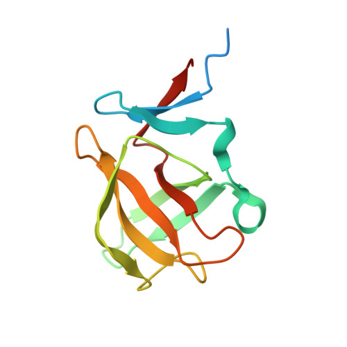
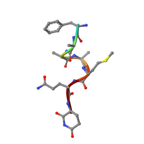
Identification and structural basis of C-terminal cyclic imides as natural degrons for cereblon.

Heim, C.Spring, A.K.Kirchgassner, S.Schwarzer, D.Hartmann, M.D.

(2022) Biochem Biophys Res Commun 637: 66-72

  • DOI: https://doi.org/10.1016/j.bbrc.2022.11.001
  • Primary Citation of Related Structures:  
    8BC6, 8BC7

  • PubMed Abstract: 

    Cereblon (CRBN) is a ubiquitously expressed E3 ligase substrate receptor and a key player in pharmaceutical targeted protein degradation. Despite substantial insight gained into its chemical ligand space that is exploited in small-molecule protein degraders, its cellular role and native mechanism of substrate recognition remained elusive so far. In this communication, we report the discovery of C-terminal aspartimide and aminoglutarimide residues as natural degron motifs that are recognized by CRBN with high specificity. These C-terminal cyclic imides are known to form in ageing proteins as a result of spontaneous chain breaks after an attack of an asparagine or glutamine side chain amide on the adjacent peptide bond, and thereby mark potentially malfunctional protein fragments. In crystal structures, we uncover that these C-terminal cyclic imides are bound in the same fashion as small-molecule CRBN modulators, and that the residues preceding the cyclic terminus contribute to the interaction with a sequence-unspecific backbone hydrogen bonding pattern with strictly conserved residues in CRBN. We postulate that C-terminal aspartimide and aminoglutarimide residues resulting from chain breaks are largely underappreciated protein damages and represent the native degrons of CRBN.


  • Organizational Affiliation

    Max Planck Institute for Biology, Tübingen, Germany; Interfaculty Institute of Biochemistry, University of Tübingen, Tübingen, Germany; NanoTemper Technologies GmbH, Munich, Germany.


Macromolecules
Find similar proteins by:  (by identity cutoff)  |  3D Structure
Entity ID: 1
MoleculeChains Sequence LengthOrganismDetailsImage
Cereblon isoform 4
A, B, C
124Magnetospirillum gryphiswaldenseMutation(s): 0 
Gene Names: MGR_0879
UniProt
Find proteins for A4TVL0 (Magnetospirillum gryphiswaldense)
Explore A4TVL0 
Go to UniProtKB:  A4TVL0
Entity Groups  
Sequence Clusters30% Identity50% Identity70% Identity90% Identity95% Identity100% Identity
UniProt GroupA4TVL0
Sequence Annotations
Expand
  • Reference Sequence

Find similar proteins by:  Sequence   |   3D Structure  

Entity ID: 2
MoleculeChains Sequence LengthOrganismDetailsImage
PHE-PHE-GLU-GLN-MET-GLN-QCI8Homo sapiensMutation(s): 0 
Sequence Annotations
Expand
  • Reference Sequence
Experimental Data & Validation

Experimental Data

  • Method: X-RAY DIFFRACTION
  • Resolution: 1.72 Å
  • R-Value Free: 0.211 
  • R-Value Work: 0.174 
  • Space Group: P 21 21 21
Unit Cell:
Length ( Å )Angle ( ˚ )
a = 56.606α = 90
b = 59.45β = 90
c = 89.059γ = 90
Software Package:
Software NamePurpose
REFMACrefinement
XDSdata reduction
XDSdata scaling

Structure Validation

View Full Validation Report


Ligand Structure Quality Assessment 


Entry History & Funding Information

Deposition Data


Funding OrganizationLocationGrant Number
Max Planck SocietyGermany--

Revision History  (Full details and data files)

  • Version 1.0: 2023-01-11
    Type: Initial release
  • Version 1.1: 2024-02-07
    Changes: Data collection, Refinement description