- Information
- AI Chat
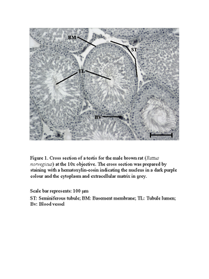
Biology-for-QLD An-Aust-Perp 3E Units 1-2 9780190310219 sample-chapter-3 low-res secure
Introduction to Cell Biology (BIO1140)
University of Ottawa
Recommended for you


Preview text
Cell structure
The basic unit of structure and function in living organisms is the cell. The cell is a self-sustaining chemical system. All the chemical reactions necessary for it to maintain its existence occur in its protoplasm. In order to maintain its chemical integrity, the cell must be physically separated from the environment and yet capable of exchanging material with it. The cell membrane forms this boundary. The efficiency of exchange decreases with an increase in size of the cell, so there is an upper limit to how large a cell can grow. Most cells, therefore, can be seen only when viewed with a microscope.
FIGURE 1 A 3D image of a eukaryotic cell cut-away shows the many organelles inside.
CHAPTER
3
OXFORD UNIVERSITY PRESS
→ Recognise that prokaryotic and eukaryotic cells have many features in common, which is a reflection of their common evolutionary past. → Recall that prokaryotic cells lack internal membrane-bound organelles, do not have a nucleus, are significantly smaller than eukaryotes, usually have a single circular chromosome and exist as single cells. → Understand that eukaryotic cells have specialised organelles to facilitate biochemical processes: - photosynthesis (chloroplasts) - cellular respiration (mitochondria) - synthesis of complex molecules including proteins (rough endoplasmic reticulum), carbohydrates, lipids and steroids (smooth endoplasmic reticulum), pigments, tannins and polyphenols (plastids) - the removal of cellular products and wastes (lysosomes). → Explain, using an example, how the arrangement of internal membranes can control biological processes (e. folding of membranes in mitochondria increases the surface area for enzyme- controlled reactions). → Identify the following structures from an electron micrograph: chloroplast, mitochondria, rough endoplasmic reticulum and lysosome. → Compare the structure of prokaryotes and eukaryotes. → Describe the structure of the cell membrane (including protein channels, phospholipids, cholesterol and glycoproteins) based on the fluid mosaic phospholipid bilayer model. → Describe how the cell membrane maintains relatively stable internal conditions via the passive movement (diffusion, osmosis) of some substances along a concentration gradient.
####### OBJECTIVES
68 BIOLOGY FOR QUEENSLAND: AN AUSTRALIAN PERSPECTIVE UNITS 1 & 2
SAMPLE
→ Explain how the cell membrane maintains relatively stable internal conditions via the process of active transport of a named substance against a concentration gradient. → Understand that endocytosis is a form of active transport that usually moves large polar molecules that cannot pass through the hydrophobic cell membrane into the cell. → Recognise that phagocytosis is a form of endocytosis. → Predict the direction of movement of materials across cell membranes based on factors such as concentration, and the physical and chemical nature of the materials. → Explain how the size of a cell is limited by the relationship between surface area to volume ratio and the rate of diffusion.
Source: Biology 2019 v1 General Senior Syllabus © Queensland Curriculum & Assessment Authority
####### PRACTICALS
MANDATORY PRACTICAL
3 The effect of surface area to volume ratio on the rate of diffusion
MANIPULATIVE SKILL
3 Use of the light microscope
MANDATORY PRACTICAL
3 Preparation and examination of wet mount specimens
SUGGESTED PRACTICAL
3 Identifying organelles from electron micrographs
SUGGESTED PRACTICAL
3 Modelling the selectively permeable cell membrane
OXFORD UNIVERSITY PRESS CHAPTER 3 CELL STRUCTURE 69
SAMPLE
He learned to grind and polish small glass spheres (a biconvex lens) of such quality that they had a magnification of 270. Leeuwenhoek mounted this lens on a flat copper plate and held it very close to his eye. The object being studied was placed on the head of a movable pin attached to the plate on the other side of the lens.
Using this simple microscope, Leeuwenhoek was the first to shine a light through living single- celled animals (protozoans) in pond water. He called them animalcules. For about 150 years microscopic work was limited to these two ‘primitive’ types of microscope. By the late 1800s many of the distortions of the lenses were corrected due to the work by German engineer Carl Zeiss and his employees, Schott and Abbe. They developed optical systems that could focus on objects smaller than a wavelength (approximately 0 μm (micrometres)). Because they used light as an imaging system, they were called light microscopes. Although a powerful and useful tool, light microscopes still could not provide a detailed observation of the internal structure of the cell. While some recent techniques have been employed in improving light microscopes, they are still limited.
As an understanding of the physical nature of matter, particularly the structure of the atom, developed, new technologies arose. The application of these advances led to the development of microscopes with higher magnification and resolution.
In the 1930s, the electron microscope was developed by Ernst Ruska and Max Knoll. Like its predecessors, this changed the possibilities for science. This microscope uses a narrow beam of electrons. The beam width is adjustable, but at its smallest is below the size of a hydrogen atom. The beam is focused with magnets and the final image is converted to light in a way similar to that on a television screen. A photograph called an electron micrograph is taken of the image. This is necessary because the electron beam prevents the magnified image from being viewed directly. For the same reason, the electron microscope is difficult to use on living material.
In summary, technological advancements in physics allowed magnification and resolution to be massively improved because the electron microscope uses a very narrow beam of electrons as opposed to the broader beam of light used previously.
FIGURE 2 Leeuwenhoek’s microscope
FIGURE 1 Hooke’s microscope
electron micrograph a photographic image of a sample magnified by an electron microscope
OXFORD UNIVERSITY PRESS CHAPTER 3 CELL STRUCTURE 71
SAMPLE
Most microscopes can be divided into another two groups depending on how the imaging beam (light or electrons) interacts with the specimen. In scanning microscopes, the beam reflects off the surface of the specimen to produce a three- dimensional image. Transmission microscopes send the beam through the specimen and a two- dimensional image is formed. For a specimen to be useful in the transmission microscope, the beam has to be able to pass through it and reveal its internal detail. Specimens need to be stained with special chemicals to produce clear detail. Coloured dyes are used in light microscopy while heavy metals are used for electron microscopy. The stain produces variations in absorption by different parts of the specimen. Large or thick specimens will not allow the beam through them. They must be sectioned or sliced, then stained. The object is embedded in a material (commonly, wax in light microscopy and plastic for electron microscopy) that will hold it together while being thinly sliced. Sequences of these sections can then be used to translate the two- dimensional information into a three- dimensional model.
Cell theory
The relationship between science and technology is clearly demonstrated in the development of the cell theory. Three hundred and fifty years ago there was no knowledge of cells since they were too small to be seen. It was only with the invention of the microscope that scientists like Leeuwenhoek were able to discover the previously unseen world of cells. Just as the telescope was a critical device for astronomers, the microscope was central to the progress of biology. While the discovery of cells was first made with the advent of the microscope in the seventeenth century, further progress was slow. In the nineteenth century two scientists
ocular lens (eyepiece)
coarse focus knob
fine focus knob
arm
condenser focus knob
base
mirror
iris diaphragm lever
condenser lens
stage
objective lens
revolving nosepiece
body tube
(a) (b)
30 cm 1 m
high voltage
electron gun magnetic condenser lens
electron beam image
fibre-optic camera
fluorescent screen
eyepiece
magnetic projector lens
magnetic objective lens
specimen
FIGURE 3 (a) A modern light microscope with inbuilt transmitted light source; (b) an electron microscope
cell theory the theory that all organisms are made up of cells and their products, and that growth and reproduction are fundamentally due to division of cells
72 BIOLOGY FOR QUEENSLAND: AN AUSTRALIAN PERSPECTIVE UNITS 1 & 2 OXFORD UNIVERSITY PRESS
SAMPLE
3.
Cells tend to be very small (0–10 μ, or micrometres, in diameter, where 1 μ = 10 – 6 m = 1 millionth of a metre) although there are a few notable exceptions, for example an emu’s egg which is 13 cm long. The smallest organisms have only one cell and are called unicellular. Although a size range exists in these organisms (the smaller cells are generally ‘simpler’), they are all microscopic. Larger organisms are composed of several to trillions of cells working together and are multicellular. In these organisms, different groups of cells perform different jobs to keep the organism alive. Each cell in the group is dependent on each other to survive. Occasionally groups of cells are closely associated with each other but are not directly connected. These groups are called colonial. Each cell is technically able to live independently, but they band together and divide up their functions.
The cell as the building block
oflife
KEYIDEAS ✚ The difference between prokaryotic and eukaryotic cells ✚ The endosymbiotic theory of the evolution of eukaryotic cells ✚ The size of cells including surface area to volume ratio
unicellular composed of a single cell
multicellular composed of many cells
colonial an animal composed of the same unicellular organisms joined into a cluster where each individual can carry out all functions necessary for life
0 nm 1 nm 10 nm 100 nm 1 μm 10 μm 100 μm 1 mm 1 cm 0 m 1 m 10 m 100 m 1 km
unaided eye light microscope electron microscope
atoms
lipids
small molecules
protein T2 phage (virus)
chloroplast
most bacteria
plant and animal cells
fish egg
white lipped tree frog
human blue whale giant pine tree
Ångstrom units (Å) 10 × 1000 × 1000 × 1000 Å = 1 m 1 Å = 10–10 m
nanometres (nm) 1000 × 1000 × 1000 nm = 1 m 1 nm = 10–9 m
micrometres (μm) 1000 × 1000 μm = 1 m 1 μm = 10–6 m
millimetres (mm) 1000 mm = 1 m 1 mm = 10–3 m
centimetres (cm) 100 cm = 1 m 1 cm = 10–2 m range of sizes for atoms, molecules and viruses range of sizes for cells range of sizes for organs and tissues
metres (m)
range of sizes for organisms
(a)
(b) Units of measurement
FIGURE 1 The size of a range of objects
74 BIOLOGY FOR QUEENSLAND: AN AUSTRALIAN PERSPECTIVE UNITS 1 & 2 OXFORD UNIVERSITY PRESS
SAMPLE
Common features
All the chemical reactions necessary for a cell to stay alive occur in its protoplasm (the colourless insides of the cell). To maintain its internal environment, the cell must be physically separated from the environment and still able to take in nutrients and remove waste. The cell membrane surrounds the outside of the cell and forms a boundary between the chemical reactions occurring inside the cell and the ever-changing outside of the cell. To be able to reproduce, all cells need a set of instructions that contain all the information that enable the cell to survive. These instructions are contained in genetic material called nucleic acids. The most important of the nucleic acids is deoxyribonucleic acid (DNA).
Prokaryotic cells
It is believed that the first life forms arose, under unique environmental conditions, about four billion years ago in a process termed abiogenesis. These first organisms were composed of a single cell – a cell membrane containing a watery mixture of organic chemicals, including a strand of nucleic acid. This type of cell is called a prokaryotic cell. All of the chemical processes required to sustain life occurred in this single cell. It is highly likely that there were many different types of nucleic acid, each coding for slightly different proteins, and so the original cells would have been slightly different to each other right from the beginning. It is thought that the early life forms arose from two different groups that are now called the Archaea and the Bacteria.
Over billions of years these cells have mutated and changed into diverse types with different physical and chemical characteristics. All of these prokaryotic organisms, known as prokaryotes, are composed of an extremely small, single cell contained within a semi-rigid cell wall. There is no compartmentalisation of the cell contents for specific functions. Some have a flagellum to help them move about in their aquatic environment (which may be the body fluids of multicellular organisms). Some die in the presence of oxygen while others cannot survive without it. Some are heterotrophs (feed on other organisms), some use inorganic molecules to provide for their energy needs (chemosynthesis) and others rely on photosynthesis.
cell membrane a selectively permeable barrier that controls the movement of substances into and out of the cell
abiogenesis the formation of a living organism from non- livingmatter
prokaryotic cell a cell which does not contain a membrane- bound nucleus or organelles
prokaryote an organism without a membrane- bound nucleus or organelles
LIGHT ENERGY SOURCE
CARBON SOURCE
EXAMPLES
some use organic matter
some oxidise organic compounds
organic matter
most bacteria including human pathogens
some oxidise inorganic substances, e. ammonia sulphides and iron
inorganic matter
hydrogen, sulphur, iron and nitrifying bacteria
some use CO 2
photosynthetic sulphur bacteria
photosynthetic non-sulphur bacteria
PHOTOSYNTHETIC CHEMOSYNTHETIC
BACTERIA
FIGURE 2 Energy and substrate requirements of bacteria
chromosome a thread- shaped body consisting of nucleic acid (usually DNA) and (except in bacteria) protein
Prokaryotic cells are very small. Their DNA or genetic material is usually found in an area of the cell called the nucleoid in the form of a single circular chromosome (see Figure 3).
multicellular organism an organism composed of many integrated cells
OXFORD UNIVERSITY PRESS CHAPTER 3 CELL STRUCTURE 75
SAMPLE
One view of nucleus formation is that the cell membrane began to infold. Not only did the membrane enclose the DNA to form a nucleus, but it also formed other membranous structures and channels within the cell.
FIGURE 6 A eukaryotic animalcell
eukaryote an organism whose cells contain membrane- bound organelles
FIGURE 5 Possible formation of the nucleus and membranous structures of the eukaryotic cell
Further mutations in these cells resulted in different types of single- celled eukaryotic organisms. As different organisms evolved, they changed the environment in which they lived and this led to further evolution. The development of the eukaryotic cell paved the way for the evolution of the multicellular organism. Different cells could become specialised for specific functions as a result of the numbers and types of organelles they possessed.
Organisms made up of eukaryotic cells are known as eukaryotes. Examples are animals, plants, protists and fungi.
infolding of plasma membrane
DNA
chromatin
plasma membrane
mitochondrion
lysosome
ribosomes
rough endoplasmic reticulum
smooth endoplasmic reticulum
pinocytic vesicle
nucleolus nuclear membrane
cilium
Golgi body centrioles
OXFORD UNIVERSITY PRESS CHAPTER 3 CELL STRUCTURE 77
SAMPLE
Surface area to volume ratio
Cells can be different shapes, making it difficult to compare their sizes. As a cell becomes larger, both the surface area and the volume of the cell increases; however, the volume increases at a faster rate than the surface area. For this reason, biologists compare the surface area to volume ratio of cells to compare the size of cells. Large cells have small surface area : volume ratios, while small cells have large surface area : volume ratios. All the chemical reactions (metabolism) required for cell survival occur within its volume. Cells need to be small to allow nutrients to move easily into the centre of a cell’s volume and for wastes to move out. Larger cells (with a large surface area and even larger volume) have more needs and wastes than can be coped with by their limited surfaces. Therefore, when a cell becomes too large, it struggles to survive. Ways around this dilemma involve a change in shape or the development of projections. Flattening or lengthening a cell or having numerous fine projections of the cell membrane (microvilli) can increase the cell's surface area : volume ratio. The largest plant cell yet discovered is found in a green alga (Valonia) and is about 1 cm in diameter (the average diameter of plant cells is 30–50 μm). This alga lives in the warm waters of the Great Barrier Reef and tidal movement ensures the rapid supply of nutrients and removal of wastes. The longest animal cell is the 2 m nerve cell of the giant squid. An increase in the internal membranes of the eukaryotic cell can also improve exchanges with its environment. The fact remains, however, that cells tend to be small and large organisms tend to be multicellular.
microvillus (plural microvilli) minute finger- like projection from a cell’s surface; can form a brush border
TABLE 1 Characteristics of prokaryotic and eukaryotic cells
Prokaryotic Eukaryotic Characteristic Bacteria Protists Fungi Plants Animals Nuclear membrane
Absent Present Present Present Present
Mitochondria Absent Present Present Present Present Chloroplasts Absent Present in some forms
Absent Present Absent
Mode of nutrition Heterotrophic or autotrophic (chemosynthesis or photosynthesis)
Photosynthesis or heterotrophic or combination of both
Heterotrophic by absorption
Photosynthesis Heterotrophic by ingestion
Multicellularity Absent Absent in many groups
Present except in yeasts
Present Present
Increasing the surface area for metabolic reactions within the cell Eukaryotic cells contain membrane-bound organelles in which specific metabolic reactions occur. In some of these organelles the membrane is highly folded, e. the inner membrane of the mitochondria and the endoplasmic reticulum. This increases the surface area for attachment of enzymes involved in the enzyme- controlled reactions. The more enzymes available, the greater the reaction rate for the available substrate.
78 BIOLOGY FOR QUEENSLAND: AN AUSTRALIAN PERSPECTIVE UNITS 1 & 2 OXFORD UNIVERSITY PRESS
SAMPLE
The cells of eukaryotic organisms (all organisms apart from archaebacteria, bacteria and cyanobacteria, which are prokaryotic) contain membrane-bound organelles. Most of these structures are not normally visible under the light microscope but can be viewed with an electron microscope. A number of organelles are found in both plant and animal cells – these are outlined in Table 1, along with an electron micrograph of each.
3.
Eukaryotic cell organelles
KEY IDEAS ✚ Organelles and their different structures and functions ✚ The difference in structure between prokaryotic and eukaryotic cells
TABLE 1 Organelles found in plant and animal cells Organelle Structure and function Electron micrograph Nucleus The total contents of the nucleus are called nucleoplasm. The nucleus controls cell activity through DNA, the genetic material that controls protein production and cell division. In the non-dividing cell, the DNA and protein form a diffuse network of threads called chromatin. These condense during cell division into chromosomes. The nucleus is surrounded by a double membrane, with pores that allow specific molecules into and out of the nucleus. The nucleus contains the nucleolus, which is composed of proteins, DNA and RNA, and which produces ribosomes.
chromatin (DNA nucleus and protin)
nucleolus
Endoplasmic reticulum
Endoplasmic reticulum is a network of flattened, membrane-bound sacs called cisternae. It is primarily involved in transporting molecules around the cell. There are two types of endoplasmic reticulum. Smooth endoplasmic reticulum is responsible for the synthesis and transport of lipids and steroids. Rough endoplasmic reticulum has ribosomes on the membrane surfaces that are involved in the synthesis of proteins. Transport of proteins occurs in their cisternae. The extensive network of the endoplasmic reticulum provides a large surface area for enzyme-mediated metabolic reactions to occur.
Rough ER – ribosomes are seen as small dots on the surface
nucleus control centre of the eukaryotic cell
chromatin the nucleoprotein of chromosomes seen as fine, dispersed threads of genetic material and found in eukaryotic cells in the interphase stage
smooth endoplasmic reticulum (smooth ER) a membranous structure with connections to both nuclear and cell membranes, involved in production, storage and transport of materials within the cells; lipids and steroids are produced here
rough endoplasmic reticulum (rough ER) a membranous structure with connections to both nuclear and cell membranes, involved in production, storage and transport of materials within the cells; proteins are synthesised on ribosomes
ribosome the site of protein synthesis in cells; composed of RNA, produced in nucleolus and found on rough ER (eukaryotes) or in cytoplasm (eukaryotes and prokaryotes)
80 BIOLOGY FOR QUEENSLAND: AN AUSTRALIAN PERSPECTIVE UNITS 1 & 2 OXFORD UNIVERSITY PRESS
SAMPLE
Organelle Structure and function Electron micrograph Mitochondria Mitochondria (singular mitochondrion) are cigar-shaped and bound by a double membrane. The inner membrane is folded into cristae, which increase the surface area for attachment of enzymes and their metabolic reactions. The membranes enclose the fluid matrix. Mitochondria are the site of chemical reactions that release energy in a useable form. Mitochondria contain special mitochondrial DNA and ribosomes that control their enzyme production, and allow them to replicate themselves. Mitochondria are crucial for the process of cellular respiration.
outer membrane
inner membrane
inter-membrane space
cytosolcytosol matrix space
cristae
Golgi body The Golgi body is composed of a stack of flattened membranous sacs or cisternae, which are continually being formed at one end and budded off as vesicles at the other. They are closely associated with the endoplasmic reticulum, receiving the enzymes, proteins, lipids, glycoproteins and polysaccharides synthesised there, enclosing them in membranes and releasing them either into the cytoplasm or outside the cell. Consequently, they serve as collecting, sorting, processing and distribution centres. Vesicle A vesicle is a membranous sac that transports molecules around a cell. There are different types of vesicles including lysosomes and vacuoles. Lysosome A lysosome is a simple spherical sac containing digestive enzymes. Lysosomes can fuse with vesicles or food vacuoles to digest their contents. Unwanted cellular products and wastes are broken down (autophagy) by lysosomes. They can also cause complete breakdown of damaged cells (autolysis) and so are sometimes termed ‘suicide sacs’. Centrioles Centrioles consist of a set of two cylindrical bodies next to the nucleus. They organise the DNA when a cell divides, and can form the whip-like flagella or hair like cilia that help some cells move.
centriole 1
centriole 2 perpendicular to 1
centriole 1
centriole 2 perpendicular to 1
mitochondrion (plural mitochondria) a membrane- bound cellular organelle; has enzymes for aerobic respiration on inner folded membrane (cristae), and a fluid matrix cristae the folds of the inner membrane of mitochondria cellular respiration cellular process in which glucose is broken down to release energy in many small, enzyme- controlled reactions Golgi body a membranous stack of cisternae involved in the packaging and secretion of cell products vacuole a cellular inclusion, or space within the cell; small in animals (food storage or digestion); large in plants (storage and osmotic control) lysosome an organelle containing digestive enzymes concerned with intracellular digestion centriole a paired tubular structure found in animals and algae; involved in formation of cilia and flagella and in cell division f lagellum (plural f lagella) a whip- like extension of the cell, used in locomotion by some Bacteria, Protozoa and spermatozoa cilium (plural cilia) a hair- like projection of the cell membrane
3 Preparation and examination of wet mount specimens Go to page 404 »
MANDATORY PRACTICAL
3 Use of the light microscope MANIPULATIVESKILL Go to page 409 »
OXFORD UNIVERSITY PRESS CHAPTER 3 CELL STRUCTURE 81
SAMPLE
grana (singular granum) stacked portions of membranes found in chloroplasts; sites of light- dependent reactions for photosynthesis
Studytip Students must be able to identify a chloroplast, mitochondria, rough endoplasmic reticulum and lysosomes from an electron micrograph.
FIGURE 2 A plant cell viewed with (a) a light microscope and (b) an electron microscope. The amount of detail seen in cells varies based on the equipment that is being used to view it.
Many organelles move within the cytoplasm in an ordered fashion. They are held in place by a series of fine filaments inside the cell called the cytoskeleton. There are three main components of the cytoskeleton, outlined in Table 3.
Structure and function Electron micrograph Plastids (chloroplasts, chromoplasts and leucoplasts)
These are double-membraned organelles of varying shape. Three types are common. Chromoplasts are non-photosynthetic, containing red, yellow and orange pigments that provide colour to flowers, fruit and leaves. Leucoplasts are colourless and involved in starch storage. Chloroplasts are large, consisting of a fluid matrix or stroma and stacks of flattened membranous discs called grana (singular stack = granum). Each disc is termed a thylakoid. They contain chlorophyll and function in photosynthesis. Like mitochondria, they also contain DNA and ribosomes and can replicate themselves. Chloroplasts are an important site for photosynthesis in plant cells.
Chromoplast
Chloroplast
Vacuole A large central vacuole contains cell sap and is responsible for storage of substances and control of water levels in the cell (osmotic balance). vacuole
(a) (b) nucleolus cytoplasm: granular appearance (whole cell minus nucleus) cell membrane (pressed against cell wall) cell wall
chloroplast
plasmodesmata
starch granules
cell walls of neighbouring cells
Golgi body
mitochondrion
nucleolus nucleoplasm nuclear membrane
vacuole, containing cell sap
diameter: about 40 μm
chromatin – fine threads
cell membrane
cell wall
mitochondrion lysosome Golgi body ribosome (on rough endoplasmic reticulum)
endoplasmic reticulum (smooth)
vacuole
grana
chloroplast
micrograph.
nucleus
nuclear membrane
OXFORD UNIVERSITY PRESS CHAPTER 3 CELL STRUCTURE 83
SAMPLE
FIGURE 3 Animal cell (a) and plant cell (b)
ANIMAL CELL
nuclear membrane chromatin nucleolus smooth endoplasmic reticulum
smooth endoplasmic reticulum
Cell nucleus and endoplasmic reticulum
ribosomes
matrixribosomes
inner membrane outer membrane
cristae mitochondrial DNA
Mitochondrion
Golgi body (apparatus) — semi-circular system of membranes
rough endoplasmic reticulum
rough endoplasmic reticulum
pore
chromatin microbody centrioles
lysosome
cytoskeleton
ribosomes
Golgi body
vesicle
cisternae
plasma membrane
nucleolus
nucleus
mitochondrion
(a)
pore
84 BIOLOGY FOR QUEENSLAND: AN AUSTRALIAN PERSPECTIVE UNITS 1 & 2 OXFORD UNIVERSITY PRESS
SAMPLE
TABLE 3 Components of the cytoskeleton
Feature Structure and function Electron micrograph Microfilaments Microfilaments are fine strands of the globular protein actin. They are able to bind with other proteins, and in so doing are able to hold organelles in position within the cytoplasm. Interactions of actin microfilaments with another protein, myosin, bring about general movement of the cytoplasm, organelles and the cell as a whole. Muscle cells have a high content of both of these proteins. The sliding action of actin and myosin results in contraction and relaxation and therefore movement. Some cells in animals (e. lining the intestine and kidney tubules) have minute finger-like projections of the membrane. These microvilli increase the cell surface area greatly without increasing volume to any great extent. Since they usually occur in large numbers and appear like the bristles of a brush, cells bearing microvilli are said to have a brush border. Actin microfilaments allow them to contract. The combined actions of increased surface area and movement facilitate uptake of materials into the cell.
MF – microfilament MT – microtubule MF
MT MT
A cell with a brush border – each projection is a microvillus Microtubules Microtubules are hollow cylinders of the protein tubulin. Being more rigid than microfilaments, they provide greater mechanical support for the cell. In plant cell walls microtubules provide the framework along which cellulose is laid down. Microtubules also provide routes along which materials in the cytoplasm can move, assisting transport within cells. During cell division microtubules form the spindle fibres that attach to chromosomes and draw them apart. Cilia (short) and flagella (long) are cellular projections covered by the cell membrane. They are composed of an exact array of microtubules. The sliding of adjacent pairs of these tubules causes the projections to move. In this way, cilia and flagella can bring about movement of a cell or of a liquid over the cell’s surface.
Transverse section of a cilium showing the arrangement of microtubules
Cilia
Salmonella bacteria with several flagella Intermediate filaments
Intermediate filaments are composed of a variety of fibrous proteins, and range in size between that of the microfilaments and microtubules. These filaments are strong, stable and resistant to stretching, thereby providing mechanical support for the cell.
86 BIOLOGY FOR QUEENSLAND: AN AUSTRALIAN PERSPECTIVE UNITS 1 & 2 OXFORD UNIVERSITY PRESS
SAMPLE
microfilament a solid rod of actin protein found in the cytoplasm, cilia and flagella of eukaryotic cells
microtubule a hollow protein strand in cytoplasm; involved in cell transport and structure
Comparing prokaryotic and eukaryotic organelles
The prokaryotes (bacteria and cyanobacteria) are the most ancient cells. They are smaller than eukaryotic cells and have a simpler structure (see Table 5). Like plant cells, bacteria have a semi- rigid cell wall surrounding the cell membrane, but it is made of murein (a protein–carbohydrate complex), and also they lack distinct membrane-bound organelles and a cytoskeleton. The genetic material is bound into a single circular DNA molecule floating within the cytoplasm. Smaller segments of DNA, called plasmids, are not part of the prokaryote’s chromosome, but can carry antibiotic resistance. Although many ribosomes are present in prokaryotes, they are not attached to any other structure and differ chemically from those of eukaryotes. Some bacteria have flagella. The flagellum, however, is outside the cell membrane and is different in structure to that found in animals.
TABLE 4 Comparison of eukaryotic cells: plant, animal and fungal
Feature Plant cell Animal cell Fungal cell Cell wall Rigid, cellulose, may be lignified
None Chitin
Vacuoles Large: up to 80% of cell volume
Small if present Small if present
Centrioles Absent in most Present Absent in most Chloroplasts Present in photosynthetic cells
Absent Absent
Granules Starch Glycogen Glycogen Cilia Absent Present in certain cells or cell types
Absent
Mode of nutrition Autotrophic Heterotrophic Heterotrophic
TABLE 5 Comparison of eukaryotic and prokaryotic cells
Structure Eukaryotic cells Prokaryotic cells Cell wall Absent in animal cells, present in plant cells (cellulose) and fungi (chitin)
Present (murein)
Cell membrane Present Present Nucleus Surrounded by membrane Not surrounded by membrane Chromosomes Multiple, composed of nucleic acid (linear DNA) and proteins
Singular, composed only of nucleic acid (circular DNA) Ribosomes Present Present but are different chemically Chloroplasts Endoplasmic reticulum Golgi body Lysosomes Membrane-bound organelles Mitochondria Vacuoles
Present (chloroplasts present only in plants)
Absent
Flagella Complex – microtubules Simple, with no microtubules (extracellular, i. not enclosed by the cell membrane)
murein a glycoprotein forming the cell wall in bacteria
antibiotic resistance the ability of bacteria to resist the effects of an antibiotic to which they were once sensitive
OXFORD UNIVERSITY PRESS CHAPTER 3 CELL STRUCTURE 87
SAMPLE
Biology-for-QLD An-Aust-Perp 3E Units 1-2 9780190310219 sample-chapter-3 low-res secure
Course: Introduction to Cell Biology (BIO1140)
University: University of Ottawa


- Discover more from: