6CAN

Prolyl oligopeptidase mutant S477C from Pyrococcus furiosus

  • Classification: HYDROLASE
  • Organism(s): Pyrococcus furiosus
  • Expression System: Escherichia coli
  • Mutation(s): Yes 

  • Deposited: 2018-01-31 Released: 2019-02-06 
  • Deposition Author(s): Ellis-Guardiola, K., Lewis, J.C., Sukumar, N.
  • Funding Organization(s): National Science Foundation (NSF, United States), Army Research Office, National Institutes of Health/National Institute of General Medical Sciences (NIH/NIGMS), Department of Energy (DOE, United States)

Experimental Data Snapshot

  • Method: X-RAY DIFFRACTION
  • Resolution: 2.20 Å
  • R-Value Free: 0.251 
  • R-Value Work: 0.196 
  • R-Value Observed: 0.197 

wwPDB Validation   3D Report Full Report


This is version 1.4 of the entry. See complete history


Literature

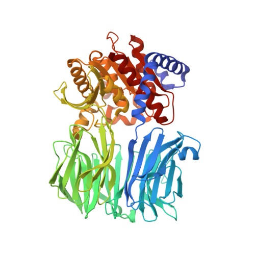
Crystal Structure and Conformational Dynamics of Pyrococcus furiosus Prolyl Oligopeptidase.

Ellis-Guardiola, K.Rui, H.Beckner, R.L.Srivastava, P.Sukumar, N.Roux, B.Lewis, J.C.

(2019) Biochemistry 58: 1616-1626

  • DOI: https://doi.org/10.1021/acs.biochem.9b00031
  • Primary Citation of Related Structures:  
    5T88, 6CAN

  • PubMed Abstract: 

    Enzymes in the prolyl oligopeptidase family possess unique structures and substrate specificities that are important for their biological activity and for potential biocatalytic applications. The crystal structures of Pyrococcus furiosus ( Pfu) prolyl oligopeptidase (POP) and the corresponding S477C mutant were determined to 1.9 and 2.2 Å resolution, respectively. The wild type enzyme crystallized in an open conformation, indicating that this state is readily accessible, and it contained bound chloride ions and a prolylproline ligand. These structures were used as starting points for molecular dynamics simulations of Pfu POP conformational dynamics. The simulations showed that large-scale domain opening and closing occurred spontaneously, providing facile substrate access to the active site. Movement of the loop containing the catalytically essential histidine into a conformation similar to those found in structures with fully formed catalytic triads also occurred. This movement was modulated by chloride binding, providing a rationale for experimentally observed activation of POP peptidase catalysis by chloride. Thus, the structures and simulations reported in this study, combined with existing biochemical data, provide a number of insights into POP catalysis.


  • Organizational Affiliation

    Department of Chemistry , University of Chicago , Chicago , Illinois 60637 , United States.


Macromolecules
Find similar proteins by:  (by identity cutoff)  |  3D Structure
Entity ID: 1
MoleculeChains Sequence LengthOrganismDetailsImage
Prolyl endopeptidase
A, B
616Pyrococcus furiosusMutation(s): 2 
UniProt
Find proteins for Q51714 (Pyrococcus furiosus)
Explore Q51714 
Go to UniProtKB:  Q51714
Entity Groups  
Sequence Clusters30% Identity50% Identity70% Identity90% Identity95% Identity100% Identity
UniProt GroupQ51714
Sequence Annotations
Expand
  • Reference Sequence
Experimental Data & Validation

Experimental Data

  • Method: X-RAY DIFFRACTION
  • Resolution: 2.20 Å
  • R-Value Free: 0.251 
  • R-Value Work: 0.196 
  • R-Value Observed: 0.197 
  • Space Group: P 1 21 1
Unit Cell:
Length ( Å )Angle ( ˚ )
a = 56.292α = 90
b = 178.764β = 104.35
c = 59.293γ = 90
Software Package:
Software NamePurpose
PHENIXrefinement
HKL-2000data reduction
HKL-2000data scaling
Cootmodel building
PHENIXphasing

Structure Validation

View Full Validation Report


Entry History & Funding Information

Deposition Data


Funding OrganizationLocationGrant Number
National Science Foundation (NSF, United States)United StatesCHE 1351991
Army Research OfficeUnited States62247-LS
National Institutes of Health/National Institute of General Medical Sciences (NIH/NIGMS)United StatesT32 GM008720
National Institutes of Health/National Institute of General Medical Sciences (NIH/NIGMS)United StatesP41 GM103403
Department of Energy (DOE, United States)United StatesDE-AC02-06CH11357

Revision History  (Full details and data files)

  • Version 1.0: 2019-02-06
    Type: Initial release
  • Version 1.1: 2019-02-20
    Changes: Author supporting evidence, Data collection
  • Version 1.2: 2019-08-21
    Changes: Data collection, Database references
  • Version 1.3: 2019-11-27
    Changes: Author supporting evidence
  • Version 1.4: 2023-10-04
    Changes: Advisory, Data collection, Database references, Refinement description