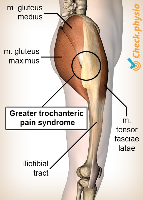
Greater trochanter pain syndrome
Tendinosis of the hip abductors / bursitis trochanterica
Pain on the outside of the hip that occurs whilst walking is a typical symptom of the greater trochanter pain syndrome. The pain is experienced around the hard bump located on the outside of the hip.
Description of the condition
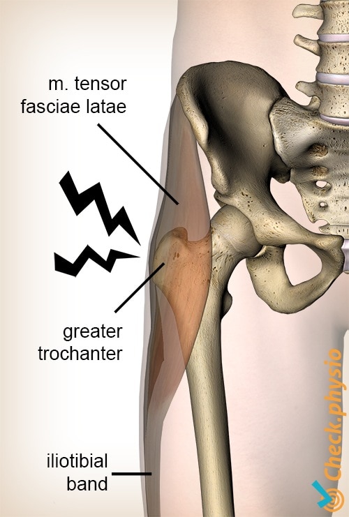
The iliotibial band is a large band of connective tissue (fascia) that runs from the pelvic ridge over the outside of the upper leg to the knee. It passes the greater trochanter along the way. This is a bump on the thigh that protrudes outwards. The pain is caused by damaged or irritated structures that move over or along the greater trochanter.
The muscles located on the outside of the hip are called the hip abductors. They raise the thigh outwards and prevent the hip joint from collapsing when we walk.
Anatomy
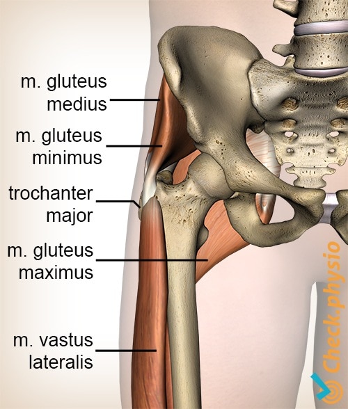
There are two muscles that attach to the iliotibial band which originate from the pelvis. These are the gluteus maximus and the tensor fasciae latae. The gluteus medius and the gluteus minimus are two other muscles that are located underneath the first two muscles;. Those muscles run from the pelvis to the greater trochanter, where they attach. The vastus lateralis also attaches to the greater trochanter, which runs from there to the knee.
All these structures move directly over each other. Each muscle is surrounded by bursae to prevent friction. Bursae are like smooth bumper pads that prevent bones and tendons from rubbing over each other.
The pain that occurs in the case of greater trochanter pain syndrome is the result of various injuries to the structures described above. Greater trochanter pain syndrome is a collective term for the symptoms that occur around the greater trochanter.
Cause and origin
The symptoms can develop either gradually or acutely. There are various causes of greater trochanter pain syndrome.
- Tendinosis of the hip abductors: the tendon structure of the muscles decreases in quality.
- Partial ruptures (tears) of the hip abductors.
- Calcification of the attachment of the tendons surrounding the greater trochanter.
- Excessive friction of the iliotibial band over the greater trochanter (snapping hip).
- Sometimes the bursae can also be inflamed (this is more commonly known as 'bursitis trochanterica' and is almost never an isolated event).
Signs & symptoms
There is pain on the outside of the hip, around the hard bump that can be located here (the greater trochanter). The pain is felt when walking and when one presses on the greater trochanter. This can also cause the patient pain during the night, when he/she lies on the affected side. The pain radiates over the outside of the upper thigh towards the knee.
The hip abductors can be weakened, resulting in the patient 'waddling', with the trunk leaning over the affected side (Trendelenburg's sign). Standing on the affected leg can provoke the symptoms, sometimes after even as little as 30 seconds.
Diagnosis
Treatment
Physiotherapy can provide relief if relative rest and alleviating the strain on the affected leg does not ease the symptoms. The treatment consists of excentrically performed muscle strengthening of the hip abductors.
Other treatment forms include pain medication, providing shoe inserts and/or a corticosteroid injection. A corticosteroid injection can be very effective, but can be counter-productive in the long run.
Exercises
Follow the specially compiled exercise programme here with exercises for greater trochanteric pain syndrome.
You can check your symptoms using the online physiotherapy check or make an appointment with a physiotherapy practice in your area.
References
Kingzett-Taylor, A., Tirman, P.F., Feller, J., McGann, W., Prieto, V., Wischer, T., Cameron, J.A., Cvitanic, O. & Genant, H.K. (1999). Tendinosis and tears of gluteus medius and minimus muscles as a cause of hip pain: MR imaging findings. AJR Am J Roentgenol. 1999 Oct;173(4):1123-6.
Nugteren, K. van & Winkel, D. (2007). Onderzoek en behandeling van de heup. Houten: Bohn Stafleu van Loghum.
Williams, B.S. & Cohen, S.P. (2009). Greater trochanteric pain syndrome: a review of anatomy, diagnosis and treatment. Anesth Analg. 2009;108:1662-70.