Article

Acanthamoeba Sequence Types and Allelic Variations in Isolates from Clinical and Different Environmental Sources in Italy

by
Federica Berrilli
1,*,
Margherita Montalbano Di Filippo
2,
Isabel Guadano-Procesi
1,*,
Marta Ciavurro
1 and
David Di Cave
1
1
Department of Clinical Sciences and Translational Medicine, University of “Tor Vergata”, 00133 Rome, Italy
2
Istituto Superiore di Sanità, 00155 Rome, Italy
*
Authors to whom correspondence should be addressed.
Microorganisms 2024, 12(3), 544; https://doi.org/10.3390/microorganisms12030544
Submission received: 23 January 2024 / Revised: 1 March 2024 / Accepted: 5 March 2024 / Published: 8 March 2024
(This article belongs to the Special Issue Advances in Acanthamoeba)

Abstract

:
The genus Acanthamoeba comprises free-living amoebae distributed in a wide variety of environments. These amoebae are clinically significant, causing opportunistic infections in humans and other animals. Despite this, limited data on Acanthamoeba sequence types and alleles are available in Italy. In the present study, we analyzed all Acanthamoeba sequences deposited from Italy with new positive Acanthamoeba clinical samples from symptomatic AK cases, to provide an overview of the genetic variants’ spatial patterns from different sources within the Italian context. A total of 137 Acanthamoeba sequences were obtained. Six sequence types were identified: T2/6, T3, T4, T11, T13, and T15. Only T4 and T15 were found in both sources. The Acanthamoeba T4 sequence type was found to be the most prevalent in all regions, accounting for 73% (100/137) of the Italian samples analyzed. The T4 sequence type demonstrated significant allelic diversity, with 30 distinct alleles from clinical and/or environmental samples. These outcomes enabled a better understanding of the distribution of Acanthamoeba isolates throughout Italy, reaffirming its well-recognized ubiquity. Acanthamoeba isolates analysis from keratitis, together with the environmental strains monitoring, might provide important information on different genotypes spreading. This might be useful to define the transmission pathways of human keratitis across different epidemiological scales.

1. Introduction

The genus Acanthamoeba (Amoebozoa, Discosea) includes ubiquitous free-living amoebae (FLA) widely distributed in environments such as soil, fresh and marine water, and air. These amoebae have the potential to cause opportunistic infections in humans and other animals, like amoebic keratitis (AK) or granulomatous amoebic encephalitis (GAE). As recently reviewed [1], morphological approaches were primarily used in classifying species/taxa within the genus. Initially, Acanthamoeba was subdivided into three distinct groups (I–III) [2]. However, the identification of species remained arduous, and the advent of molecular tools, especially methods of analysis based on DNA differences, provided new insights into the relationships between the isolates [3,4,5].
The classification of Acanthamoeba is now mainly based on the analysis of the nuclear SSU rRNA gene (18S rDNA) variability—only partially confirming the morphological classification—and 23 sequence types, also named genotypes (from T1 to T23), have been identified so far, phylogenetically grouped into four major lineages [6]. In particular, the widely employed ASA.S1 region, a ~430 bp fragment matching the most variable segment of the 18S rDNA as proposed by Schroeder et al. [7], has proved to be a helpful fragment of the gene in the study of Acanthamoeba genetic variability, in the evolutionary relationships between the isolates, and in the definition of the different genotypes within the genus [1].
Along with the increased data on the sequence differences within this hypervariable segment, a different degree of intra-genotype variation was also observed among Acanthamoeba populations. Specifically, numerous studies have been conducted by examining polymorphisms in the DF3 region within the amplimer ASA.S1, yielding a sequence approximately 200–250 bases in size that includes most of the highly variable portions of this region, in order to better describe and categorize the genetic variation that exists in this region. These studies allowed the identification of numerous “alleles” within different sequence types, with the T4 genotype being the most variable. In addition to the T4, data on allele identification from a small number of genotypes—T3, T5, T11, T15, and supergroup T2/6—are now also available [1]. The introduction of the ‘allele’ concept in 2002 by Booton et al. [8]. marked a fundamental step in examining Acanthamoeba in both clinical and environmental contexts.
As stated, Acanthamoeba can act as an opportunistic parasite in humans, causing different infections, including granulomatous amoebic encephalitis, cutaneous lesions and sight-threatening amoebic keratitis. From a public health perspective, the molecular identification of Acanthamoeba isolates from clinical and environmental samples is therefore of great interest. Overall, the majority of isolates in clinical or environmental studies belong to the T4 genotype, which was further divided into seven main subtypes, labeled T4A to T4G based on phylogenetic analyses of 18S rRNA sequences for complete or almost complete sequences (with a length exceeding 2000 bases in length) [1,9]. Recently, a new nuclear lineage within the Acanthamoeba T4 genotype, labeled T4H, has been described [10].
The clustering of Acanthamoeba isolates from clinical cases coupled with the tracking of strains in the environment through high-resolution genotyping at the sequence type and allele levels could provide insights into the spread of different genetic variants, which would be useful in better identifying the transmission pathways of human keratitis at different epidemiological scales.
In Italy, few and geographically fragmented data are available on Acanthamoeba sequence types distribution from clinical [11,12,13] and environmental samples [14,15,16,17]. In the present study, sequence data retrieved from GenBank were compared with new positive Acanthamoeba clinical samples from symptomatic AK cases identified by phylogenetic analyses, to investigate the genetic variation within the ASA.S1 fragment in isolates from different sources in Italy. The specific objectives included: (i) adding new data on the distribution of Acanthamoeba sequence types from humans; (ii) conducting for the first time a comprehensive analysis of allelic variations in sequences obtained from all clinical and environmental isolates accessible in Italy; and (iii) providing a wide picture of the spatial patterns of genetic variants in isolates from various sources within the Italian context.

2. Materials and Methods

2.1. New Clinical Sample Collection and Molecular Characterization

Before conducting the analysis of the allelic variations in Acanthamoeba sequences from all clinical and environmental isolates accessible in Italy, the present work contributed to producing new original clinical sequences. In particular, the study involved corneal scraping specimens from symptomatic patients with suspected AK collected between 2015 and 2022 in the Unit of Parasitology of the Azienda Ospedaliera Universitaria Policlinico Tor Vergata (PTV) of Rome, Central Italy. To accurately identify the isolates and obtain information about Acanthamoeba sequence types and allelic variations, amplification of a fragment within the 18S rRNA gene containing the ASA.S1 region was performed. DNA was extracted from each sample using the Starlet extraction automate (SeeGene, Seoul, Republic of Korea) and used for molecular characterization. The PCR reaction mix encompassed a total volume of 25 μL, which contained 12.5 μL PCR master mix 2X (Promega, Milano, Italy), 5 μL of template DNA, and 0.5 μL of the primer pair JDP1 (5′-GGCCCAGATCGTTTACCGTGAA) and JDP2 (5′-TCTCACAAGCTGCTAGGGAGTCA–3′) to amplify a PCR product of ca. 423 to 551 bp [7]. The cycling conditions included one step at 96 °C for 2 min, followed by 35 cycles of denaturing for 1 min at 96 °C, annealing for 1 min at 60 °C, and extension for 1 min at 72 °C, with a final extension for 7 min at 72 °C, performed in a T100 PCR thermal cycler (BioRad, Segrate, Italy). Negative and positive controls were included in each batch of DNA extraction and PCR reaction. PCR products were examined by electrophoresis on 1% agarose gel stained by SYBR™ Safe DNA Gel Stain (Invitrogen™, Waltham, MA, USA). Amplicons were purified by the mi-PCR Purification Kit (Metabion GmbH, Steinkirchen, Germany) according to the manufacturer’s instructions, and then sent to an independent laboratory for sequencing (Bio-Fab Research, Rome, Italy). Forward and reverse Sanger sequences from each PCR product were examined by DNA chromatograms using FinchTV 1.4 (Geospiza, Inc, Seattle, WA, USA), and the obtained consensus sequences were aligned by the latest version of MEGA 11 [18], with representative reference sequences used as provided on the website: “https://u.osu.edu/acanthamoeba/acanthamoeba-sequence-types/ (accessed on 15 December 2023)”. To identify isolates at the sequence type level, a Maximum Likelihood (ML) phylogenetic tree was generated. The construction of the phylogenetic tree was conducted after testing for the best evolutionary models explaining the data [18]. Bootstrapping with 1000 replicates was used to determine support for the genetic distances.

2.2. Data Deposition

The new clinical sequences obtained in the present study are available at NCBI GenBank with the accession numbers PP126046–PP126065.

2.3. Database Development

For the purposes of this study, a search in PubMed Nucleotide of all Acanthamoeba sequences deposited from Italy was performed using a multistep strategy matching the following keywords: Acanthamoeba AND Italy OR Italian; FLA AND Italy OR Italian; and free-living amoeba AND Italy OR Italian. The full check of GenBank records was performed paying attention to the “country” and “isolation_source” features. This allowed the retrieval of all sequences referring to environmental and clinical samples described in Italy and suitable for the analysis conducted in this study.

2.4. Allelic Identification

Alleles variability is found within the 18S rRNA region of the sequences named ASA.S1. Hence, to properly identify the isolates at allele level, all Acanthamoeba sequences recovered by the search described above plus those obtained in the present study from new clinical specimens (see Section 2.1) were appropriately aligned and trimmed by both MEGA 11 and AliView [19], with alleles representative sequences provided as lists on the website: “https://u.osu.edu/acanthamoeba/alleles-within-sequence-types-2/ (updated October 2023) (accessed on 15 December 2023)”.

3. Results

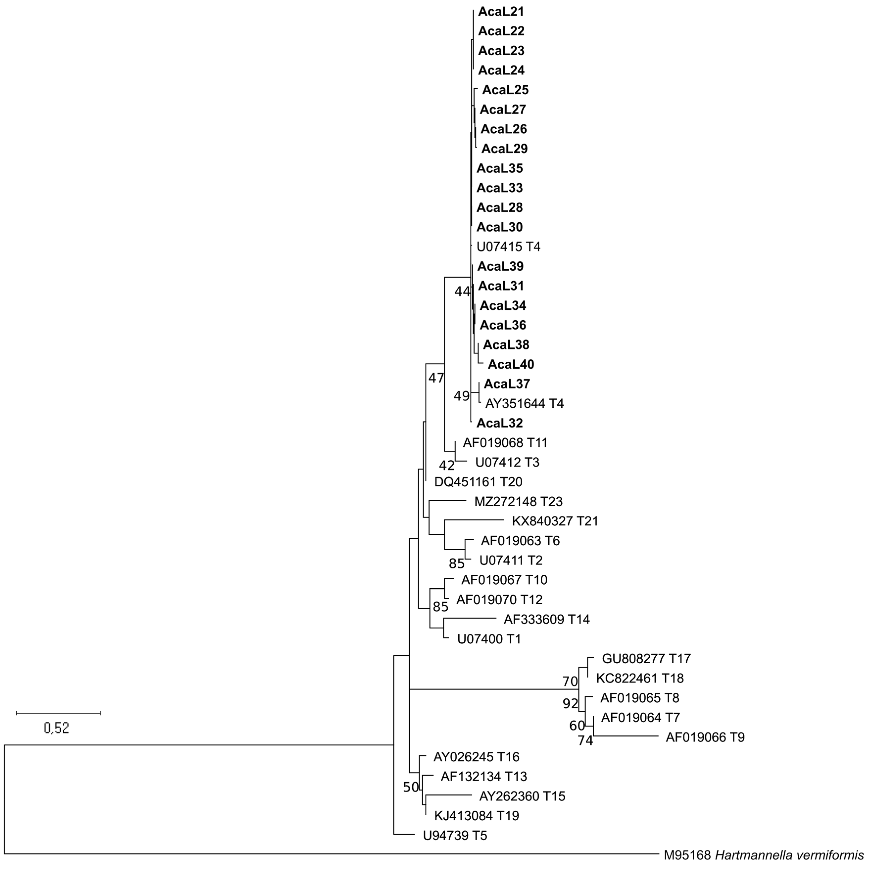
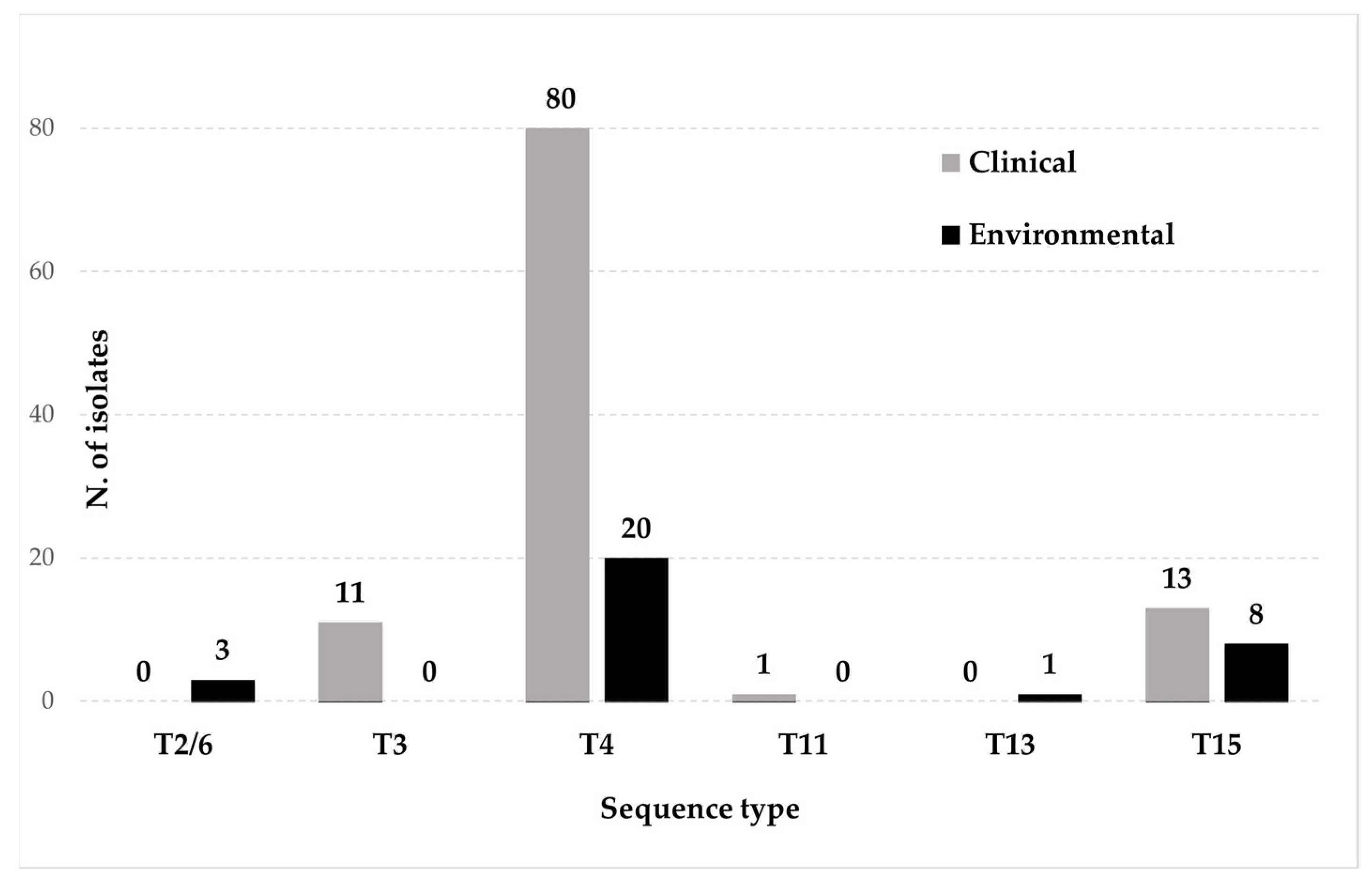
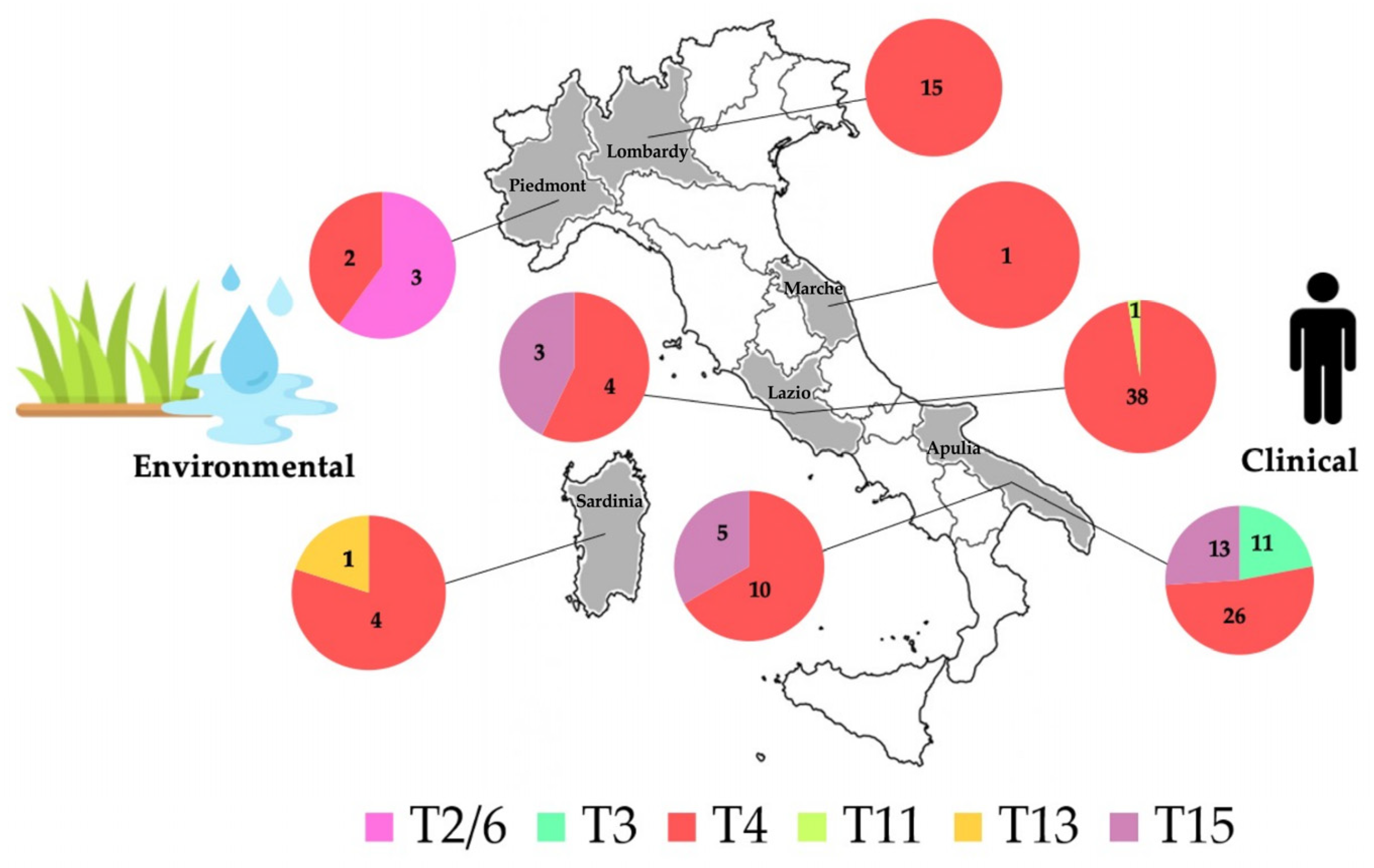
3.1. Identification of New Clinical Isolates and Sequence Types Analysis

In the present study, twenty Acanthamoeba isolates from suspected AK patients were obtained and successfully PCR-amplified. Good-quality sequences of ~405-bp length were achieved for all samples. The topology of the ML phylogenetic tree showed all the isolates analyzed here clustering in the composite T4 sequence type clade (Figure 1).
Through the search strategy applied as described in Section 2.3, coupled with the acquisition of 20 new clinical sequences from this study, a total of 137 Acanthamoeba sequences linked to isolates from Italy were obtained. Details for each sequence are presented in Table 1, encompassing all sequences related to clinical isolates, and in Table 2, including sequences derived from environmental samples.
Overall, six different sequence types were detected in Italy: T2/6 (3 isolates; 2.2%), T3 (11 isolates; 8%), T4 (100 isolates; 73%), T11 (1 isolate; 0.7%), T13 (1 isolate; 0.7%), and T15 (21 isolates; 15.4%). Acanthamoeba T3, T4, T11, and T15 have been identified in humans, while T2/6, T4, T13, and T15 were from environmental samples, with only T4 and T15 shared in both sources (Table 3, Figure 2).
Data on the geographic distribution of Acanthamoeba genotypes were from six Italian regions. The majority of samples originated from Apulia (65/137; 47.4%), followed by Lazio (46/137; 33.6%), Lombardy (15/137; 10.9%), and Piedmont and Sardinia (5/137; 3.7% each). Additionally, Marche had only one sequence available (1/137; 0.7%) (Table 1 and Table 2, Figure 3). The distribution of sequence types varies across regions and sources; however, ST4 consistently predominates in all analyzed areas of the country, regardless of the source type, except in the Piedmont region where a slight dominance of isolates belonging to the T2/6 supergroup was observed (Figure 3).
Reports from both clinical and environmental samples were only from two regions, Lazio and Apulia, where a partial overlapping of sequence types was observed. Specifically, in the Lazio Region, three genotypes were identified: T4 (42/46; 91.3%), T11 (1/46; 2.2%), and T15 (3/46; 6.5%). Among these, T4 exhibited a higher prevalence and was shared between samples from human AK (38/42; 90.5%) and water sources (4/42; 9.5%). The specimen assigned to genotype T11 was recovered from one patient, while the three samples recognized as T15 were obtained from thermal water.
In Apulia, sequence types T3 (11/65; 16.9%), T4 (36/65; 55.4%), and T15 (18/65; 27.7%) were detected. Sequence types T4 and T15 were isolated in both clinical and water samples, whereas T3 was uniquely identified in AK patients.
In Sardinia, genotypes T13 (1/5; 20%) and T4 (4/5, 80%) were described in one sample harvested in soil from a grassland site, while the T2/6 supergroup (3/5, 60%) and T4 sequence type (2/5, 40%) were recognized in Piedmont from soil collected from a rice field. Only T4 sequences were identified in Lombardy (N = 15) and Marche (N = 1), exclusively in clinical samples. Details on the sequence types, isolate IDs, geographical regions, sources, and references are provided in Table 1 and Table 2.

3.2. Allele Identification and Analysis

To provide a more precise depiction of the variation degree present within the Acanthamoeba isolates, each sequence from all sequence types, excluding T13 (as allele analysis for this genotype is not yet available), was identified at the allele level, following the procedures outlined in Section 2.4.
As illustrated in Figure 4, allele analysis revealed the presence of a single variant within the sequence types T3 (allele T3/03) and T11 (allele T11/08), which both originated from AK cases. Three isolates from soil samples in Piedmont belonging to the T2/6 supergroup were identified as alleles T26A/01 (one isolate) and T26B/01 (two isolates).
Two alleles, T15/01 and T15/02, were also identified within the T15 sequence type. The T15/01 allele was assigned to the majority of the T15 isolates (18/21; 85.7%) and obtained from both human and water sources, all from the Apulia Region. On the other hand, the T15/02 allele was derived from three hot spring water samples from Lazio (3/21; 14.3%).
In contrast, isolates grouped in the T4 sequence type exhibited a substantial allelic diversity, with 30 different alleles assigned to isolates from clinical and/or environmental samples. Noteworthy, three environmental isolates, one from thermal water in Lazio, one from tap water collected in Apulia, and one from soil in Sardinia, exhibited a mixed allele combination (T4/01-T4A + MT4/25-T4B; OT4/114-T4A + OT4/143-T4A; and T4/07-T4A + T4/16-T4A, respectively). These combinations were identified based on the polymorphisms reported in the FASTA sequences.
The most commonly identified allele was T4/01-T4A (22/100; 22%), which was predominantly present in AK specimens (20/80; 25%) and only encountered twice in environmental samples (2/20; 10%). The AK isolates were derived from various studies, including the present one, conducted on clinical samples from Lazio and Apulia. The two environmental samples were collected from thermal water sources in Lazio; one of the two samples showed a mixed allele combination.
The other two allele types founded most frequently were T4/13-T4A (9/100; 9%) and AKT4/22 T4A (8/100; 8%). The first, identified in clinical samples from Lazio (n = 6) and Lombardy (n = 2), has been also assigned to one isolate from tap water collected in the Lazio region. The second allele was identified in AK patients from Lombardy (n = 2), Apulia (n = 1), and Lazio (n = 2), and in ornamental fountain water and tap water from Apulia (n = 3). Three alleles, each with a frequency of 6%, have been identified: T4/09–T4B, predominantly observed in clinical samples; ZT4/24-T4D, largely assigned to environmental samples; and T4/06-T4B, exclusively found in AK isolates, including some obtained in the present study. The remaining 24 alleles, each with a frequency less than or equal to 5%, comprised 6 alleles shared between human and environmental samples, 14 alleles exclusively found in clinical isolates, and 4 alleles detected solely in environmental samples.
Lastly, for three Acanthamoeba isolates (two obtained from soil in Sardinia and one from AK in Lazio), it was not possible to assign the allele, as the sequence did not match with any deposited allele segment thus far.

4. Discussion

Over the past decades, an increasing number of studies on Acanthamoeba distribution and genetic diversity have been conducted worldwide [5,7,8,15,20,21,22]. These studies primarily contributed to advancements regarding classification and phylogenetic relationships between Acanthamoeba isolates, but they also provided insight into different aspects of ecology and spatial distribution of these organisms, useful for determining potential threats to human health. In fact, despite being free-living organisms, several amoebae within the genus Acanthamoeba exhibit an “amphizoic” behavior. This means they are capable of transforming into an endozoic parasitic stage, causing serious and sometimes fatal diseases in humans [23]. Transmission to humans or other animals typically begins through exposure to contaminated water or soil containing the trophozoite stage [24]. Therefore, a comprehensive knowledge of the distribution of this organism in both environmental and clinical samples is crucial for accurately assessing the risk of infection.
In Italy, since 2008, studies on the molecular characterization of Acanthamoeba allowed the identification of various genotypes in isolates from different sources. However, there are no investigations that simultaneously analyzed isolates of both clinical and environmental origin within the same geographic area. Due to the limited and fragmented data so far available, this study is designed to serve as a comprehensive large-scale survey regarding sequence type distribution and allelic variations of Acanthamoeba in isolates from various sources and different geographic locations across Italy, aiming to give a more comprehensive overview of their diffusion and to offer insight into the potential transmission dynamics of Acanthamoeba within the country.
As summarized in Table 1 and Table 2, the presence of Acanthamoeba in Italy has been documented in clinical specimens and/or in different environments with six sequence types, T2/6 (supergroup), T3, T4, T11, T13, and T15 detected so far. It is noteworthy that all these genotypes have been described to cause AK worldwide [25].
The twenty new clinical samples identified in this study were all assigned to T4, thus providing further support to the evidence that the T4 genotype plays a prominent role in the epidemiology of human infections, mainly AK. Taking into account these new clinical cases, as evidenced in Table 1 and Table 2, the T4 genotype represents 73% (100/137) of the counted Italian samples, 58.4% (80/137) from humans, and 14.6% (20/137) from the environment, distributed across all the examined regions. This finding is in agreement with data from the literature, emphasizing that the majority of isolates in clinical or environmental studies belong to the T4 sequence type [1,7,20,21]. This genotype, also defined as A. castellanii complex, encompasses several morphological species, including the type species of the genus A. castellanii. High intra-genotypic variation is therefore observed, leading to its subdivision into eight main subtypes and to the definition of a high number of alleles accepted to date (https://u.osu.edu/acanthamoeba/alleles-within-sequence-types-2/; updated October 2023) (accessed on 15 December 2023).
The high level of polymorphism here detected within the T4 sequence type, as evidenced by the identification of thirty different alleles with different patterns of SNPs, is therefore not unexpected. Among these, twelve alleles were documented to be shared between clinical and environmental samples. The most frequently observed was T4/01-T4A (22%). In the literature, the T4/01-T4A allele type was initially identified in Acanthamoeba keratitis patients in Hong Kong [8]. Some years later, it has also been found in samples collected from drinking water treatment plants in Spain [22]. However, in contrast to Italian data, this allele is detected with low frequency worldwide, as recently reviewed [1]. The other two frequently encountered allele types in both human and environmental isolates were T4/13 and AKT4/22. Both of these alleles have been reported in over 100 deposited isolates worldwide, with AKT4/22 being the most prevalent, found in more than 240 isolates from many countries [1].
Notably, twenty-four low-frequency T4 allele types, each occurring at less than or equal to 5% frequency, dominate the distribution in Italy. They were predominantly observed as single cases in AK patients or, in four cases, assigned to mixed Acanthamoeba samples from grassland and rice field soil and from tap water. As previously mentioned [1,21], these ambiguous sequencing reads (mixed) may derive from a multi-cyst/multi-trophozoite extraction, representing an unrecognized mixture of isolates. Additionally, this ambiguity could be attributed to intracellular polymorphism in the 18S rRNA gene.
Among clinical reports, the only no-AK sample derived from a case of disseminated Acanthamoeba infection in an immunocompromised patient was attributed to the allele OT4/39. This allele, labeled since 2015, has significant importance as it identifies the type isolate for the genus Acanthamoeba (A. castellanii ATCC 30011) [1]. Unfortunately, no further information is available on this case as the data are unpublished.
The second most abundant genotype described in Italy was T15. Like T4, this sequence type was found in both clinical and environmental samples in two regions, Lazio and Apulia. Until 2009, the T15 genotype had only been detected in a few isolates attributed to the species A. jacobsi, isolated from different environments [5]. The first case of keratitis associated with this genotype was identified in Italy [11], thus enlarging the spectrum of Acanthamoeba sequence types capable of causing pathogenic effects in humans. Currently, the T15 genotype represents the third most involved genotype in keratitis cases after T4 and T3 [25] and has a notable worldwide environmental presence, as recently reviewed [26]. Remarkably, the majority of T15 Acanthamoeba isolates occurred in Italy in both clinical and environmental isolates from Apulia and shared the identical allele sequence T15/01. This allele is reported to have the highest frequency (56%) among the alleles found within T15 sequences in the DNA databases:“https://u.osu.edu/acanthamoeba/alleles-within-sequence-types-t15/; updated October 2023 (accessed on 15 December 2023)”. Conversely, three isolates from Lazio thermal water, showing identical sequences to each other, were identified as T15/02. It is worth noting that the almost complete Rns sequence obtained subsequently for one of these isolates (Pool-4-37; Accession number KY513796) revealed a group I intron in the 18S ribosomal region, expanding the recognized genotype of Acanthamoeba with nuclear introns [27].
The remaining four sequence types, T2/6, T3, T11, and T13, were reported with lower frequency in various Italian regions, either in clinical or environmental isolates.
The three isolates from the soil, identified here as subtypes T2/6A (allele T26A/01) and T2/6B (allele T26B/01), belong to the T2/6 supergroup. Within this supergroup are classified all isolates clustering between the two closely related sequence types T2 and T6 [9], as thoroughly analyzed by Fuerst and Booton [1].
Sequence type T3, reported at 8%, was identified in 11 ocular isolates, all originating from Apulia and presenting the same allele T3/03. In the recent systematic review of Acanthamoeba in keratitis by Diehl et al. [25], T3 represents the second most prevalent genotype detected worldwide. It was detected for the first time by Gast et al. [4] by a strain identified in the marine amoeba A. griffini [28] and described primarily as a human pathogen associated with AK in a patient from the United Kingdom by Ledee et al. [29]. In Italy, sequence type T3 was also previously described in one patient presenting with a wide corneal ulcer in Tuscany [30]. However, since no deposited sequence is available, this Acanthamoeba isolate was excluded from our analysis.
Finally, two sequence types have been sporadically identified in Italy. The first, the T13 sequence type, refers to a single isolate found in one environmental sample from Sardinia but also isolated by the same authors in two samples from soils at high altitudes in Tibet [15]. This genotype was only encountered once in a case of amoebic keratitis in South Africa [31] and seems to be a rarely identified form globally [1]. The second one was the T11 sequence type, reported in the present study in one AK patient from Lazio. This genotype, closely related to T3, is also found with a low prevalence (2.07%) in AK cases from Europe, America, and Asia [25].
The results obtained in the present study allowed a better understanding of the extensive circulation of Acanthamoeba sequence types in Italy, confirming the well-established nature of these organisms as ubiquitous protozoans.
The identification of isolates from both humans and the environment at the allele level seems to suggest that, despite the genetic diversity observed within genotypes, this variability does not appear necessarily correlated to a differential isolates’ ability to act as potential sources of infection for humans. This consideration is particularly applicable to isolates belonging to the sequence type T4, which, along with their specific properties (e.g., greater virulence and transmissibility) are the most widespread in natural habitats, thus having a higher likelihood of coming into contact with humans.
As for the other sequence types and alleles reported in Italy so far, the results here obtained indicate a widespread presence of less frequent or rare variants of Acanthamoeba isolates in the environment and in clinical cases. The real biogeographic distribution and consequent potential contribution to human infections of each of these genotypes in Italy remains unclear and can only be elucidated through the analysis of a more extensive and suitably diverse set of samples from both environmental sources and clinical cases.
Studying genetic variations within and among Acanthamoeba will help in formulating hypotheses about potential host–amoeba interactions, identifying possible pathways for the transmission of these free-living organisms, and exploring pathogenic aspects associated with distinct sequence types.

Author Contributions

Conceptualization, F.B., M.M.D.F., and D.D.C.; methodology, F.B., M.M.D.F., and I.G.-P.; formal analysis, I.G.-P. and M.M.D.F.; investigation and laboratory work, M.M.D.F. and M.C.; software, I.G.-P. and M.C.; data curation, F.B., I.G.-P., and M.C.; validation, all authors; writing—original draft preparation, F.B. and I.G.-P.; review and editing, all authors; supervision, F.B., M.M.D.F., I.G.-P., and D.D.C. All authors have read and agreed to the published version of the manuscript.

Funding

This research received no external funding.

Data Availability Statement

The data discussed are provided in the text. Sequences obtained in this work have been deposited at GenBank, NCBI, with the accession numbers PP126046-PP126065.

Conflicts of Interest

The authors declare no conflicts of interest.

References

  1. Fuerst, P.A.; Booton, G.C. Species, Sequence Types and Alleles: Dissecting Genetic Variation in Acanthamoeba. Pathogens 2020, 9, 534. [Google Scholar] [CrossRef]
  2. Pussard, M.; Pons, R. Morphologie de la paroi kystique et taxonomie du genre Acanthamoeba (protozoa, amoebida). Protistologica 1977, 13, 557–598. [Google Scholar]
  3. Johnson, A.M.; Fielke, R.; Christy, P.E.; Robinson, B.; Baverstock, P.R. Small subunit ribosomal RNA evolution in the genus Acanthamoeba. Microbiology 1990, 136, 1689–1698. [Google Scholar] [CrossRef] [PubMed]
  4. Gast, R.J.; Ledee, D.R.; Fuerst, P.A.; Byers, T.J. Subgenus systematics of Acanthamoeba: Four nuclear 18S rDNA sequence types. J. Eukaryot. Microbiol. 1996, 43, 498–504. [Google Scholar] [CrossRef] [PubMed]
  5. Hewett, M.K.; Robinson, B.S.; Monis, P.T.; Saint, C.P. Identification of a new Acanthamoeba 18S rRNA gene sequence type, corresponding to the species Acanthamoeba jacobsi Sawyer, Nerad and Visvesvara, 1992 (Lobosea: Acanthamoebidae). Acta Protozool. 2003, 42, 325–329. [Google Scholar]
  6. Corsaro, D. Exploring LSU and ITS rDNA Sequences for Acanthamoeba identification and phylogeny. Microorganisms 2022, 10, 1776. [Google Scholar] [CrossRef] [PubMed]
  7. Schroeder, J.M.; Booton, G.C.; Hay, J.; Niszl, I.A.; Seal, D.V.; Markus, M.B.; Fuerst, P.A.; Byers, T.J. Use of subgenic 18S ribosomal DNA PCR and sequencing for genus and genotype identification of Acanthamoebae from humans with keratitis and from sewage sludge. J. Clin. Microbiol. 2001, 39, 1903–1911. [Google Scholar] [CrossRef]
  8. Booton, G.C.; Kelly, D.J.; Chu, Y.W.; Seal, D.V.; Houang, E.; Lam, D.S.; Byers, T.J.; Fuerst, P.A. 18S ribosomal DNA typing and tracking of Acanthamoeba species isolates from corneal scrape specimens, contact lenses, lens cases, and home water supplies of Acanthamoeba keratitis patients in Hong Kong. J. Clin. Microbiol. 2002, 40, 1621–1625. [Google Scholar] [CrossRef]
  9. Corsaro, D. Update on Acanthamoeba phylogeny. Parasitol. Res. 2020, 119, 3327–3338, Erratum in Parasitol. Res. 2021, 120, 1927–1928. [Google Scholar] [CrossRef]
  10. Corsaro, D.; Venditti, D. Molecular evidence for a new lineage within the Acanthamoeba T4 genotype. Parasitol. Res. 2023, 122, 1445–1450. [Google Scholar] [CrossRef]
  11. Di Cave, D.; Monno, R.; Bottalico, P.; Guerriero, S.; D’Amelio, S.; D’Orazi, C.; Berrilli, F. Acanthamoeba T4 and T15 genotypes associated with keratitis infections in Italy. Eur. J. Clin. Microbiol. Infect. Dis. 2009, 28, 607–612. [Google Scholar] [CrossRef] [PubMed]
  12. Gatti, S.; Rama, P.; Matuska, S.; Berrilli, F.; Cavallero, A.; Carletti, S.; Bruno, A.; Maserati, R.; Di Cave, D. Isolation and genotyping of Acanthamoeba strains from corneal infections in Italy. J. Med. Microbiol. 2010, 59, 1324–1330. [Google Scholar] [CrossRef] [PubMed]
  13. Di Cave, D.; D’Alfonso, R.; Dussey Comlavi, K.A.; D’Orazi, C.; Monno, R.; Berrilli, F. Genotypic heterogeneity based on 18S-rRNA gene sequences among Acanthamoeba isolates from clinical samples in Italy. Exp. Parasitol. 2014, 145 (Suppl. S4), 6–9. [Google Scholar] [CrossRef] [PubMed]
  14. Murase, J.; Frenzel, P. Selective grazing of methanotrophs by protozoa in a rice field soil. FEMS Microbiol. Ecol. 2008, 65, 408–414. [Google Scholar] [CrossRef] [PubMed]
  15. Geisen, S.; Fiore-Donno, A.M.; Walochnik, J.; Bonkowski, M. Acanthamoeba everywhere: High diversity of Acanthamoeba in soils. Parasitol. Res. 2014, 113, 3151–3158. [Google Scholar] [CrossRef] [PubMed]
  16. Montalbano Di Filippo, M.; Santoro, M.; Lovreglio, P.; Monno, R.; Capolongo, C.; Calia, C.; Fumarola, L.; D’Alfonso, R.; Berrilli, F.; Di Cave, D. Isolation and molecular characterization of free-living amoebae from different water sources in Italy. Int. J. Environ. Res. Public Health 2015, 12, 3417–3427. [Google Scholar] [CrossRef] [PubMed]
  17. Berrilli, F.; Di Cave, D.; Novelletto, A.; Montalbano Di Filippo, M. PCR-based identification of thermotolerant free-living amoebae in Italian hot springs. Eur. J. Protistol. 2021, 80, 125812. [Google Scholar] [CrossRef] [PubMed]
  18. Tamura, K.; Stecher, G.; Kumar, S. MEGA11: Molecular evolutionary genetics analysis version 11. Mol. Biol. Evol. 2021, 38, 3022–3027. [Google Scholar] [CrossRef]
  19. Larsson, A. AliView: A fast and lightweight alignment viewer and editor for large data sets. Bioinformatics 2014, 30, 3276–3278. [Google Scholar] [CrossRef]
  20. Page, F.C. A New Key to Freshwater and Soil Gymnamoebae; Freshwater Biological Association: Ambleside, UK, 1988. [Google Scholar]
  21. Stothard, D.R.; Schroeder-Diedrich, J.M.; Awwad, M.H.; Gast, R.J.; Ledee, D.R.; Rodriguez-Zaragoza, S.; Dean, C.L.; Fuerst, P.A.; Byers, T.J. The evolutionary history of the genus Acanthamoeba and the identification of eight new 18S rRNA gene sequence types. J. Eukaryot. Microbiol. 1998, 45, 45–54. [Google Scholar] [CrossRef]
  22. Magnet, A.; Fenoy, S.; Galván, A.L.; Izquierdo, F.; Rueda, C.; Fernandez Vadillo, C.; Del Aguila, C. A year long study of the presence of free living amoeba in Spain. Water Res. 2013, 47, 6966–6972. [Google Scholar] [CrossRef] [PubMed]
  23. Page, F.C. Rosculus ithacus Hawes, 1963 (Amoebida, Flabellidae) and the amphizoic tendency in amoebae. Acta Protozool. 1974, 13, 143–154. [Google Scholar]
  24. Khan, N.A. Acanthamoeba: Biology and increasing importance in human health. FEMS Microbiol. Rev. 2006, 30, 564–595. [Google Scholar] [CrossRef]
  25. Diehl, M.L.N.; Paes, J.; Rott, M.B. Genotype distribution of Acanthamoeba in keratitis: A systematic review. Parasitol. Res. 2021, 120, 3051–3063. [Google Scholar] [CrossRef] [PubMed]
  26. Otero-Ruiz, A.; Gonzalez-Zuñiga, L.D.; Rodriguez-Anaya, L.Z.; Lares-Jiménez, L.F.; Gonzalez-Galaviz, J.R.; Lares-Villa, F. Distribution and current state of molecular genetic characterization in pathogenic free-living amoebae. Pathogens 2022, 11, 1199. [Google Scholar] [CrossRef]
  27. Corsaro, D.; Köhsler, M.; Montalbano Di Filippo, M.; Venditti, D.; Monno, R.; Di Cave, D.; Berrilli, F.; Walochnik, J. Update on Acanthamoeba jacobsi genotype T15, including full-length 18S rDNA molecular phylogeny. Parasitol. Res. 2017, 116, 1273–1284. [Google Scholar] [CrossRef] [PubMed]
  28. Sawyer, T. Acanthamoeba griffini a new species of marine amoeba. J. Protozool. 1971, 18, 650–654. [Google Scholar] [CrossRef]
  29. Ledee, D.R.; Hay, J.; Byers, T.J.; Seal, D.V.; Kirkness, C.M. Acanthamoeba griffini. Molecular characterization of a new corneal pathogen. Investig. Ophthalmol. Vis. Sci. 1996, 37, 544–550. [Google Scholar]
  30. Antonelli, A.; Favuzza, E.; Galano, A.; Montalbano Di Filippo, M.; Ciccone, N.; Berrilli, F.; Mencucci, R.; Di Cave, D.; Rossolini, G.M. Regional spread of contact lens-related Acanthamoeba keratitis in Italy. New Microbiol. 2018, 41, 83–85. [Google Scholar]
  31. Grün, A.L.; Stemplewitz, B.; Scheid, P. First report of an Acanthamoeba genotype T13 isolate as etiological agent of a keratitis in humans. Parasitol. Res. 2014, 113, 2395–2400. [Google Scholar] [CrossRef]
Figure 1. Phylogenetic tree based on Acanthamoeba 18S rRNA partial sequences generated in the present study (represented in bold) and reference sequences representing different Acanthamoeba sequence types (Tn) selected as indicated in “https://u.osu.edu/acanthamoeba/acanthamoeba-sequence-types/ (accessed on 15 December 2023)” and retrieved from GenBank. Analysis was inferred by using the Maximum likelihood method (ML). Genetic distances were calculated using the Kimura 2-parameter model + G. Numbers on the tree nodes indicate bootstrap values >40%.
Microorganisms 12 00544 g001
Figure 2. Sequence type distribution of Acanthamoeba isolates in clinical and environmental samples from Italy.
Microorganisms 12 00544 g002
Figure 3. Geographical distribution of Acanthamoeba genotypes in Italy. Both clinical and environmental isolates are represented. Values indicate the number of sequence types calculated for each region.
Microorganisms 12 00544 g003
Figure 4. Acanthamoeba alleles within the different sequence types detected in Italy. Different colors represent different alleles: T26B/01 (2); T26A/01 (1); T3/03 (11); T4/01-T4A (21); T4/02-T4C (2); T4/06-T4B (6); T4/08-T4A (3); T4/09-T4B (6); T4/10-T4B (5); T4/12-T4E (1); T4/13-T4A (9); T4/16-T4A (1); T4/17-T4C (1); T4/21-T4C (1); AK T4/22-T4A (8); MT4/25-TB (4); RT4/33-T4D (2); ZT4/24-T4D (6); OT4/39-T4A (1); OT4/40-T4A (3); OT4/42-T4A (1); OT4/48-T4Neff (3); OT4/54-T4D (1); OT4/56-T4E (2); OT4/70-T4Neff (1); OT4/96-T4A (1); OT4/108-T4A (3); OT4/136-T4A (1); OT4/139-T4B (1); OT4/142-T4D (1); T11/08 (1); T15/01 (18); and T15/02 (3).
Microorganisms 12 00544 g004
Table 1. Acanthamoeba database of clinical sequences obtained through a multistep strategy search in GenBank. Isolates from present study are included. Accession number, genotype, allele, isolate, and region of sampling are indicated. Results are presented in order by genotype, according to the region. “ indicates as above.
Acc. NumberGenotypeAlleleIsolateRegionReference
KJ094639T3T3/03AcaP1Apulia[13]
KJ094641T3T3/03AcaP3[13]
KJ094643T3T3/03AcaP6[13]
KJ094646T3T3/03AcaP9[13]
KJ094647T3T3/03AcaP10[13]
KJ094649T3T3/03AcaP12[13]
KJ094654T3T3/03AcaP18[13]
KJ094655T3T3/03AcaP19[13]
KJ094665T3T3/03AcaP29[13]
KJ094666T3T3/03AcaP30[13]
KJ094669T3T3/03AcaP33[13]
EF654665T4OT4/108-T4AAca1Apulia[11]
EF654666T4OT4/108-T4AAca2[11]
EF654667T4OT4/108-T4AAca3[11]
EU741255T4OT4/40-T4AAca4[11]
EU741250T4T4/09-T4BAca6[11]
EU741257T4T4/01-T4AAca7[11]
EU741251T4T4/01-T4AAca8[11]
EU741252T4OT4/56-T4EAca9[11]
EU741253T4T4/09-T4BAca10[11]
EU741254T4OT4/42-T4AAca11[11]
FJ195368T4T4/01-T4A Aca13[11]
KT735324T4MT4/25-T4BMIPV1Lombardy[12]
KT735325T4MT4/25-T4BMIPV2[12]
KT735326T4AK T4/22-T4AMIPV3[12]
KT735327T4T4/13-T4AMIPV4[12]
KT735328T4T4/01-T4AMIPV5[12]
KT735329T4MT4/25-T4BMIPV6[12]
KT735330T4T4/06-T4BMIPV7[12]
KT735331T4T4/08-T4AMIPV8[12]
KT735332T4T4/02-T4CMIPV9[12]
KT735333T4T4/06-T4BMIPV10[12]
KT735334T4AK T4/22-T4AMIPV11[12]
KT735335T4T4/06-T4BMIPV12[12]
KT735336T4T4/13-T4AMIPV13[12]
KT735337T4T4/01-T4AMIPV14[12]
KT735338T4T4/01-T4AMIPV15[12]
KJ094640T4T4/01-T4AAcaP2Apulia[13]
KJ094653T4RT4/33-T4DAcaP17[13]
KJ094656T4T4/01-T4AAcaP20[13]
KJ094657T4T4/09-T4BAcaP21[13]
KJ094658T4T4/01-T4AAcaP22[13]
KJ094661T4T4/01-T4AAcaP25[13]
KJ094662T4ZT4/24-T4DAcaP26[13]
KJ094663T4T4/01-T4AAcaP27[13]
KJ094664T4T4/01-T4AAcaP28[13]
KJ094667T4T4/09-T4BAcaP31[13]
KJ094668T4T4/01-T4AAcaP32[13]
KJ094670T4T4/01-T4AAcaP34[13]
KJ094672T4AK T4/22-T4AAcaP36[13]
KJ094673T4T4/01-T4AAcaP37[13]
KJ094674T4T4/01-T4AAcaP38[13]
KJ094675T4OT4/48-T4NeffAcaL1Lazio[13]
KJ094676T4RT4/33-T4DAcaL2[13]
KJ094677T4T4/13-T4AAcaL3[13]
KJ094678T4AK T4/22-T4AAcaL4[13]
KJ094679T4T4/13-T4AAcaL6[13]
KJ094680T4T4/17-T4CAcaL7[13]
KJ094681T4OT4/139-T4BAcaL8[13]
KJ094682T4T4/01-T4AAcaL9[13]
KJ094684T4Not assignedAcaL11[13]
KJ094685T4OT4/40-T4AAcaL12[13]
KJ094686T4T4/21-T4CAcaL13[13]
KJ094687T4OT4/136-T4AAcaL14[13]
KJ094688T4T4/09-T4BAcaL15[13]
KJ094689T4T4/13-T4AAcaL16[13]
KJ094690T4T4/01-T4AAcaL17[13]
KJ094691T4OT4/48-T4NeffAcaL18[13]
KJ094692T4T4/01-T4AAcaL19[13]
KJ094693T4T4/16-T4AAcaL20[13]
JQ031557T4OT4/39-T4AAcaKM01MarcheUnpublished
PP126046T4T4/10-T4BAcaL21LazioPresent study
PP126047T4T4/10-T4BAcaL22
PP126048T4T4/10-T4BAcaL23
PP126049T4T4/10-T4BAcaL24
PP126050T4T4/02-T4CAcaL25
PP126051T4T4/01-T4AAcaL26
PP126052T4T4/08-T4AAcaL27
PP126053T4T4/06-T4BAcaL28
PP126054T4T4/13-T4AAcaL29
PP126055T4MT4/25-TBAcaL30
PP126056T4OT4/96-T4AAcaL31
PP126057T4T4/10-T4BAcaL32
PP126058T4T4/06-T4BAcaL33
PP126059T4T4/13-T4AAcaL34
PP126060T4T4/06-T4BAcaL35
PP126061T4T4/13-T4AAcaL36
PP126062T4OT4/142-T4DAcaL37
PP126063T4OT4/56-T4EAcaL38
PP126064T4AK T4/22-T4AAcaL39
PP126065T4OT4/54-T4DAcaL40
KJ094683T11T11/08AcaL10Lazio[13]
EU741256T15T15/01Aca5Apulia[11]
FJ195367T15T15/01Aca12[11]
FJ195369T15T15/01Aca14[11]
KJ094642T15T15/01AcaP5[13]
KJ094644T15T15/01AcaP7[13]
KJ094645T15T15/01AcaP8[13]
KJ094648T15T15/01AcaP11[13]
KJ094650T15T15/01AcaP13[13]
KJ094651T15T15/01AcaP15[13]
KJ094652T15T15/01AcaP16[13]
KJ094659T15T15/01AcaP23[13]
KJ094660T15T15/01AcaP24[13]
KJ094671T15T15/01AcaP35[13]
Table 2. Acanthamoeba database of environmental sequences obtained through a multistep strategy search in GenBank. Accession number, genotype, allele, isolate, region of sampling, and source are indicated. Results are presented in order by genotype, according to the region. “ indicates as above.
Acc. NumberGenotypeAlleleIsolateRegionSourceReference
AB425949T2/6T26A/01SE2_6FPiedmontSoil [14]
AB425945T2/6T26B/01OB3b_3A[14]
AB425955T2/6T26B/01E_5C[14]
AB425948T4T4/12-T4ESM6_6APiedmontSoil [14]
AB425952T4T4/09-T4BMbc_3E[14]
KF928945T4Not assignedSar43SardiniaSoil[15]
KF928946T4OT4/48-T4NeffSar44[15]
KF928947T4OT4/70-T4NeffSar45[15]
KF928949T4Mixed (T4/07-T4A; T4/16-T4A)Sar63[15]
KP756942T4T4/01-T4ALaz12TLazioThermal water[16]
KP756943T4Mixed (T4/01-T4A; MT4/25-T4B)Laz17T[16]
KP756944T4T4/13-T4ALaz3TWTap Water[16]
KP756950T4ZT4/24-T4DPugl74FApuliaOrnamental fountain water[16]
KP756951T4AK T4/22-T4APugl76F[16]
KP756952T4ZT4/24-T4DPugl77F[16]
KP756953T4ZT4/24-T4DPugl80F[16]
KP756954T4ZT4/24-T4DPugl85F[16]
KP756955T4AK T4/22-T4APugl86F[16]
KP756956T4T4/08-T4APugl88GGroundwater[16]
KP756957T4Mixed (OT4/114-T4A; OT4/143-T4A)Pugl89TWTap Water[16]
KP756958T4OT4/40-T4APugl100TW[16]
KP756959T4AK T4/22-T4APugl101TW[16]
MT109098T4ZT4/24-T4DTB:04/16 PB37_A4LazioHot water of natural pools[17]
KF928948T13Not assigned Sar48SardiniaSoil[15]
KP756945T15T15/01Pugl67WApuliaWell Water[16]
KP756946T15T15/01Pugl69GGroundwater[16]
KP756947T15T15/01Pugl70G[16]
KP756948T15T15/01Pugl71G[16]
KP756949T15T15/01Pugl72G[16]
Not depositedT15T15/02PC:07/16 P445_A15LazioHot water of natural pools[17]
MT109099T15T15/02PC:07/16 P437_A15[17]
Not depositedT15T15/02PC:07/16 P137_A15[17]
Table 3. Sequence type distribution of Acanthamoeba in clinical and environmental samples from Italy based on the present study or retrieved from GenBank.
SourceT2/6T3T4T11T13T15Reference
Clinical--11--3[11]
--15---[12]
--1---Unpublished
-11331-10[13]
--20---Present study
Environmental3-2---[14]
--4-1-[15]
--13--5[16]
--1--3[17]
Total3111001121
Disclaimer/Publisher’s Note: The statements, opinions and data contained in all publications are solely those of the individual author(s) and contributor(s) and not of MDPI and/or the editor(s). MDPI and/or the editor(s) disclaim responsibility for any injury to people or property resulting from any ideas, methods, instructions or products referred to in the content.

