Quick Facts
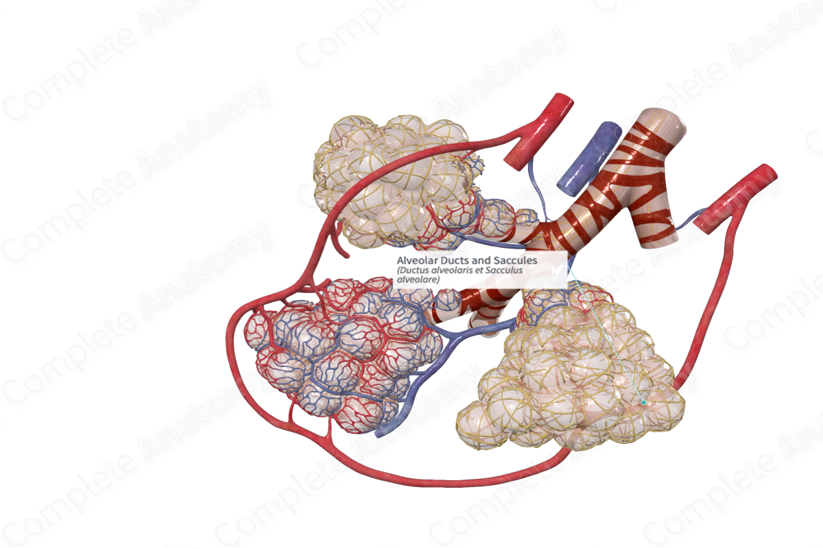
Alveolar ducts are the small passages connecting the respiratory bronchioles and the alveolar sacs. The alveolar saccules are the spaces into which the alveolar ducts open distally, and with which the alveoli communicate (Dorland, 2011).
Related parts of the anatomy
Structure/Morphology
The terminal part of the respiratory bronchioles give rise to alveolar ducts and saccules, or sacs. The walls of these structures are extremely thin, which allows respiration to occur.
The alveolar sacs are made of numerous alveoli, separated from each other by a thin interalveolar septum. A communication remains open between the alveoli through small alveolar pores, or pores of Köhn.
The cellular component of the alveoli is of a simple squamous type supported on a complete basal lamina. Type I pneumocytes are the most abundant cell type lining the alveolar walls. They are in close contact with the endothelial lining of the blood capillaries which facilitates gas exchange.
Interspersed between the type I pneumocytes are the type II pneumocytes, which may be in isolation, or found in small clusters. This cell population are secretory cells that synthesize and secrete surfactant. Surfactant spreads over the alveolar surface, reducing the surface tension within the alveoli. In addition, type II pneumocytes act as stem cells, replacing the type I pneumocytes where necessary (Eroschenko, 2008).
Scavenging macrophages (or dust cells) are found in the connective tissue of the alveolar walls and interalveolar septa. They remove debris from the alveoli and are often called dust cells because of the carbon deposits visible in their cytoplasm after ingestion (Ovalle et al., 2013).
Function
This is the point where maximum gas exchange occurs between the blood and the respiratory system. The capillaries form a special structure with the alveoli, the blood-air barrier, or alveolar-capillary membrane. The basal lamina of the squamous epithelium of the alveoli and the endothelium of the capillaries are fused, thus forming a membrane that allows gas to diffuse across it. This region is less than 2 μm wide (Ovalle et al., 2013, Simionescu, 2001).
References
Dorland, W. (2011) Dorland's Illustrated Medical Dictionary. 32nd edn. Philadelphia, USA: Elsevier Saunders.
Eroschenko, V. P. (2008) DiFiore's Atlas of Histology with Functional Correlations. Point (Lippincott Williams and Wilkins) Series: Wolters Kluwer Health/Lippincott Williams & Wilkins.
Ovalle, W. K., Nahirney, P. C. and Netter, F. H. (2013) Netter's Essential Histology. ClinicalKey 2012: Elsevier Saunders.
Simionescu, M. (2001) 'Cellular components of the air-blood barrier', J Cell Mol Med, 5(3), pp. 320-1.
