
Deep Anterior Lamellar Keratoplasty (DALK)
Home / External Disease and Cornea / Clinical Approach to Corneal Transplantation
Title: Deep anterior lamellar keratoplasty (DALK)
Author (s): Rhett Thomson, MS4, and Mark Mifflin, MD
Photographer: Mark Mifflin, MD, and Ethan Peterson
Date: 8/29/20
Video:
Keywords/Main Subjects: Deep anterior lamellar keratoplasty; DALK; Corneal Transplantation
Overview:
- Utilized for anterior corneal pathology (eg, keratoconus)1,2
- Procedure involves replacing anterior lamellae while keeping Descemets membrane (DM) and endothelium
- Outcomes are often compared to those of penetrating keratoplasty (PK)3-5
- Advantages of DALK:
- Endothelial rejection risk avoided6
- Ideal in young, healthy patients with active immune systems
- High quality donor tissue not required (helpful in resource poor areas)
- Disadvantages of DALK:
- Historically, non-smooth contour from residual lamellar dissection resulted in poorer visual outcomes
- Rupturing DM is a common complication7
- Complications often necessitate conversion to a full PK8
- Recent improvements:
- “Big bubble” technique increases smooth contour leading to better visual outcomes9,10
- “Big bubble” technique involves separating anterior lamella from Descemets using air, decreasing the chance of rupturing Descemets
- Manual intrastromal dissection can be performed as an alternative to “big bubble” technique
Description of video: Steps of DALK explained11
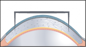
- The geographic center of the cornea is marked
- An appropriately sized vacuum trephine is selected and set to about ⅔ corneal thickness
- Cautery of the anterior surface is generally avoided in DALK

- Cautery of the anterior surface is generally avoided in DALK
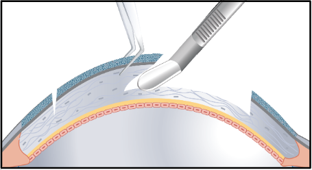
- A stainless-steel crescent blade is used to remove about half of the anterior stroma, making later removal of more posterior tissue easier


- The anterior chamber is decompressed slightly
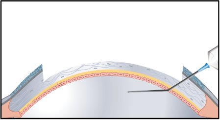
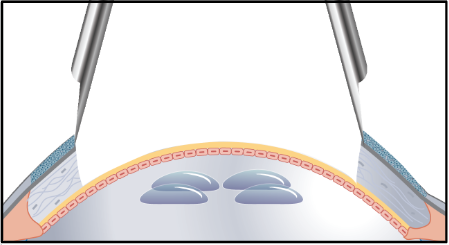
- Small indicator air bubbles are injected into the anterior chamber


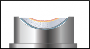
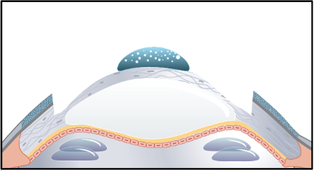
- Create a “big bubble” to separate Descemets membrane and endothelium from the anterior stroma


- As Descemets is pushed deeper, the air bubbles in the anterior chamber are often seen getting moved to the periphery

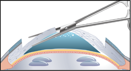
- Apply dispersive viscoelastic in advance of puncturing the lamella covering the “big bubble”

- Puncture the remaining stroma while avoiding perforation of Descemets

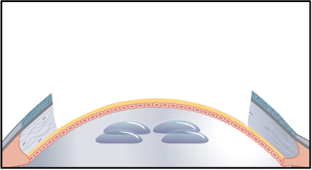
- Following the puncture of the “big bubble”, Descemets will once again move to its more natural anterior position
- This allows the air bubbles in the anterior chamber to return from the periphery to the center

- This allows the air bubbles in the anterior chamber to return from the periphery to the center
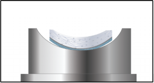
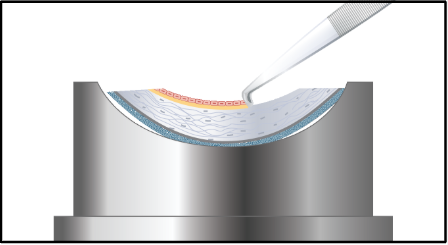
- Between Descemets and remaining anterior lamella, apply dispersive OVD (ophthalmic viscosurgical device)

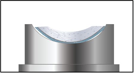
- Cut remaining stroma into four quadrants and manually cut away, often using curved cornea scissors


- Calipers are used to verify bed diameter prior to preparing and punching donor tissue

- Trypan blue is use to stain donor Descemets which is then removed
- Remove Descemets and endothelium from donor tissue


- Trephination of donor cornea

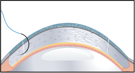
- Place donor graft on host
- Donor tissue is typically cut to the same diameter, or smaller if needed to decrease myopia in a keratoconus patient


- Donor tissue is typically cut to the same diameter, or smaller if needed to decrease myopia in a keratoconus patient
- Suture graft using standard techniques


References:
- Al-Torbak AA, Al-Motowa S, Al-Assiri Aet al. Deep anterior lamellar keratoplasty for keratoconus. Cornea 2006; 25:408-412.
- Mohammadpour M, Heidari Z, Hashemi H. Updates on Managements for Keratoconus. Journal of current ophthalmology 2018; 30:110-124.
- Song Y, Zhang J, Pan Z. Systematic Review and Meta-Analysis of Clinical Outcomes of Penetrating Keratoplasty Versus Deep Anterior Lamellar Keratoplasty for Keratoconus. Experimental and clinical transplantation : official journal of the Middle East Society for Organ Transplantation 2020; 18:417-428.
- Keane M, Coster D, Ziaei M, Williams K. Deep anterior lamellar keratoplasty versus penetrating keratoplasty for treating keratoconus. The Cochrane database of systematic reviews 2014:Cd009700.
- Pedrotti E, Passilongo M, Fasolo A, Ficial S, Ferrari S, Marchini G. Refractive outcomes of penetrating keratoplasty and deep anterior lamellar keratoplasty in fellow eyes for keratoconus. International ophthalmology 2017; 37:911-919.
- Hos D, Matthaei M, Bock Fet al. Immune reactions after modern lamellar (DALK, DSAEK, DMEK) versus conventional penetrating corneal transplantation. Progress in retinal and eye research 2019; 73:100768.
- Huang OS, Htoon HM, Chan AM, Tan D, Mehta JS. Incidence and Outcomes of Intraoperative Descemet Membrane Perforations During Deep Anterior Lamellar Keratoplasty. American journal of ophthalmology 2019; 199:9-18.
- Donoso R, Díaz C, Villavicencio P. Comparative study of keratoconus between Anwar’s deep anterior lamellar keratoplasty versus converted penetrating keratoplasty. Archivos de la Sociedad Espanola de Oftalmologia 2015; 90:257-263.
- DUA HS. Know your Bubbles. Acta ophthalmologica 2016; 94.
- Dua HS, Faraj LA, Kenawy MBet al. Dynamics of big bubble formation in deep anterior lamellar keratoplasty by the big bubble technique: in vitro studies. Acta ophthalmologica 2018; 96:69-76.
- Nanavaty MA, Vijjan KS, Yvon C. Deep anterior lamellar keratoplasty: A surgeon’s guide. Journal of current ophthalmology 2018; 30:297-310.
Faculty Approval by: Mark Mifflin, M.D.
Footer:
- Copyright statement: Copyright Author Name, ©2016. For further information regarding the rights to this collection, please visit: URL to copyright information page on Moran CORE
- Identifier: Moran_CORE_29794
Disclosure (Financial or other):