Hamilton TK, Baughman RD, Perry AE. Persistent pruritic plaque of the ear. Arch Dermatol. 1999 Apr. 135(4):464-5, 467-8. [QxMD MEDLINE Link].
Arnold M, Geilen CC, Coupland SE, Krengel S, Dippel E, Spröder J, et al. Unilateral angiolymphoid hyperplasia with eosinophilia involving the left arm and hand. J Cutan Pathol. 1999 Oct. 26(9):436-40. [QxMD MEDLINE Link].
Chen JF, Gao HW, Wu BY, Tsai WC, Chiang CP. Angiolymphoid hyperplasia with eosinophilia affecting the scrotum: a rare case report with molecular evidence of T-cell clonality. J Dermatol. 2010 Apr. 37(4):355-9. [QxMD MEDLINE Link].
Chan JK, Hui PK, Ng CS, Yuen NW, Kung IT, Gwi E. Epithelioid haemangioma (angiolymphoid hyperplasia with eosinophilia) and Kimura's disease in Chinese. Histopathology. 1989 Dec. 15(6):557-74. [QxMD MEDLINE Link].
Googe PB, Harris NL, Mihm MC Jr. Kimura's disease and angiolymphoid hyperplasia with eosinophilia: two distinct histopathological entities. J Cutan Pathol. 1987 Oct. 14(5):263-71. [QxMD MEDLINE Link].
Tsuboi H, Masuzawa M, Katsuoka K. Angiolymphoid hyperplasia with eosinophilia affecting the nail bed and underlying bone. J Dermatol. 2006 Jun. 33(6):399-402. [QxMD MEDLINE Link].
Esmaili DD, Chang EL, O'Hearn TM, Smith RE, Rao NA. Simultaneous presentation of Kimura disease and angiolymphoid hyperplasia with eosinophilia. Ophthal Plast Reconstr Surg. 2008 Jul-Aug. 24(4):310-1. [QxMD MEDLINE Link].
Zarrin-Khameh N, Spoden JE, Tran RM. Angiolymphoid hyperplasia with eosinophilia associated with pregnancy: a case report and review of the literature. Arch Pathol Lab Med. 2005 Sep. 129(9):1168-71. [QxMD MEDLINE Link].
Marcum CB, Zager JS, Bélongie IP, Messina JL, Fenske NA. Profound proliferating angiolymphoid hyperplasia with eosinophilia of pregnancy mimicking angiosarcoma. Cutis. 2011 Sep. 88(3):122-8. [QxMD MEDLINE Link].
Angiolymphoid Hyperplasia with Eosinophilia and Kimura Disease. Wolff, Goldsmith, Katz, Gilchrest, Paller, Leffell, eds. Fitzpatrick's Dermatology in General Medicine. 7th ed. New York, NY: McGraw-Hill Medical; 2008. Vol 1: 313-14.
Joshi R. Angiolymphoid hyperplasia with follicular mucinosis. Indian J Dermatol Venereol Leprol. 2007 Sep-Oct. 73(5):346-7. [QxMD MEDLINE Link].
Kempf W, Haeffner AC, Zepter K, et al. Angiolymphoid hyperplasia with eosinophilia: evidence for a T-cell lymphoproliferative origin. Hum Pathol. 2002 Oct. 33(10):1023-9. [QxMD MEDLINE Link].
Andreae J, Galle C, Magdorf K, et al. Severe atherosclerosis of the aorta and development of peripheral T-cell lymphoma in an adolescent with angiolymphoid hyperplasia with eosinophilia. Br J Dermatol. 2005 May. 152(5):1033-8. [QxMD MEDLINE Link].
Gonzalez-Cuyar LF, Tavora F, Zhao XF, Wang G, Auerbach A, Aguilera N, et al. Angiolymphoid hyperplasia with eosinophilia developing in a patient with history of peripheral T-cell lymphoma: evidence for multicentric T-cell lymphoproliferative process. Diagn Pathol. 2008 May 29. 3:22. [QxMD MEDLINE Link].
Jeon EK, Cho AY, Kim MY, Lee Y, Seo YJ, Park JK, et al. Angiolymphoid hyperplasia with eosinophilia that was possibly induced by vaccination in a child. Ann Dermatol. 2009 Feb. 21(1):71-4. [QxMD MEDLINE Link]. [Full Text].
Korekawa A, Kaneko T, Hagiwara C, et al. Angiolymphoid hyperplasia with eosinophilia in infancy. J Dermatol. 2012 May 17. [QxMD MEDLINE Link].
Adler BL, Krausz AE, Minuti A, Silverberg JI, Lev-Tov H. Epidemiology and treatment of angiolymphoid hyperplasia with eosinophilia (ALHE): A systematic review. J Am Acad Dermatol. 2016 Mar. 74 (3):506-12.e11. [QxMD MEDLINE Link]. [Full Text].
Wang YH, Yin HF. [One patient with Kimura's disease and angiolymphoid hyperplasia with eosinophilia also suffers from kidney injury]. Beijing Da Xue Xue Bao. 2008 Aug 18. 40(4):405-7. [QxMD MEDLINE Link].
Dewan P, Francis ND, Lear JT, Bunker CB. Angiolymphoid hyperplasia with eosinophilia affecting the penis. Br J Dermatol. Sept 2008. 159(3):755-7. [QxMD MEDLINE Link].
Huang M, Lloyd WC 3rd, O'Hara M. Angiolymphoid hyperplasia with eosinophilia: an unusual presentation in a child. J AAPOS. Jun 2008. 12(3):302-4. [QxMD MEDLINE Link].
Hamaguchi Y, Fujimoto M, Matsushita Y, Kitamura-Sawada S, Kawano M, Takehara K. IgG4-related skin disease, a mimic of angiolymphoid hyperplasia with eosinophilia. Dermatology. 2011. 223(4):301-5. [QxMD MEDLINE Link].
Llamas-Velasco M, Kempf W, Cota C, Fernández-Figueras MT, Lee J, Ferrara G, et al. Multiple Eruptive Epithelioid Hemangiomas: A Subset of Cutaneous Cellular Epithelioid Hemangioma With Expression of FOS-B. Am J Surg Pathol. 2019 Jan. 43 (1):26-34. [QxMD MEDLINE Link].
Ortins-Pina A, Llamas-Velasco M, Turpin S, Soares-de-Almeida L, Filipe P, Kutzner H. FOSB immunoreactivity in endothelia of epithelioid hemangioma (angiolymphoid hyperplasia with eosinophilia). J Cutan Pathol. 2018 Jun. 45 (6):395-402. [QxMD MEDLINE Link].
Lembo S, Balato A, Cirillo T, Balato N. A Long-Term Follow-Up of Angiolymphoid Hyperplasia with Eosinophilia Treated by Corticosteroids: When a Traditional Therapy is Still Up-to-Date. Case Rep Dermatol. 2011 Mar 5. 3(1):64-7. [QxMD MEDLINE Link]. [Full Text].
Horst C, Kapur N. Propranolol: a novel treatment for angiolymphoid hyperplasia with eosinophilia. Clin Exp Dermatol. 2014 Oct. 39(7):810-2. [QxMD MEDLINE Link].
Chacon AH, Mercer J. Successful management of angiolymphoid hyperplasia with eosinophilia in a split-face trial of topical tacrolimus and timolol solution. G Ital Dermatol Venereol. 2014 Jul 11. [QxMD MEDLINE Link].
Isohisa T, Masuda K, Nakai N, Takenaka H, Katoh N. Angiolymphoid hyperplasia with eosinophilia treated successfully with imiquimod. Int J Dermatol. 2012 May 16. [QxMD MEDLINE Link].
Shenefelt PD, Rinker M, Caradonna S. A case of angiolymphoid hyperplasia with eosinophilia treated with intralesional interferon alfa-2a. Arch Dermatol. 2000 Jul. 136(7):837-9. [QxMD MEDLINE Link].
Harada S, Nomura T, Miyauchi T, Shinkuma S, Fujita Y, Arita K, et al. Complete remission of angiolymphoid hyperplasia with eosinophilia using topical tacrolimus. Eur J Dermatol. 2017 Apr 1. 27 (2):194-196. [QxMD MEDLINE Link].
Braun-Falco M, Fischer S, Plötz SG, Ring J. Angiolymphoid hyperplasia with eosinophilia treated with anti-interleukin-5 antibody (mepolizumab). Br J Dermatol. 2004 Nov. 151(5):1103-4. [QxMD MEDLINE Link].
Carlesimo M, Mari E, Tammaro A, Persechino S, Camplone G. Angiolymphoid hyperplasia with eosinophilia treated with isotretinoin. Eur J Dermatol. 2007 Nov-Dec. 17(6):554-5. [QxMD MEDLINE Link].
Abrahamson TG, Davis DA. Angiolymphoid hyperplasia with eosinophilia responsive to pulsed dye laser. J Am Acad Dermatol. 2003 Aug. 49(2 Suppl Case Reports):S195-6. [QxMD MEDLINE Link].
Sotiriou E, Apalla Z, Patsatsi A, Panagiotidou DD, Ioannides D. Angiolymphoid hyperplasia with eosinophilia: good response to photodynamic therapy. Clin Exp Dermatol. 2009 Dec. 34(8):e629-31. [QxMD MEDLINE Link].
Wozniacka A, Omulecki A, Torzecka JD. Cryotherapy in the treatment of angiolymphoid hyperplasia with eosinophilia. Med Sci Monit. 2003 Jan. 9(1):CS1-4. [QxMD MEDLINE Link].
Singh S, Dayal M, Walia R, Arava S, Sharma R, Gupta S. Intralesional radiofrequency ablation for nodular angiolymphoid hyperplasia on forehead: A minimally invasive approach. Indian J Dermatol Venereol Leprol. 2014 Sep-Oct. 80(5):419-21. [QxMD MEDLINE Link].
Tambe SA, Nayak CS. Successful Management of Angiolymphoid Hyperplasia with Eosinophilia by Radiofrequency. J Cutan Aesthet Surg. 2017 Apr-Jun. 10 (2):116-118. [QxMD MEDLINE Link].
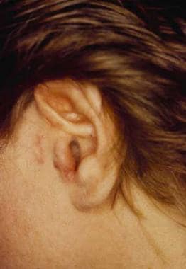 Angiolymphoid hyperplasia with eosinophilia typically exhibits flesh-color to erythematous nodules in the vicinity of the ear.
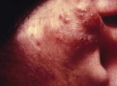 Pronounced erythema and nodularity due to angiolymphoid hyperplasia with eosinophilia.


